- Featured Product
- KD/KO Validated
Cytokeratin 8 Polyclonal antibody
Cytokeratin 8 Polyclonal Antibody for FC, IF/ICC, IHC, IP, WB, ELISA
Host / Isotype
Rabbit / IgG
Reactivity
human, mouse, rat and More (3)
Applications
FC, IF/ICC, IHC, IP, WB, ELISA
Conjugate
Unconjugated
验证数据展示
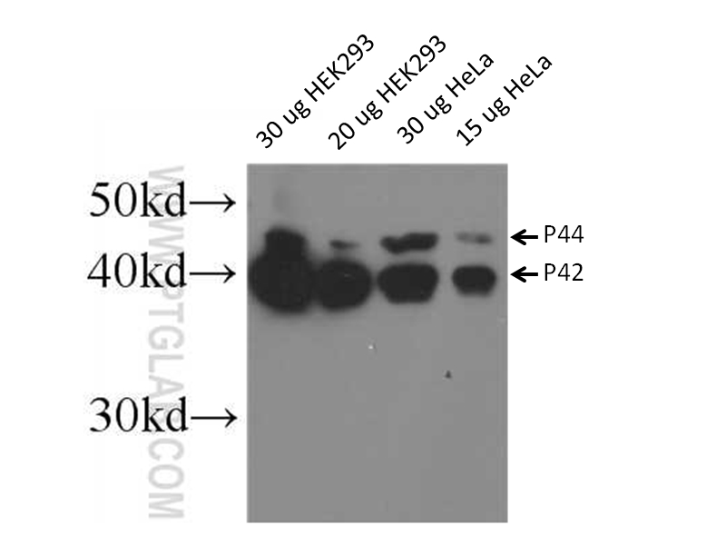
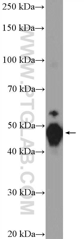
 at dilution of 1:30000 incubated at room temperature for 1.5 hours.")
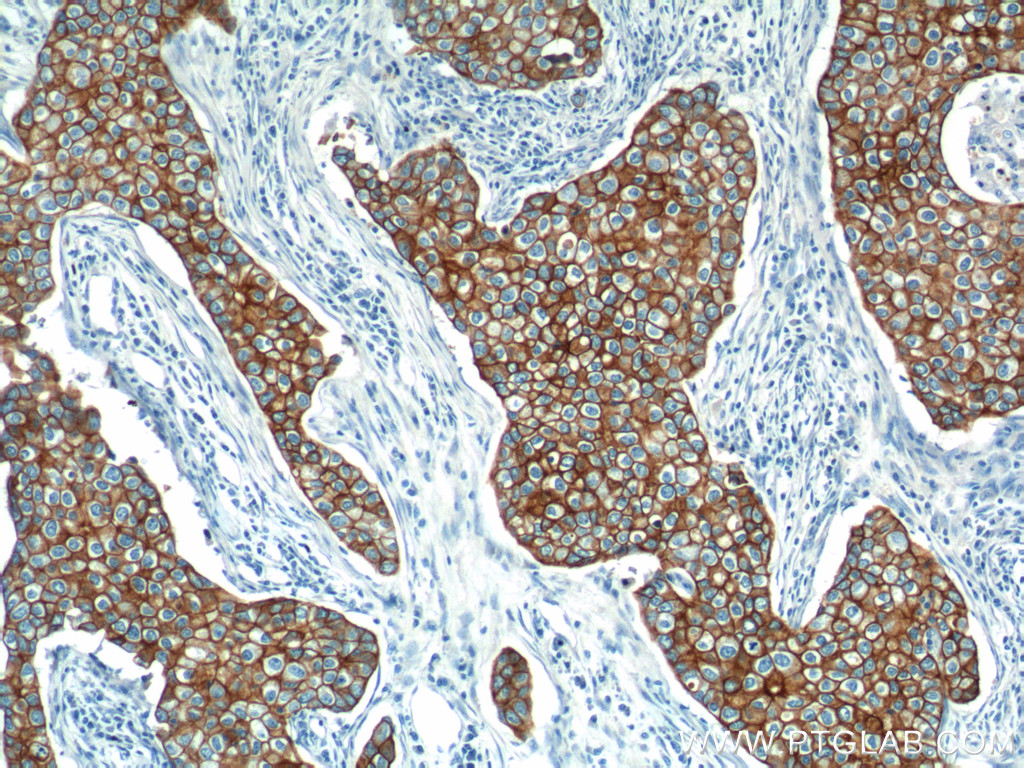
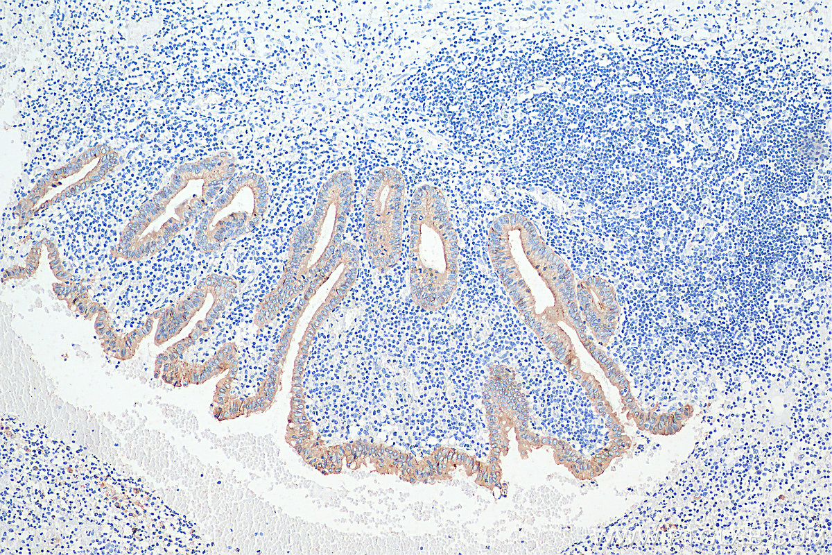
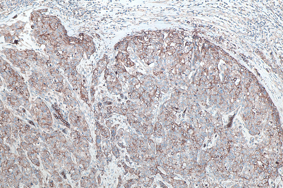
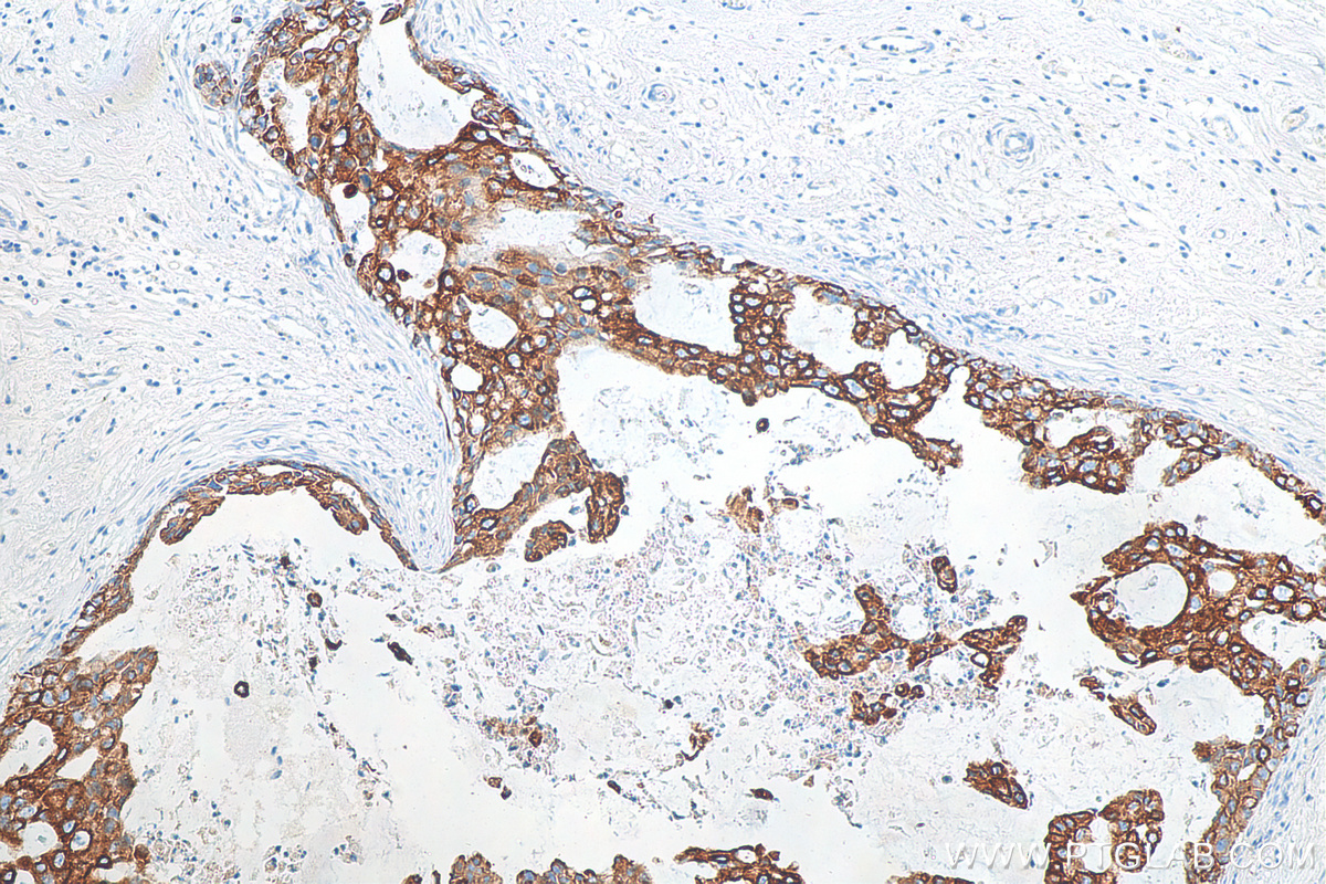
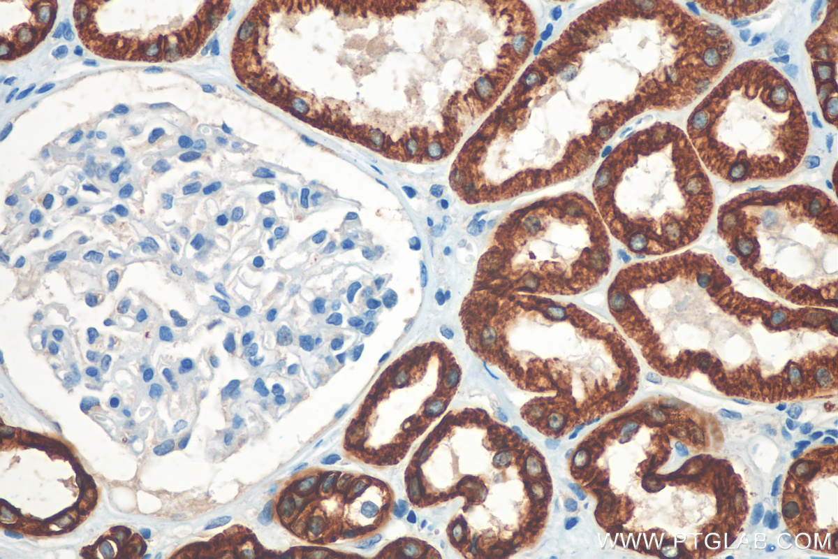
 at dilution of 1:2000 (under 40x lens). Heat mediated antigen retrieval with Tris-EDTA buffer (pH 9.0).")
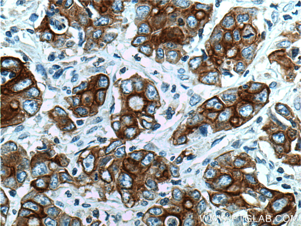
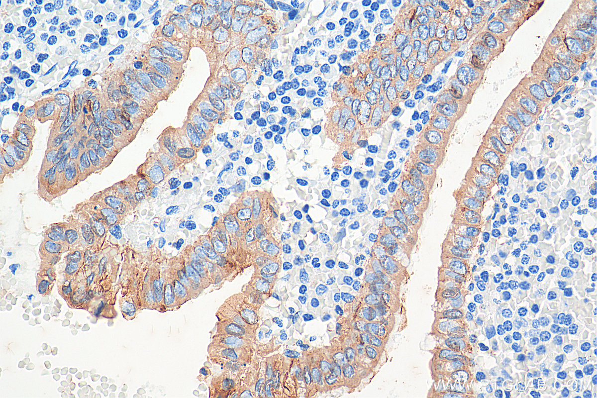
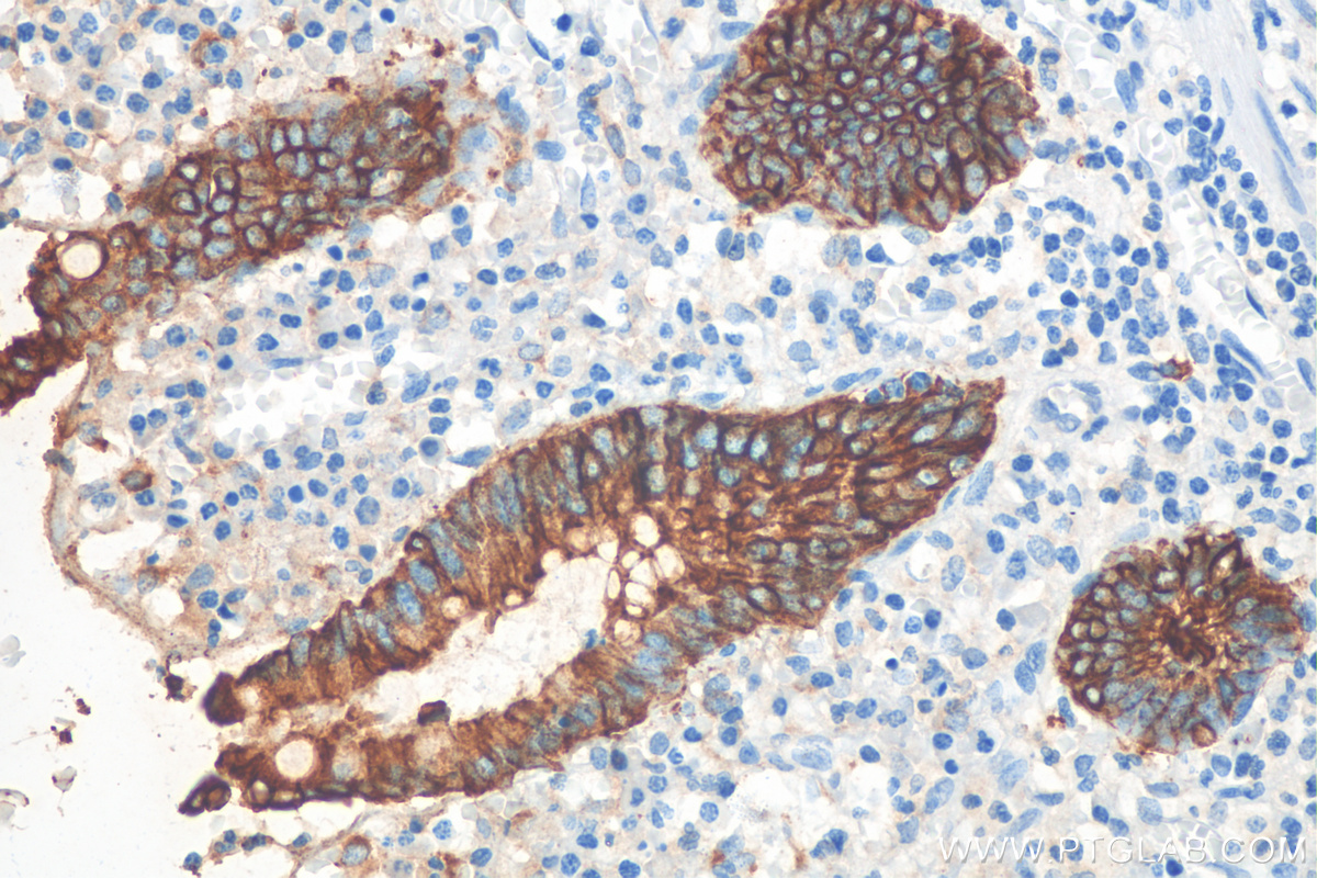
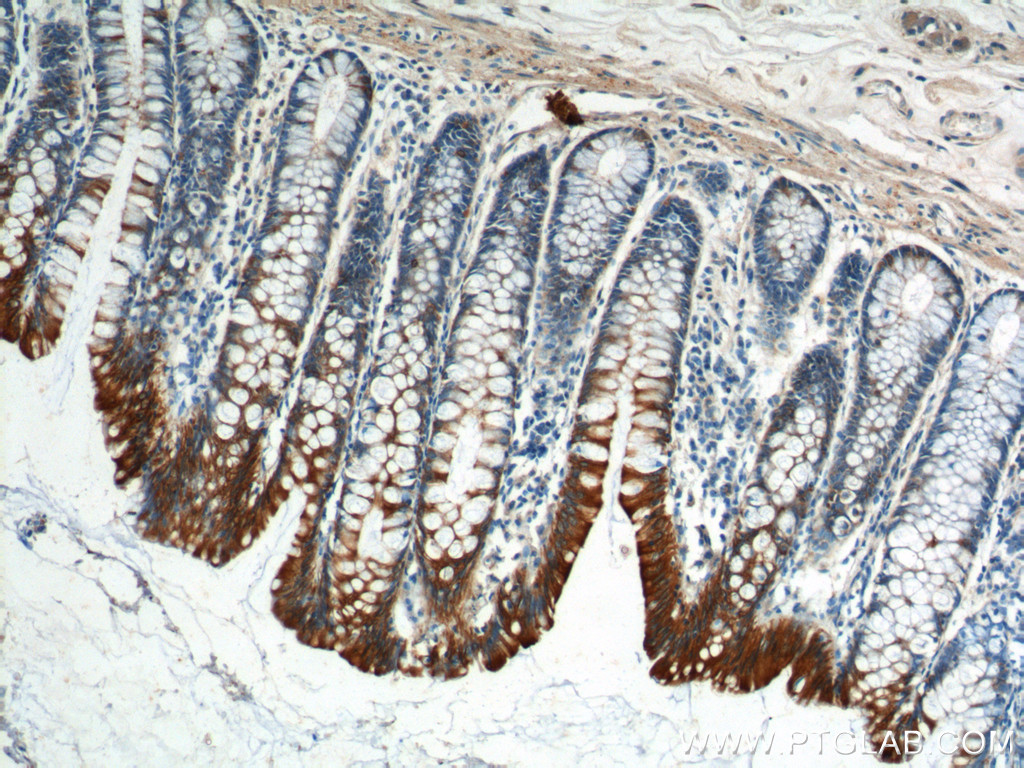
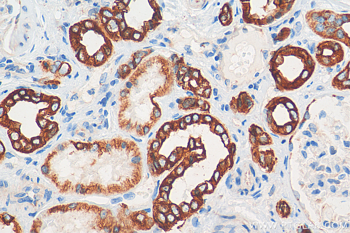
 at dilution of 1:2000 (under 10x lens). Heat mediated antigen retrieval with Tris-EDTA buffer (pH 9.0).")
 fixed <a class='green' href='/productredirect?CatalogNo=A431' target='_blank'>A431</a> cells using Cytokeratin 8 antibody (<a class='green' href='/productredirect?CatalogNo=10384-1-AP' target='_blank'>10384-1-AP</a>) at dilution of 1:400 and CoraLite®488-Conjugated AffiniPure Goat Anti-Rabbit IgG(H+L), CoraLite®594 Beta Actin antibody (<a class='green' href='/productredirect?CatalogNo=CL594-66009' target='_blank'>CL594-66009</a>, Clone: 2D4H5, red).")
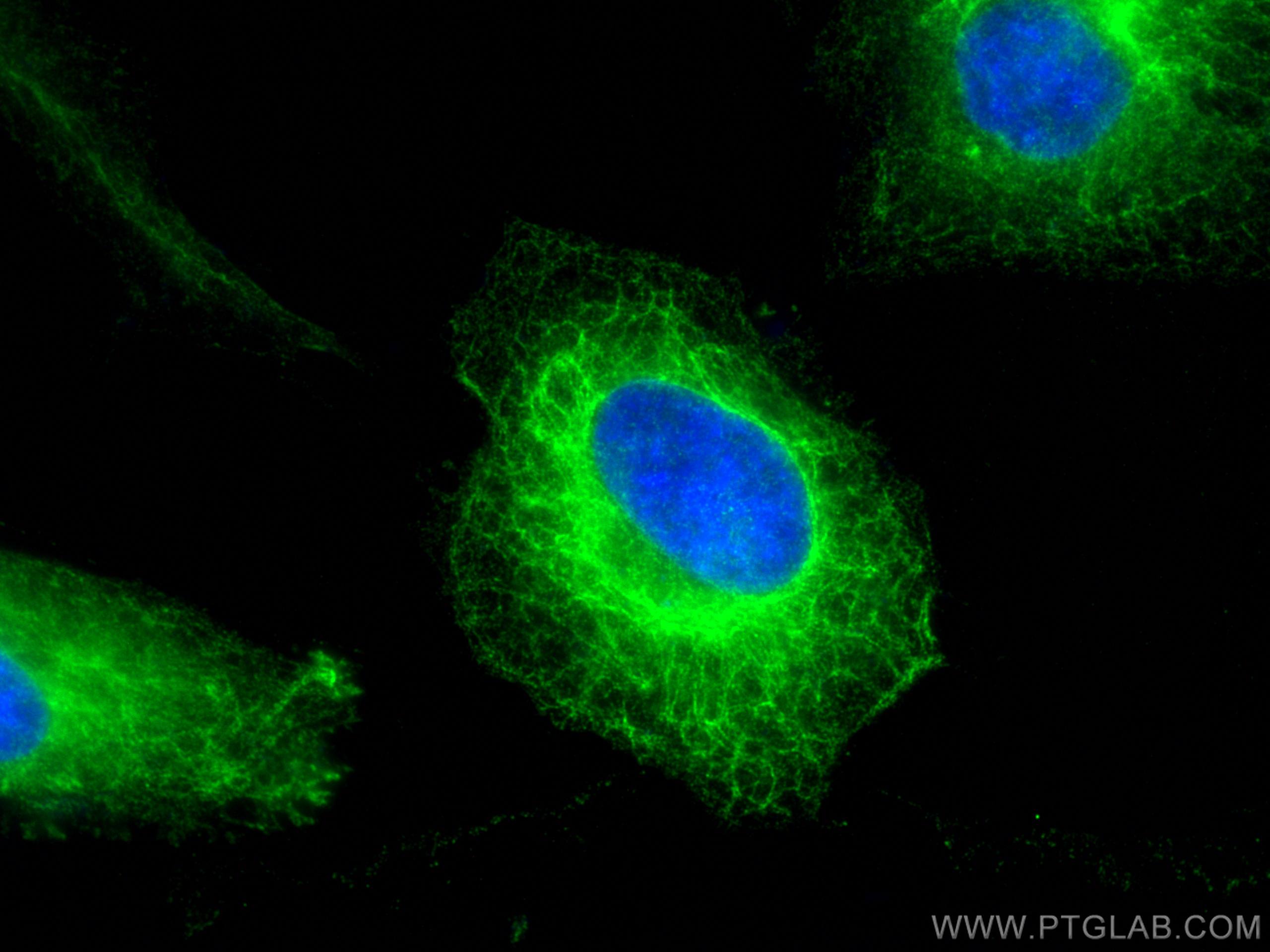
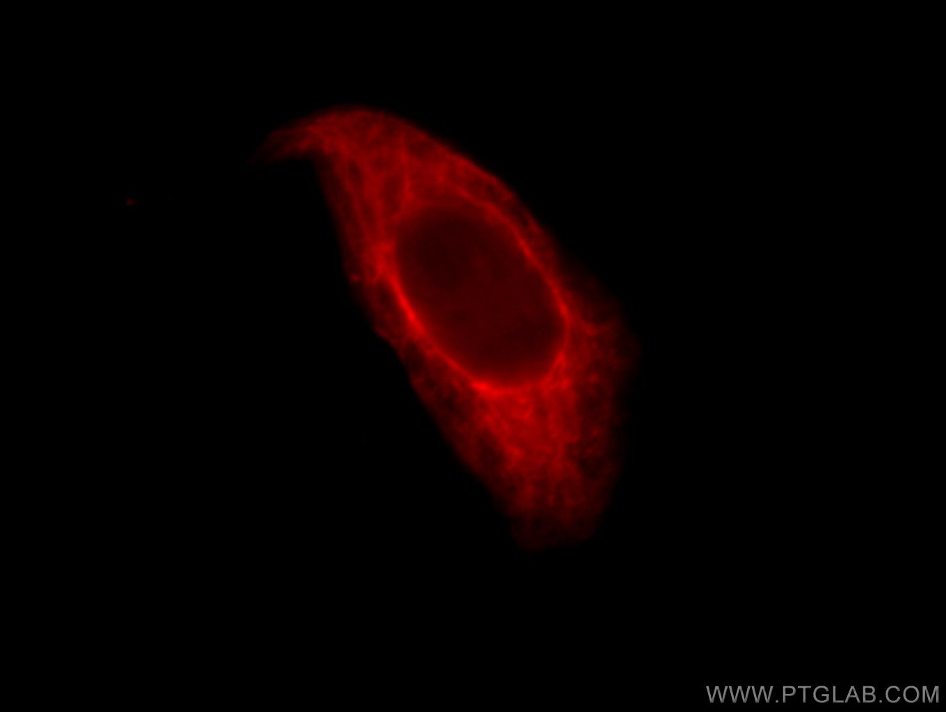
 and control antibody (blue). Fixed with 90% MeOH blocked with 3% BSA (30 min). Alexa Fluor 488-conjugated AffiniPure Goat Anti-Rabbit IgG(H+L) with dilution 1:1000.")
 fixed human breast cancer tissue using <a class='green' href='/productredirect?CatalogNo=10384-1-AP' target='_blank'>10384-1-AP</a> (Cytokeratin 8 antibody) at dilution of 1:50 and Alexa Fluor 488-conjugated AffiniPure Goat Anti-Rabbit IgG(H+L).")
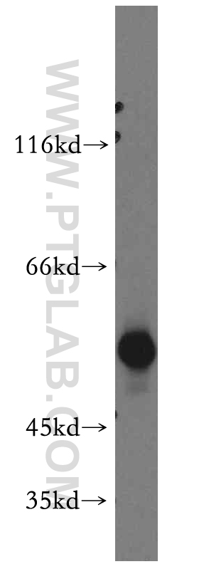
 with HeLa cells lysate 2000ug.")
经过测试的应用
| Positive WB detected in | HeLa cells, HaCaT cells, COLO 320 cells, A431 cells, A549 cells, MCF-7 cells, mouse skin tissue, rat skin tissue |
| Positive IP detected in | HeLa cells |
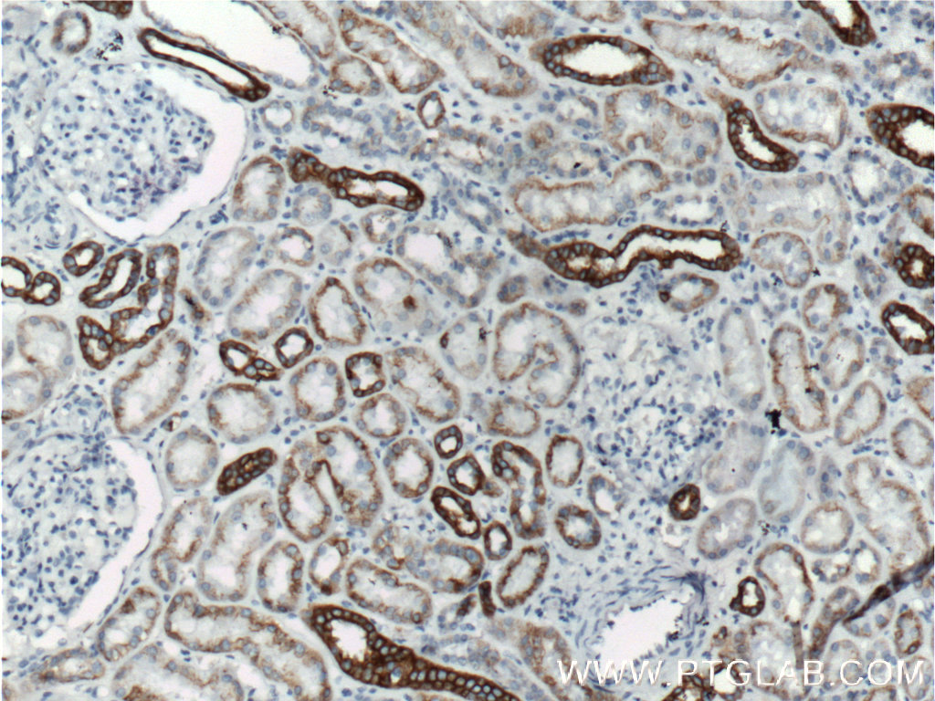
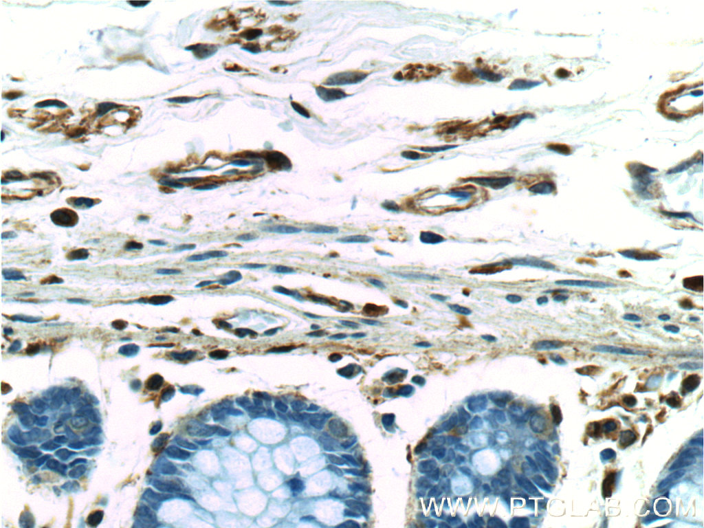
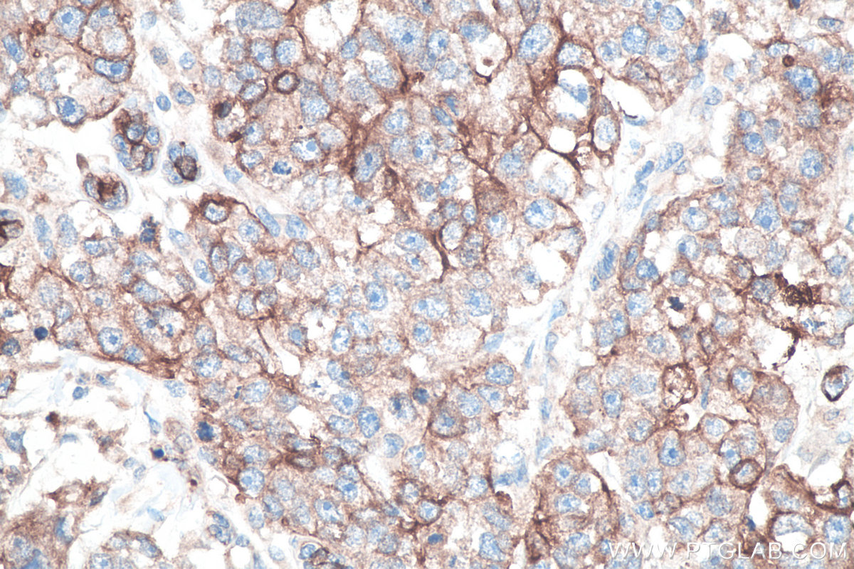
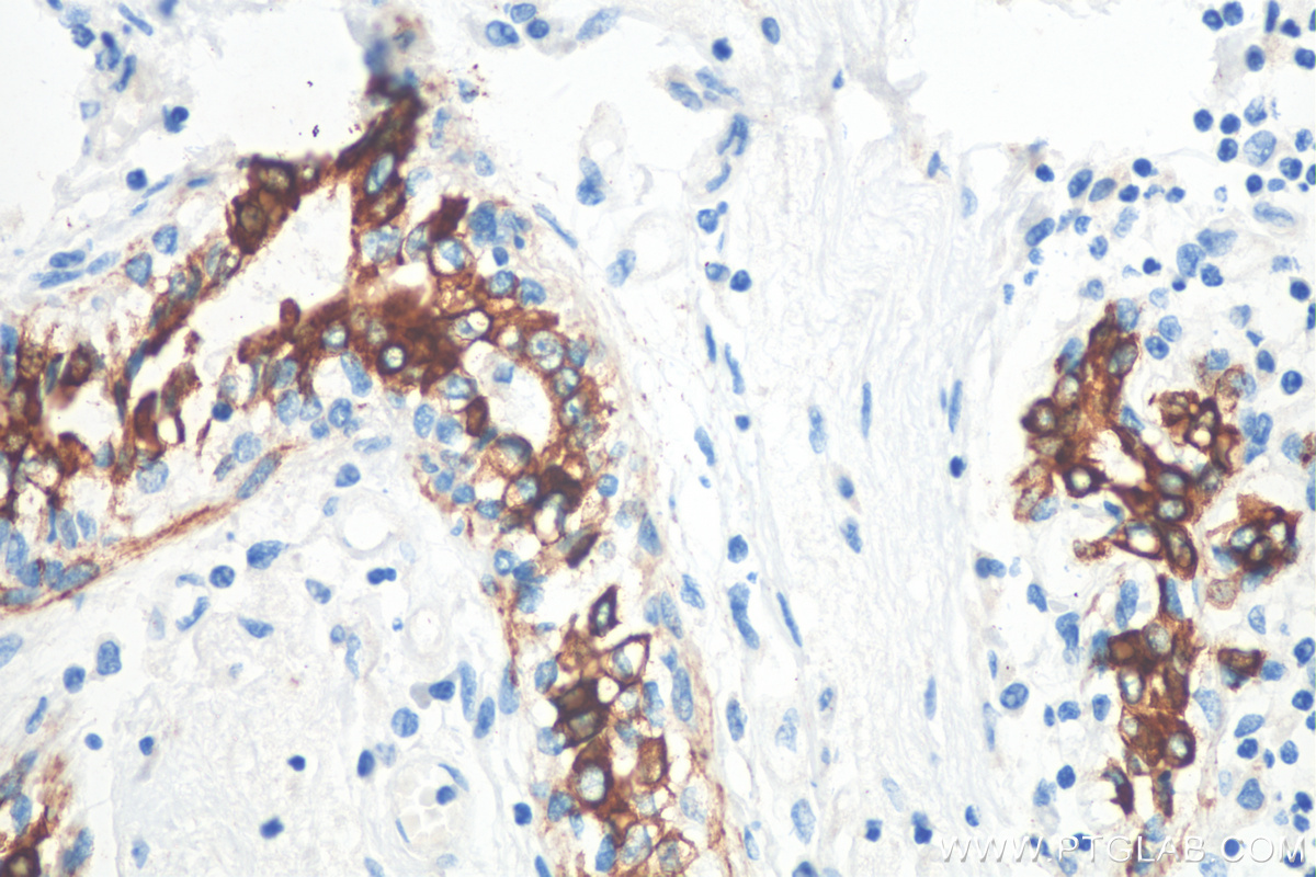
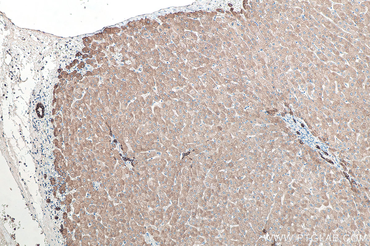
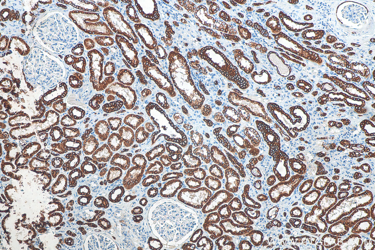
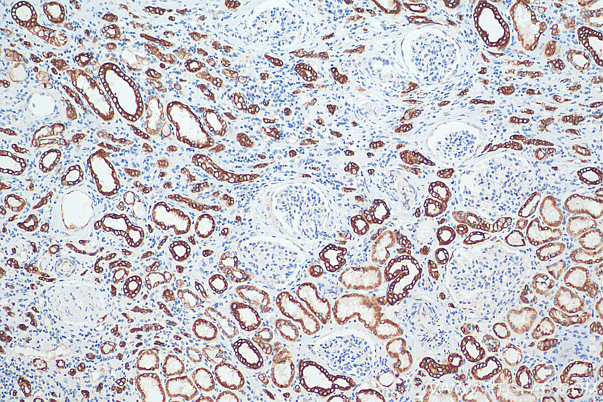
| Positive IHC detected in | human tonsillitis tissue, human colon tissue, human appendicitis tissue, human breast cancer tissue, human renal cell carcinoma tissue, human liver tissue Note: suggested antigen retrieval with TE buffer pH 9.0; (*) Alternatively, antigen retrieval may be performed with citrate buffer pH 6.0 |
| Positive IF detected in | A431 cells, HepG2 cells, human breast cancer tissue, HeLa cells |
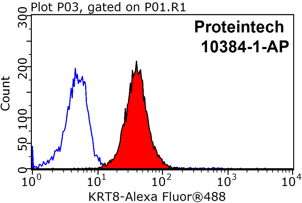
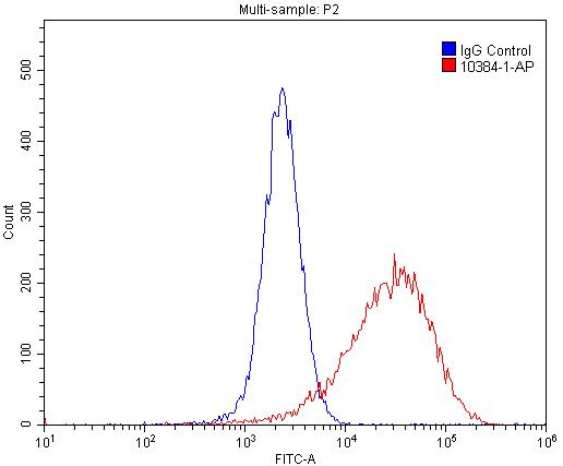
| Positive FC detected in | HepG2 cells, MCF-7 cells |
Planning an IHC experiment? We recommend our IHCeasy CK8 Ready-To-Use IHC Kit. CK8 primary antibody included.
Planning an IF experiment? We recommend our CoraLite® Plus 488 conjugated versions of this antibody.
推荐稀释比
| Application | Dilution |
|---|---|
| Western Blot (WB) | WB : 1:5000-1:40000 |
| Immunoprecipitation (IP) | IP : 0.5-4.0 ug for 1.0-3.0 mg of total protein lysate |
| Immunohistochemistry (IHC) | IHC : 1:1000-1:4000 |
| Immunofluorescence (IF) | IF : 1:200-1:800 |
| Flow Cytometry (FC) | FC : 0.20 ug per 10^6 cells in a 100 µl suspension |
| It is recommended that this reagent should be titrated in each testing system to obtain optimal results. | |
| Sample-dependent, Check data in validation data gallery. | |
产品信息
10384-1-AP targets Cytokeratin 8 in WB, IP, IHC, IF, FC, ELISA applications and shows reactivity with human, mouse, rat samples.
| Tested Applications | FC, IF/ICC, IHC, IP, WB, ELISA |
| Cited Applications | IF, IHC, IP, WB |
| Tested Reactivity | human, mouse, rat |
| Cited Reactivity | human, mouse, rat, dog, goat, pig |
| Immunogen | Cytokeratin 8 fusion protein Ag0488 种属同源性预测 |
| Host / Isotype | Rabbit / IgG |
| Class | Polyclonal |
| Type | Antibody |
| Full Name | keratin 8 |
| Synonyms | CARD2, Cell and organelle markers, CK 8, CK8, CYK8, Cytokeratin 8, Cytoskeleton Marker, K2C8, K8, keratin 8, KO, KRT8, Type II keratin Kb8 |
| Calculated Molecular Weight | 54 kDa |
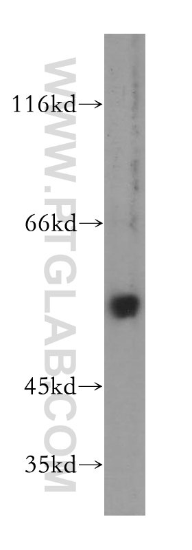
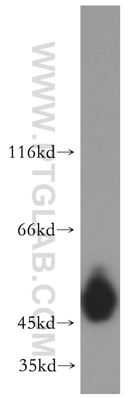
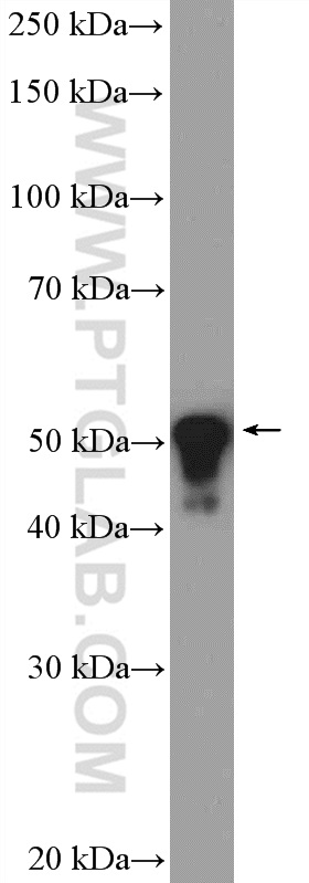
| Observed Molecular Weight | 52 kDa |
| GenBank Accession Number | BC000654 |
| Gene Symbol | KRT8 |
| Gene ID (NCBI) | 3856 |
| RRID | AB_10638912 |
| Conjugate | Unconjugated |
| Form | Liquid |
| Purification Method | Antigen affinity purification |
| UNIPROT ID | P05787 |
| Storage Buffer | PBS with 0.02% sodium azide and 50% glycerol pH 7.3. |
| Storage Conditions | Store at -20°C. Stable for one year after shipment. Aliquoting is unnecessary for -20oC storage. |
背景介绍
Keratins are a large family of proteins that form the intermediate filament cytoskeleton of epithelial cells, which are classified into two major sequence types. Type I keratins are a group of acidic intermediate filament proteins, including K9-K23, and the hair keratins Ha1-Ha8. Type II keratins are the basic or neutral courterparts to the acidic type I keratins, including K1-K8, and the hair keratins, Hb1-Hb6. KRT8 is often paired with keratin 18 in vivo.
实验方案
| Product Specific Protocols | |
|---|---|
| WB protocol for Cytokeratin 8 antibody 10384-1-AP | Download protocol |
| IHC protocol for Cytokeratin 8 antibody 10384-1-AP | Download protocol |
| IF protocol for Cytokeratin 8 antibody 10384-1-AP | Download protocol |
| IP protocol for Cytokeratin 8 antibody 10384-1-AP | Download protocol |
| Standard Protocols | |
|---|---|
| Click here to view our Standard Protocols |
发表文章
| Species | Application | Title |
|---|---|---|
J Cell Sci SNAP29 mediates the assembly of histidine-induced CTP synthase filaments in proximity to the cytokeratin network
| ||
Front Cell Dev Biol ESRRB Facilitates the Conversion of Trophoblast-Like Stem Cells From Induced Pluripotent Stem Cells by Directly Regulating CDX2. | ||
J Cell Sci SNAP29 mediates the assembly of histidine-induced CTP synthase filaments in proximity to the cytokeratin network.
| ||
Anat Rec (Hoboken) Comparative study of the external auditory canal in humans and large mammals. | ||
Oncotarget Alternative promotion and suppression of metastasis by JNK2 governed by its phosphorylation. | ||
Oncoscience The helix-loop-helix transcriptional regulator Id4 is required for terminal differentiation of luminal epithelial cells in the prostate. |