- Featured Product
- KD/KO Validated
CoraLite®594-conjugated GFAP Polyclonal antibody
GFAP Polyclonal Antibody for IF-P
Host / Isotype
Rabbit / IgG
Reactivity
human, mouse, rat
Applications
IF-P
Conjugate
CoraLite®594 Fluorescent Dye
验证数据展示
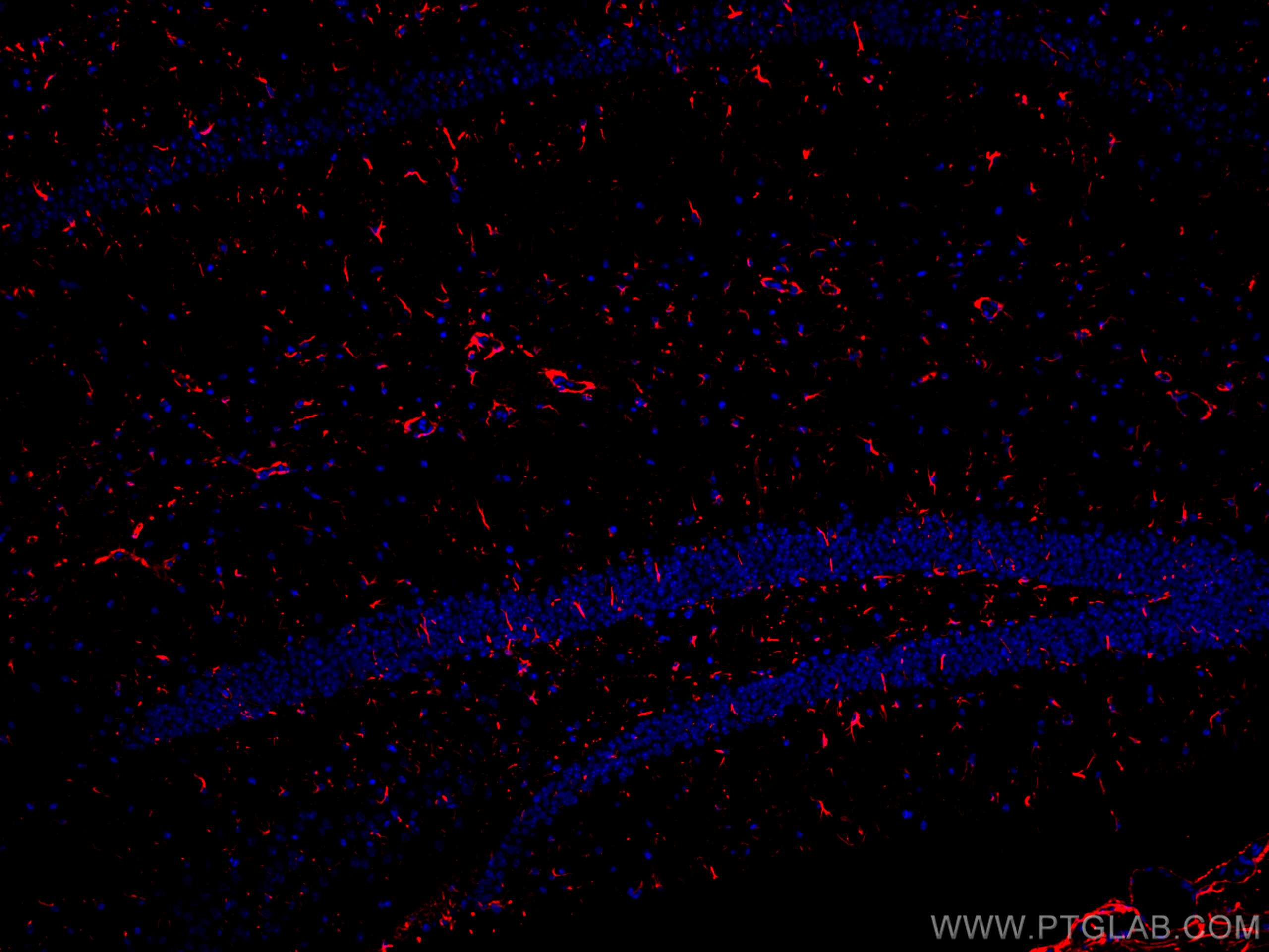
 fixed rat brain tissue using CoraLite®594 GFAP antibody (<a class='green' href='/productredirect?CatalogNo=CL594-16825' target='_blank'>CL594-16825</a>) at dilution of 1:200, CoraLite®488 MAP2 antibody (<a class='green' href='/productredirect?CatalogNo=CL488-17490' target='_blank'>CL488-17490</a>, green). DAPI (blue).")
经过测试的应用
| Positive IF detected in | rat brain tissue, mouse brain tissue |
Planning an IHC experiment? We recommend our IHCeasy GFAP Ready-To-Use IHC Kit. GFAP primary antibody included.
For other applications, we recommend the unconjugated version of this antibody, 16825-1-AP
推荐稀释比
| Application | Dilution |
|---|---|
| Immunofluorescence (IF) | IF : 1:50-1:500 |
| It is recommended that this reagent should be titrated in each testing system to obtain optimal results. | |
| Sample-dependent, Check data in validation data gallery. | |
产品信息
CL594-16825 targets GFAP in IF applications and shows reactivity with human, mouse, rat samples.
| Tested Applications | IF-P |
| Tested Reactivity | human, mouse, rat |
| Immunogen | GFAP fusion protein Ag10423 种属同源性预测 |
| Host / Isotype | Rabbit / IgG |
| Class | Polyclonal |
| Type | Antibody |
| Full Name | glial fibrillary acidic protein |
| Synonyms | FLJ45472, GFAP |
| Calculated Molecular Weight | 432 aa, 50 kDa |
| Observed Molecular Weight | 45-50 kDa |
| GenBank Accession Number | BC013596 |
| Gene Symbol | GFAP |
| Gene ID (NCBI) | 2670 |
| RRID | AB_2919838 |
| Conjugate | CoraLite®594 Fluorescent Dye |
| Excitation/Emission Maxima Wavelengths | 588 nm / 604 nm |
| Form | Liquid |
| Purification Method | Antigen affinity purification |
| UNIPROT ID | P14136 |
| Storage Buffer | PBS with 50% Glycerol, 0.05% Proclin300, 0.5% BSA, pH 7.3. |
| Storage Conditions | Store at -20°C. Avoid exposure to light. Aliquoting is unnecessary for -20oC storage. |
背景介绍
Function
GFAP (Glial fibrillary acidic protein) is a type III intermediate filament (IF) protein specific to the central nervous system (CNS). GFAP is one of the main components of the intermediate filament network in astrocytes and has been proposed as playing a role in cell migration, cell motility, maintaining mechanical strength, and in mitosis.Tissue specificity
GFAP is expressed in central nervous system cells, predominantly in astrocytes. GFAP is commonly used as an astrocyte marker. However, GFAP is also present in peripheral glia and in non-CNS cells, including fibroblasts, chondrocytes, lymphocytes, and liver stellate cells (PMID: 21219963).Involvement in disease
- Mutations in GFAP lead to Alexander disease (OMIM: 203450), an autosomal dominant CNS disorder. The mutations present in affected individuals are thought to be gain-of-function.
- Upregulation of GFAP is a hallmark of reactive astrocytes, in which GFAP is present in hypertrophic cellular processes. Reactive astrogliosis is present in many neurological disorders, such as stroke, various neurodegenerative diseases (including Alzheimer's and Parkinson's disease), and neurotrauma.
Isoforms
Astrocytes express 10 different isoforms of GFAP that differ in the rod and tail domains (PMID: 25726916), which means that they differ in molecular size. Isoform expression varies during the development and across different subtypes of astrocytes. Not all isoforms are upregulated in reactive astrocytes.Post-translational modifications
Intermediate filament proteins are regulated by phosphorylation. Six phosphorylation sites have been identified in GFAP protein, at least some of which are reported to control filament assembly (PMID: 21219963).Cellular localization
GFAP localizes to intermediate filaments and stains well in astrocyte cellular processes.实验方案
| Product Specific Protocols | |
|---|---|
| IF protocol for CL594 GFAP antibody CL594-16825 | Download protocol |
| Standard Protocols | |
|---|---|
| Click here to view our Standard Protocols |