Recombinant Human PDGFR beta/CD140b protein (Myc Tag, His Tag)
种属
Human
纯度
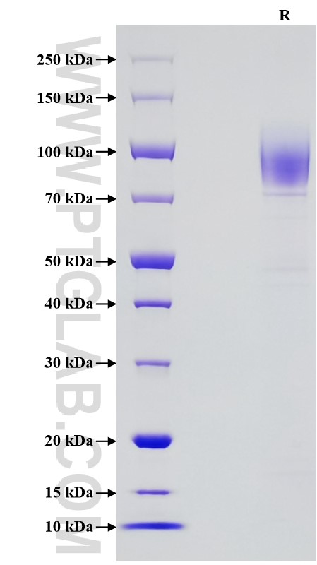
>90 %, SDS-PAGE
标签
Myc Tag, His Tag
生物活性
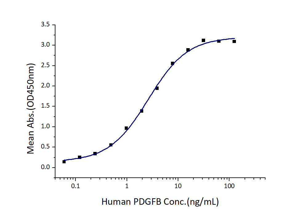
EC50: 1-5 ng/mL
验证数据展示
产品信息
| 纯度 | >90 %, SDS-PAGE |
| 内毒素 | <0.1 EU/μg protein, LAL method |
| 生物活性 | Immobilized Human PDGFR beta (Myc tag, His tag) at 0.5 μg/mL (100 μL/well) can bind Human PDGFB (hFc tag) with a linear range of 1-5 ng/mL. |
| 来源 | HEK293-derived Human PDGFR beta protein Leu33-Lys531 (Accession# P09619-1(S180F)) with a Myc tag and a His tag at the C-terminus. |
| 基因ID | 5159 |
| 蛋白编号 | P09619-1 |
| 预测分子量 | 61.6 kDa |
| SDS-PAGE | 75-110 kDa, reducing (R) conditions |
| 组分 | Lyophilized from 0.22 μm filtered solution in PBS, pH 7.4. Normally 5% trehalose and 5% mannitol are added as protectants before lyophilization. |
| 复溶 | Briefly centrifuge the tube before opening. Reconstitute at 0.1-0.5 mg/mL in sterile water. |
| 储存条件 |
It is recommended that the protein be aliquoted for optimal storage. Avoid repeated freeze-thaw cycles.
|
| 运输条件 | The product is shipped at ambient temperature. Upon receipt, store it immediately at the recommended temperature. |
背景信息
Multiple receptor tyrosine kinases and their growth factor ligands have been implicated in cancer progression and metastasis. Among these are the platelet-derived growth factor receptors (PDGFRs). Expression of PDGFRs is mainly restricted to mesenchymal cell types. PDGF is composed of homo-dimers or hetero-dimers of two polypeptide chains, denoted A and B. The two receptor subtypes show different affinities for the dimeric PDGF isoforms. In epithelial tumors, the platelet-derived growth factor receptor B (PDGFRB, also known as PDGFR beta) is mainly expressed by stromal cells of mesenchymal origin. it has also been shown that PDGFRB is also associated with the aggressive behavior of several types of tumors. The 60% of colon cancer patients express high levels of this gene and the PDGFRB expression correlates with lymphatic dissemination of this cancer. Furthermore, PDGFRB signaling in mesenchymal-like tumor cells (as colorectal cancer cells) contributes to invasion and liver metastasis formation.
参考文献:
1.Ernst J A Steller. et al. (2013). Neoplasia. 15(2):204-217.
2.Arne Ostman. et al. (2007). Adv Cancer Res. 97:247-274.
3.Ombretta Melaiu. et al. (2017). Genes Cancer. 8(1-2):438-452.
4.C E Hart. et al. (1988). Science. 240(4858):1529-1531.
5.Thomas C Wehler. et al. (2008). Oncol Rep. 19(3):697-704.

