Uni-rAb™
重组兔单克隆抗体
Uni-rAb™ 系列重组兔单克隆抗体是Proteintech最新一代抗体产品,基于ABCE™(Antigen-Specific B-Cell Cloning & Engineering)单个 B 细胞抗体发现技术平台而开发。该平台通过将兔源特异性B细胞中的抗体重链和轻链基因克隆至拥有自主产权的高表达哺乳动物细胞载体中,在体外实现重组单克隆抗体的稳定高表达。
Uni-rAb™ antibodies are the latest generation of recombinant monoclonal antibodies developed using Proteintech’s Antigen-Specific B-Cell Cloning & Engineering (ABCE™) platform. The ABCE™ platform achieves stable and high expression of recombinant monoclonal antibodies by cloning antibody heavy and light chain sequences derived from antigen-specific B cells into high-yield expression vectors followed by their introduction into suitable mammalian expression systems.
-

好抗体来自好抗原
-
高纯度,超低背景
-
高特异性
-
高批次间一致性
-
高亲和力
-
上新速度快
Uni-rAb™ 类别
ABCE™ 平台
凭借20多年的抗体开发经验,Proteintech的 ABCE™ 平台将我们研发团队的技术专长与抗体技术的最新进展和尖端自动化相结合,以确保快速、高质量地生产各种重组抗体。该平台技术路线如下:
ABCE™单个B细胞抗体发现技术平台技术路线
ABCE™单个B细胞抗体发现技术平台优势
ABCE™单个B细胞抗体发现技术平台,是Proteintech基于20余年的抗体开发经验,采用新一代技术路线,结合新一代的仪器设备而搭建。

ABCE™单个B细胞抗体发现技术平台仪器展示
相较于其他早年开发的抗体发现和生产平台,ABCE™平台作为新一代平台,具有独特优势:
1. 抗原和标记抗体优势
首先,抗体的开发离不开优质抗原的制备。Proteintech作为一家全球知名抗体生产商,拥有几乎覆盖全人类基因组的蛋白,这些蛋白抗原已经在Proteintech兔多抗和小鼠单抗的开发中得到有效验证,质量可靠。
此外,重组兔单抗技术路线中,需要利用B细胞表面抗原抗体来标记并分选B细胞,这一步针对B细胞表面抗原的优秀抗体是关键,也恰好是Proteintech的优势领域。
2. 高亲和力和高特异性的抗原特异性B细胞分选优势
在抗原特异性B细胞的分选过程中,ABCE™平台采用多色组合染色,抗原双标,通过独特的染色孵育和流式圈门,可以有效地把低亲和力B细胞和非特异性B细胞排除;

ELISA实验验证分选结果数据展示
3. 多维度筛选体系优势
Uni-rAb™ 重组重组抗体经过严格筛选和验证,以确保在多种应用中具有最高水平的性能。所有Uni-rAb™ 重组兔单克隆抗体在开发过程中,会经过高通量表达上清初筛、高通量纯化抗体再确认和抗体上线前最终检测三道筛选,通过免疫印迹(WB)、免疫荧光(IF)、流式(FC)等免疫学实验进行测试,三重数据环环相扣并一一对应,从特异性、稀释比、内源性样本检测及应用评判,确保Uni-rAb™ 重组抗体的高品质。
同时,我们还会进一步通过非标记生物分子互作分析仪进一步筛选,该仪器采用生物膜干涉技术(BLI),能够直接检测抗体与抗原之间的亲和力指数;经测试大多数Uni-rAb™ 重组兔单抗的解离常数都小于10-10。

使用生物膜干涉法(BLI)测量 Uni-rAb™ 抗体亲和力
4. 两步法纯化优势
Uni-rAb™ 重组重组兔单克隆抗体均采用Protein A和混合模式阳/阴离子交换介质两步精纯工艺,能够有效减少核酸酶、宿主蛋白、脱落Protein A、抗体聚集体和无效抗体片段等杂质残留对抗体品质的影响,确保不同批次的 Uni-rAb™ 抗体之间的高纯度和一致性。
ABCE™平台抗体精纯数据展示
5. 高批次间一致性
由于 Uni-rAb™ 重组单克隆抗体的开发依赖于特异性B细胞中确定的重链和轻链序列的体外克隆和表达,因此一旦确定抗体序列,就可以对整个生产过程进行标准化和复制。生物学定义消除了传统杂交瘤来源的单克隆抗体中常见的遗传漂变和不稳定性风险,从而确保了实验数据的高批次间一致性和可重复性。
使用三个独立批次的 Uni-rAb™ β 肌动蛋白重组抗体(81115-1-RR)对各种细胞裂解物进行蛋白质印迹分析
Validation Data
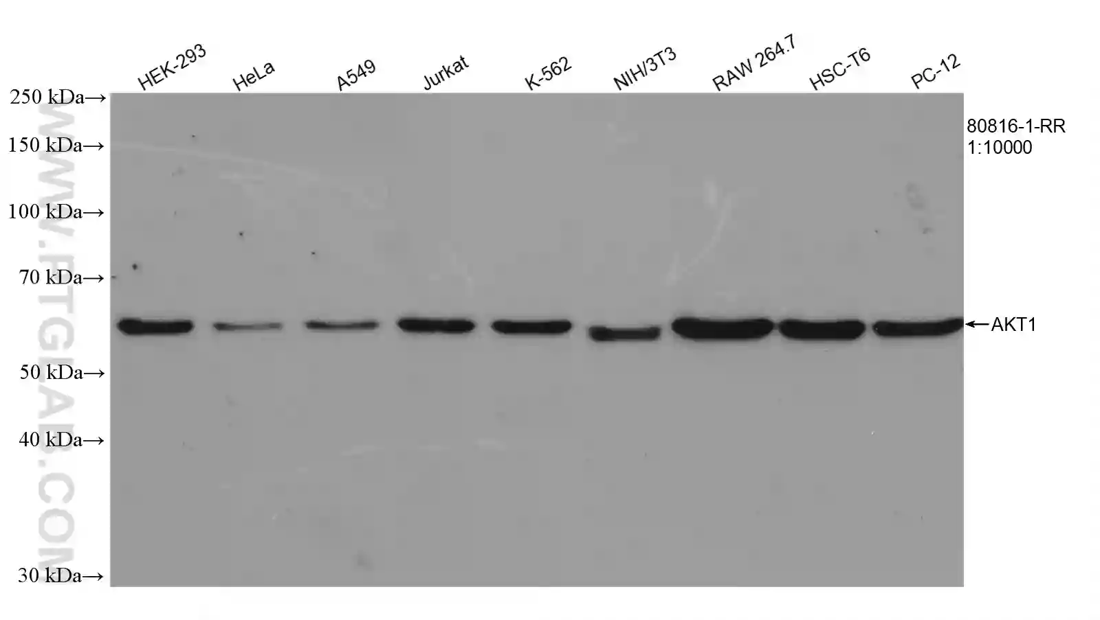
Various lysates were subjected to SDS PAGE followed by western blot with Uni-rAb™ AKT1 Recombinant Antibody (80816-1-RR).
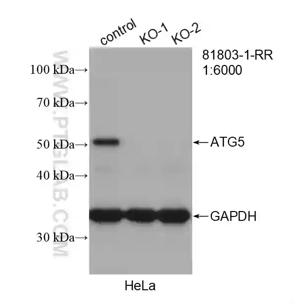
Wild-type and ATG5 knockout Hela cells were subjected to SDS PAGE followed by western blot with Uni-rAb™ ATG5 Recombinant Antibody (81803-1-RR).
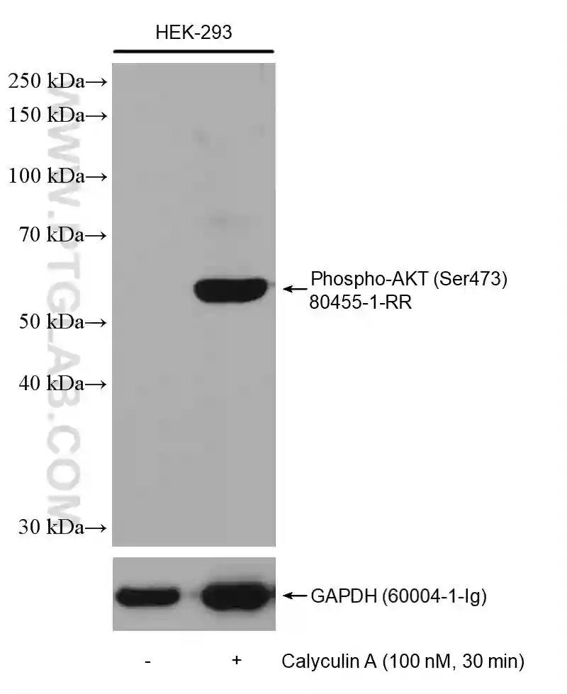
Untreated and Calyculin A treated HEK-293 cells were subjected to SDS PAGE followed by western blot with Uni-rAb™ Phospho-AKT (Ser473) Recombinant Antibody (80455-1-RR). The membrane was stripped and re-blotted with GAPDH Antibody as loading control.
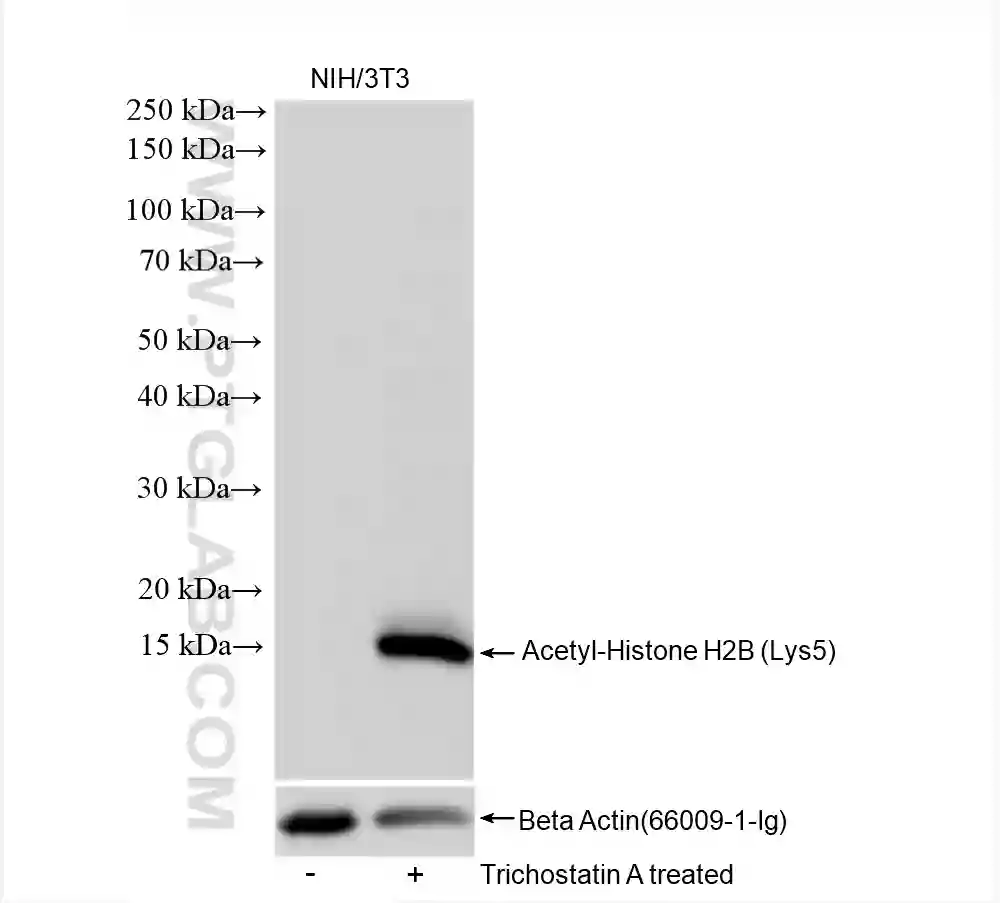
Untreated and Trichostatin A treated NIH/3T3 cells were subjected to SDS PAGE followed by western blot with Uni-rAb™ Acetyl-Histone H2B (Lys5) Recombinant Antibody (83171-4-RR). The membrane was stripped and re-blotted with Beta Actin Antibody as loading control.
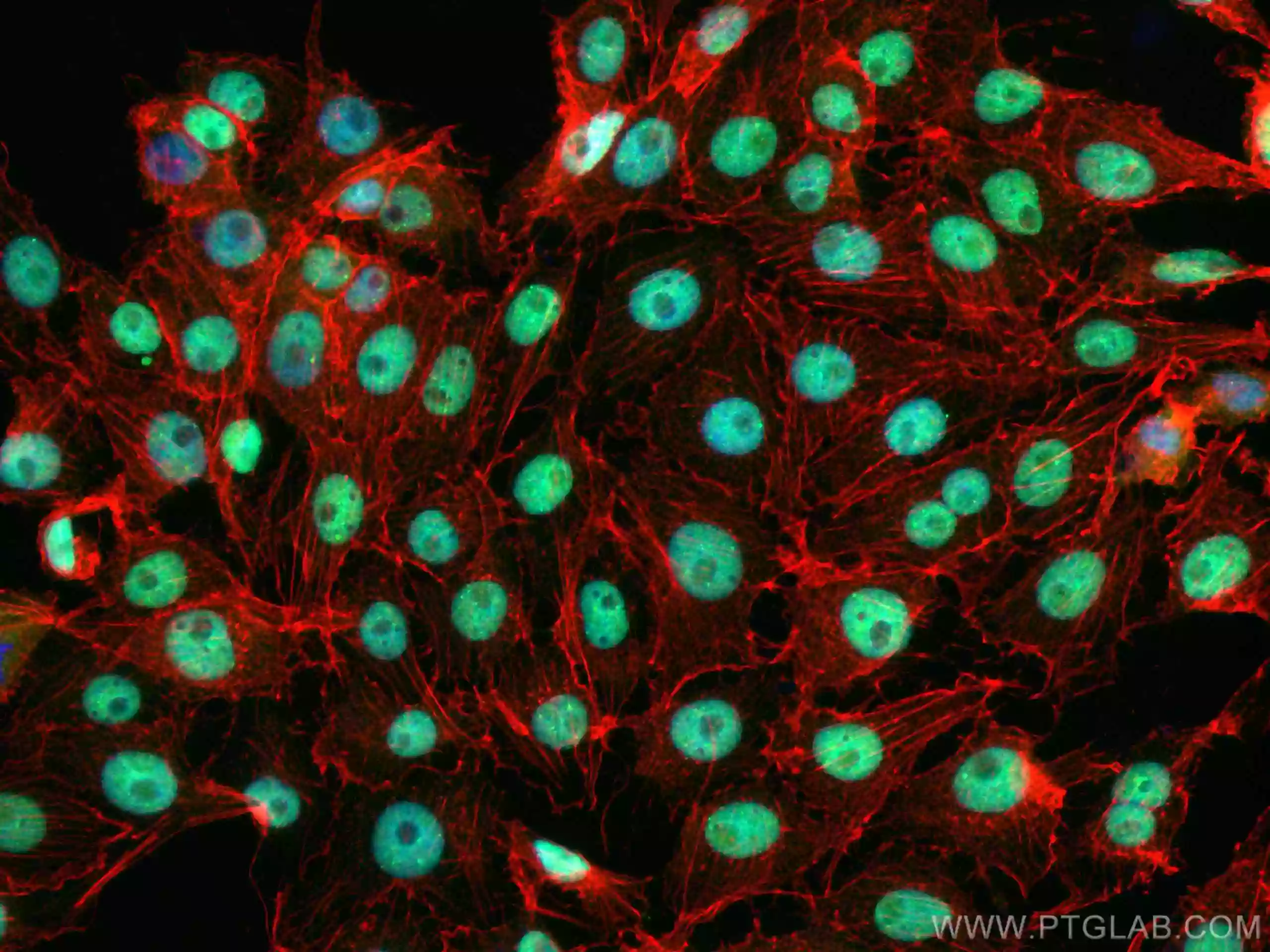
IF analysis of (4% PFA) fixed SH-SY5Y cells using Uni-rAb™ TDP-43 Recombinant Antibody (80002-1-RR, green) and CoraLite®488-Conjugated Goat Anti-Rabbit IgG (H+L). Cells were co-stained with CoraLite555-Phalloidin (red).
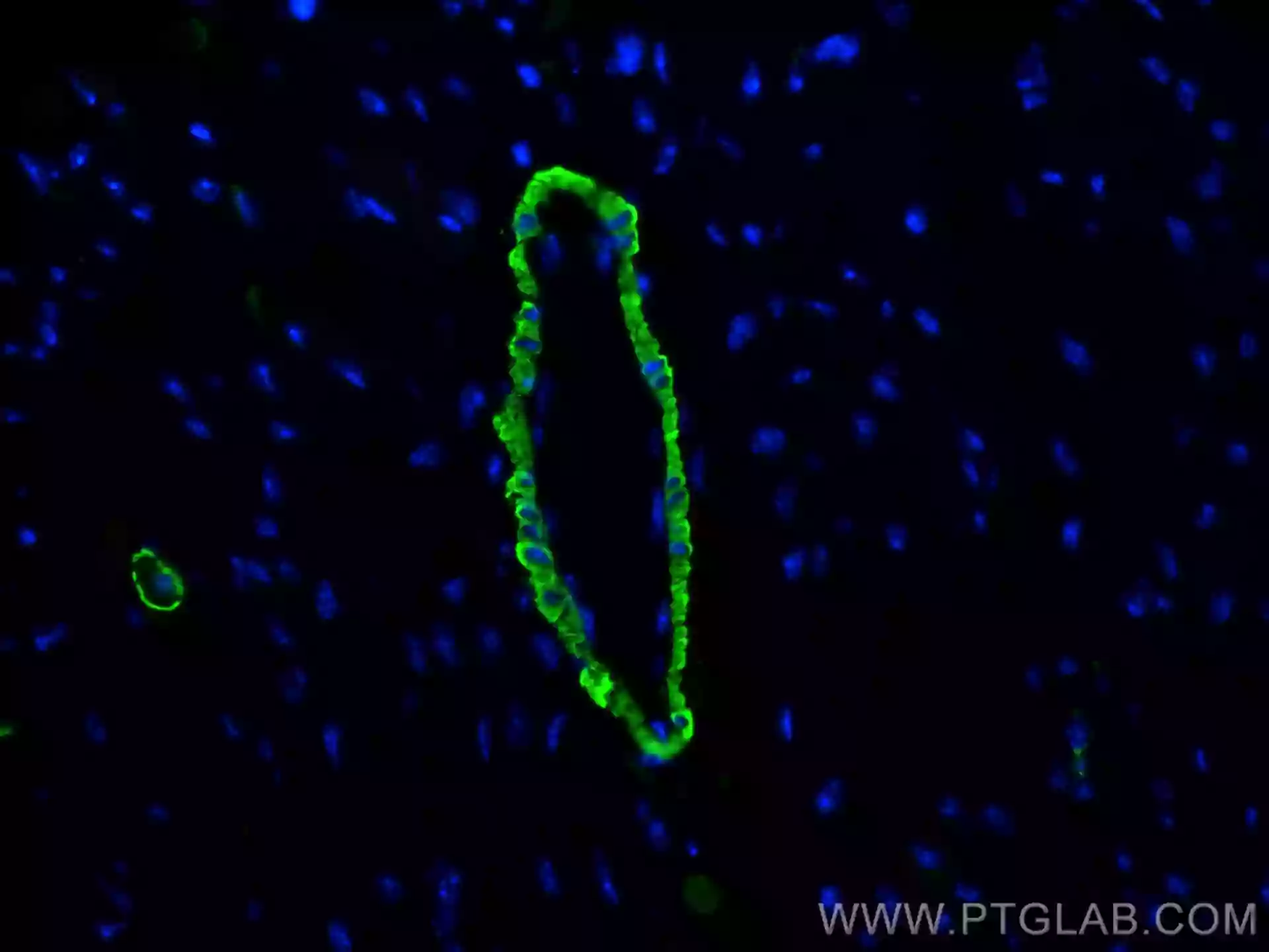
IF analysis of (4% PFA) fixed mouse heart tissue using Uni-rAb™ CoraLite® Plus 488 Smooth Muscle Actin-specific Recombinant Antibody (CL488-80008). Nucei were stained with DAPI.
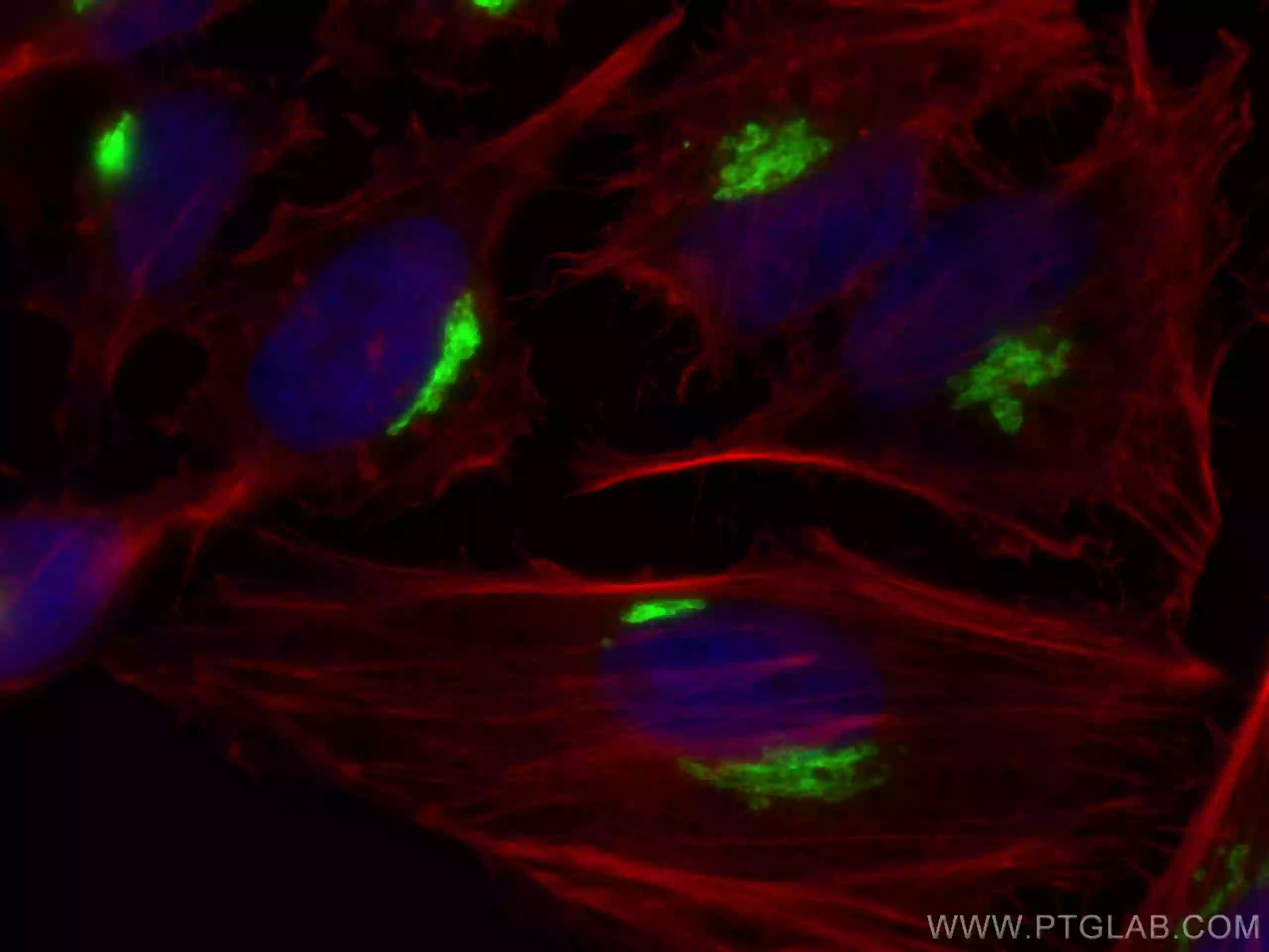
IF analysis of (4% PFA) fixed HeLa cells using Uni-rAb™ GP73/GOLPH2 Recombinant Antibody (81893-1-RR, green) and CoraLite®488-Conjugated Goat Anti-Rabbit IgG(H+L). Cells were co-stained with CL594-Phalloidin (red).
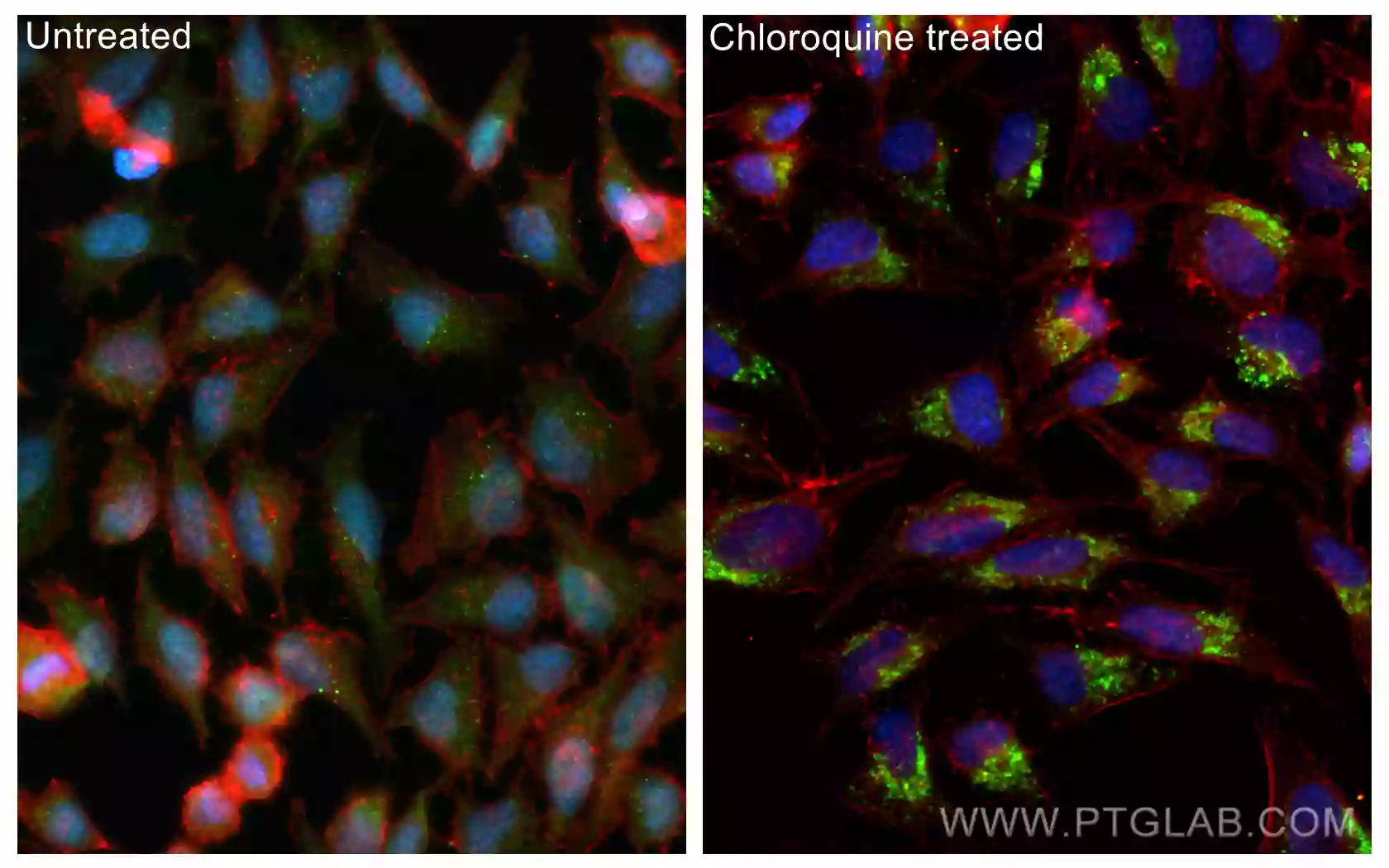
IF analysis of (-20°C Methanol) fixed untreated and Chloroquine treated HeLa cells using Uni-rAb™ LC3 Recombinant Antibody (81004-1-RR, green), CoraLite®488-Conjugated Goat Anti-Rabbit IgG (H+L) and CoraLite®594-Conjugated Beta Actin Monoclonal Antibody (CL594-66009).
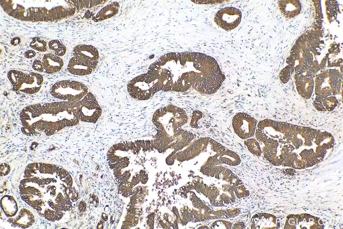
IHC analysis of FFPE human pancreas cancer tissue using Uni-rAb™ NRF2, NFE2L2 Recombinant Antibody (80593-1-RR). Heat mediated antigen retrieval with Tris-EDTA buffer (pH 9.0).
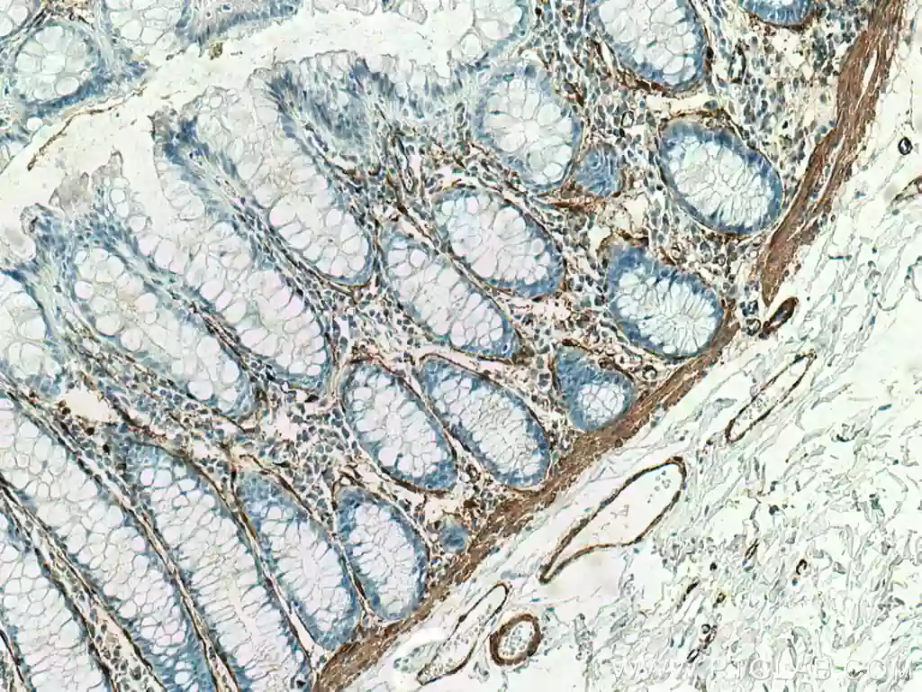
IHC analysis of FFPE human colon tissue using Uni-rAb™ a-SMA-specific Recombinant Antibody (80008-1-RR). Heat mediated antigen retrieval with Tris-EDTA buffer (pH 9.0).
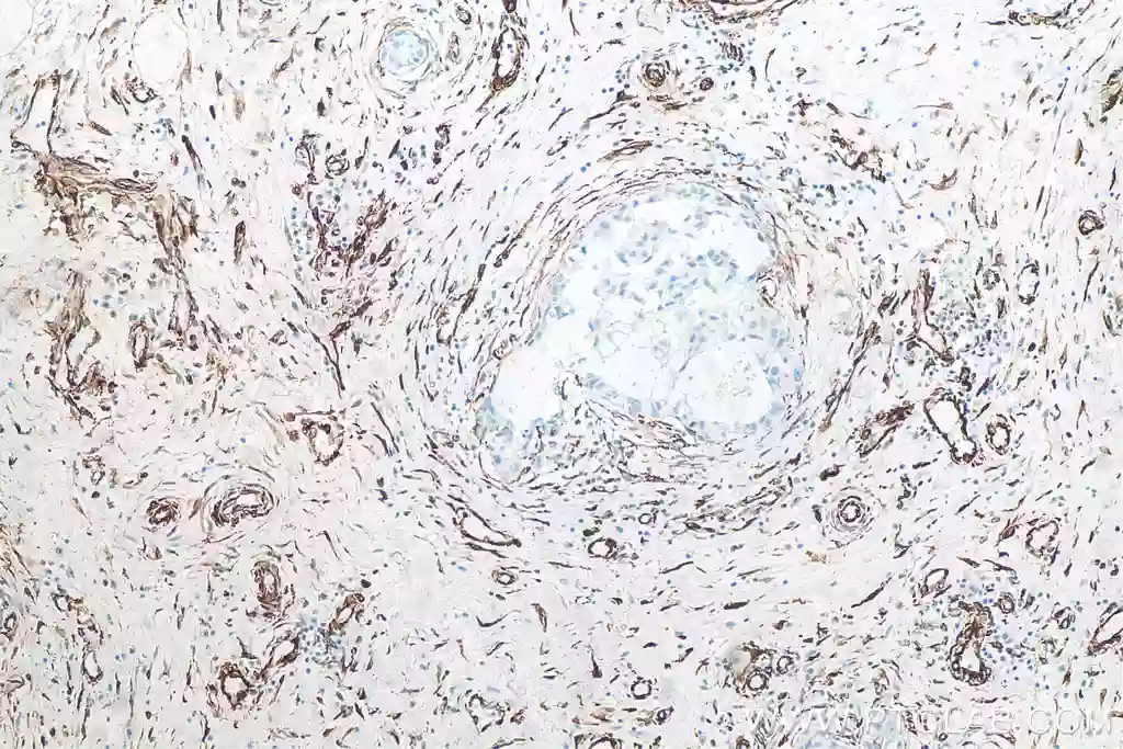
IHC analysis of FFPE human breast cancer tissue using Uni-rAb™ Vimentin Recombinant Antibody (80232-1-RR). Heat mediated antigen retrieval with Tris-EDTA buffer (pH 9.0).
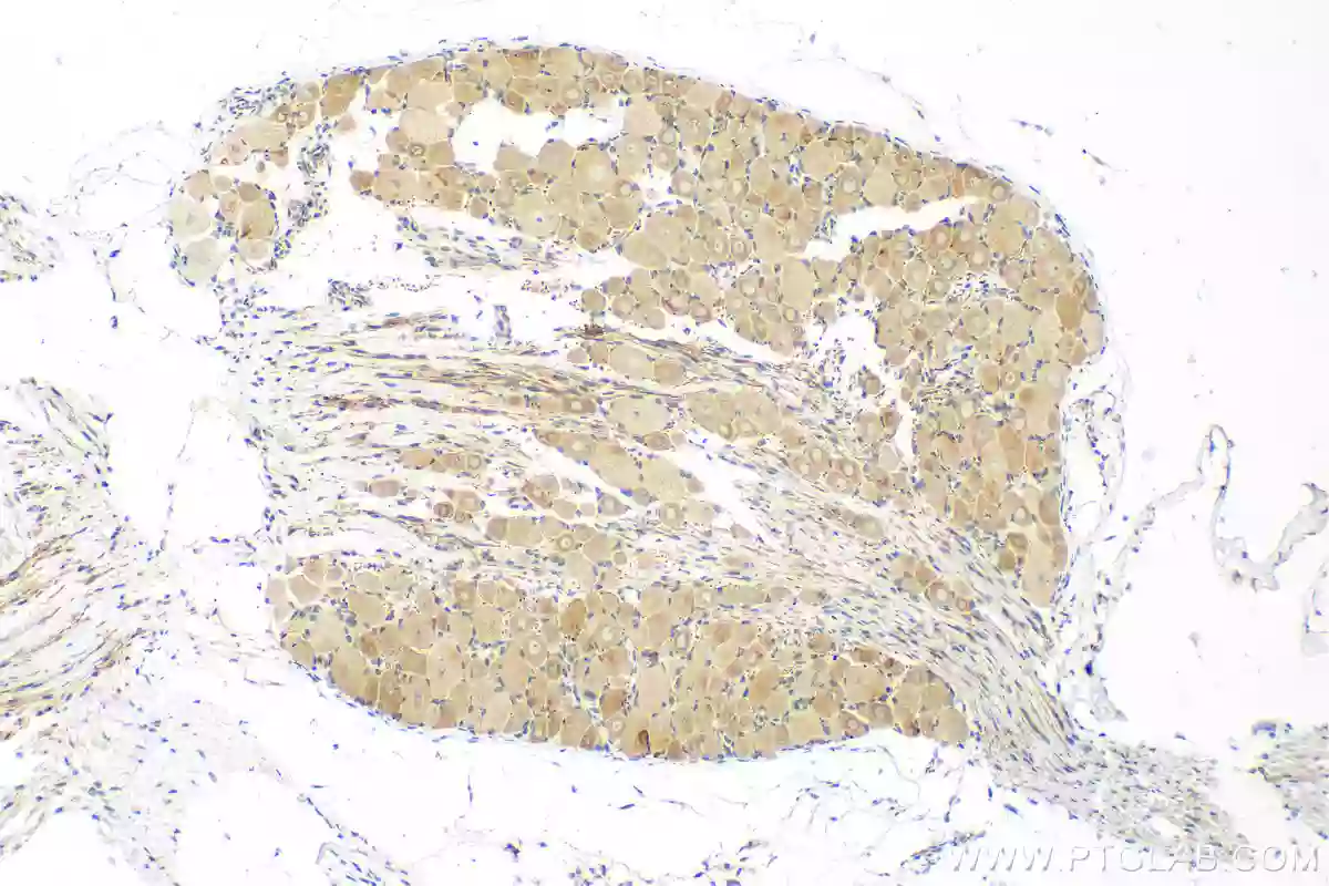
IHC analysis of FFPE rat dorsal root ganglion tissue slide using Uni-rAb™ Piezo1 (extracellular domain) Recombinant Antibody (82625-4-RR). Heat mediated antigen retrieval with Tris-EDTA buffer (pH 9.0).
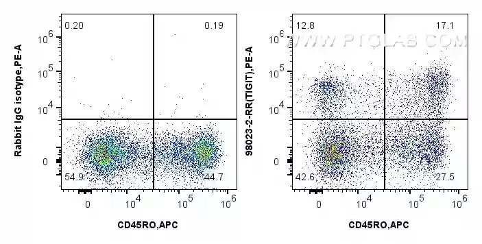
1x10^6 human PBMCs were surface stained with 0.25 ug of Uni-rAb™ Anti-Human TIGIT Rabbit Recombinant Antibody (98023-2-RR) or Isotype Control, and PE-conjugated Goat Anti-Rabbit IgG (H+L). Cells were then stained with APC Anti-Human CD45RO. Cells were not fixed. Lymphocytes were gated.
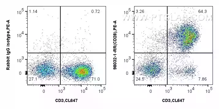
1x10^6 human PBMCs were surface stained with 0.25 ug of Uni-rAb™ Anti-Human CD28 Rabbit Recombinant Antibody (98032-1-RR) or Isotype Control, and PE-Conjugated Goat Anti-Rabbit IgG(H+L). Cell were then stained with CoraLite® Plus 647 Anti-Human CD3. Cells were not fixed. Lymphocytes were gated.
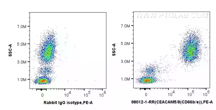
1x10^6 human peripheral blood leukocytes were surface stained with 0.25 ug of Uni-rAb™ Anti-Human CEACAM8/CD66b Rabbit Recombinant Antibody (98012-1-RR) or Isotype Control, and PE-conjugated Goat Anti-Rabbit IgG (H+L). Cells were not fixed.
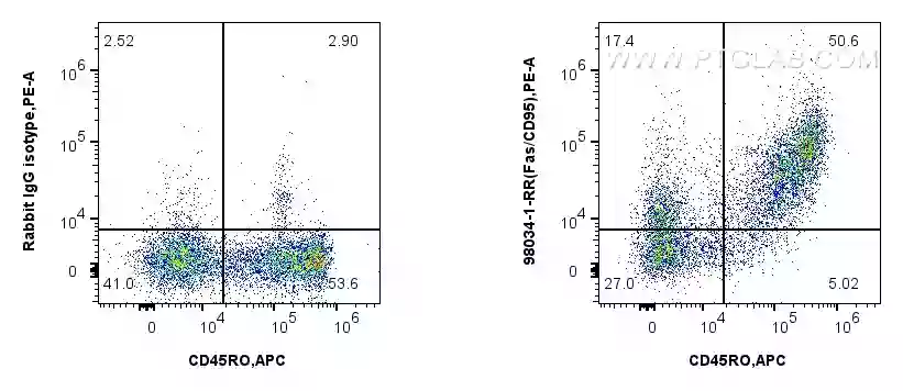
1x10^6 human PBMCs were surface stained with 0.25 ug Anti-Human Fas/CD95 Rabbit Recombinant Antibody (98034-1-RR) or Isotype Control, and PE-Conjugated Goat Anti-Rabbit IgG (H+L). Cells were then stained with APC Anti-Human CD45RO (UCHL1) (APC-65150, Clone: UCHL1). Cells were not fixed.


