Recombinant Human VEGFR1/FLT-1 protein (His Tag)
种属
Human
纯度
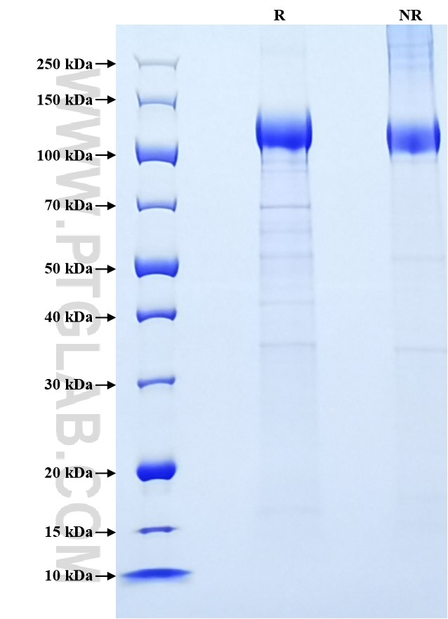
>90 %, SDS-PAGE
标签
His Tag
生物活性
未测试
验证数据展示
产品信息
| 纯度 | >90 %, SDS-PAGE |
| 内毒素 | <0.1 EU/μg protein, LAL method |
| 生物活性 | Not tested |
| 来源 | HEK293-derived Human VEGFR1 protein Ser27-Asn756 (Accession# P17948-1) with a His tag at the C-terminus. |
| 基因ID | 2321 |
| 蛋白编号 | P17948-1 |
| 预测分子量 | 86 kDa |
| SDS-PAGE | 100-140 kDa, reducing (R) conditions |
| 组分 | Lyophilized from 0.22 μm filtered solution in PBS, pH 7.4. Normally 5% trehalose and 5% mannitol are added as protectants before lyophilization. |
| 复溶 | Briefly centrifuge the tube before opening. Reconstitute at 0.1-0.5 mg/mL in sterile water. |
| 储存条件 |
It is recommended that the protein be aliquoted for optimal storage. Avoid repeated freeze-thaw cycles.
|
| 运输条件 | The product is shipped at ambient temperature. Upon receipt, store it immediately at the recommended temperature. |
背景信息
Vascular endothelial growth factor receptor-1 (VEGFR-1, FLT-1) is a receptor tyrosine kinase belonging to the VEGFR family. VEGF is a key regulator of physiological angiogenesis and has also been implicated in pathological angiogenesis associated with tumors, intraocular neovascular disorders and other conditions. The biological effects of VEGF are mediated by VEGFR-1 and VEGFR-2. Both the two receptors have seven immunoglobulin-like repeats in the extracellular domain, a single transmembrane region and a tyrosine kinase domain. VEGFR-1 binds VEGFA, PIGF and VEGFB, and plays an essential role in the development of embryonic vasculature, the regulation of angiogenesis, cell survival, cell migration, macrophage function, chemotaxis, and cancer cell invasion. Soluble VEGF receptor-1 (sVEGFR-1, sFlt-1), a truncated version of the cell membrane-spanning VEGFR-1, has the potential to act as a decoy receptor for VEGFA. Elevated serum sVEGFR-1 have been found in women with preeclampsia. Excess placental sVEGFR-1 may contribute to endothelial dysfunction, hypertension, and proteinuria in preeclampsia.
参考文献:
1. Kendall, R L et al. (1996) Biochem Biophys Res Commun.226(2):324-328.
2. Koga, Kaori et al. (2003)  J Clin Endocrinol Metab. 88(5):2348-2351.
3. Shibuya, Masaubmi. (2006) Angiogenesis.9(4):225-231.
4. Wu, Florence T H et al. (2010)  J Cell Mol Med.14(3):528-552.
