Recombinant Human CD86 protein (His Tag)
种属
Human
纯度
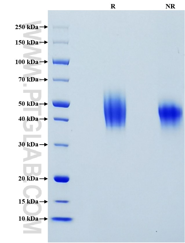
>95 %, SDS-PAGE
标签
His Tag
生物活性
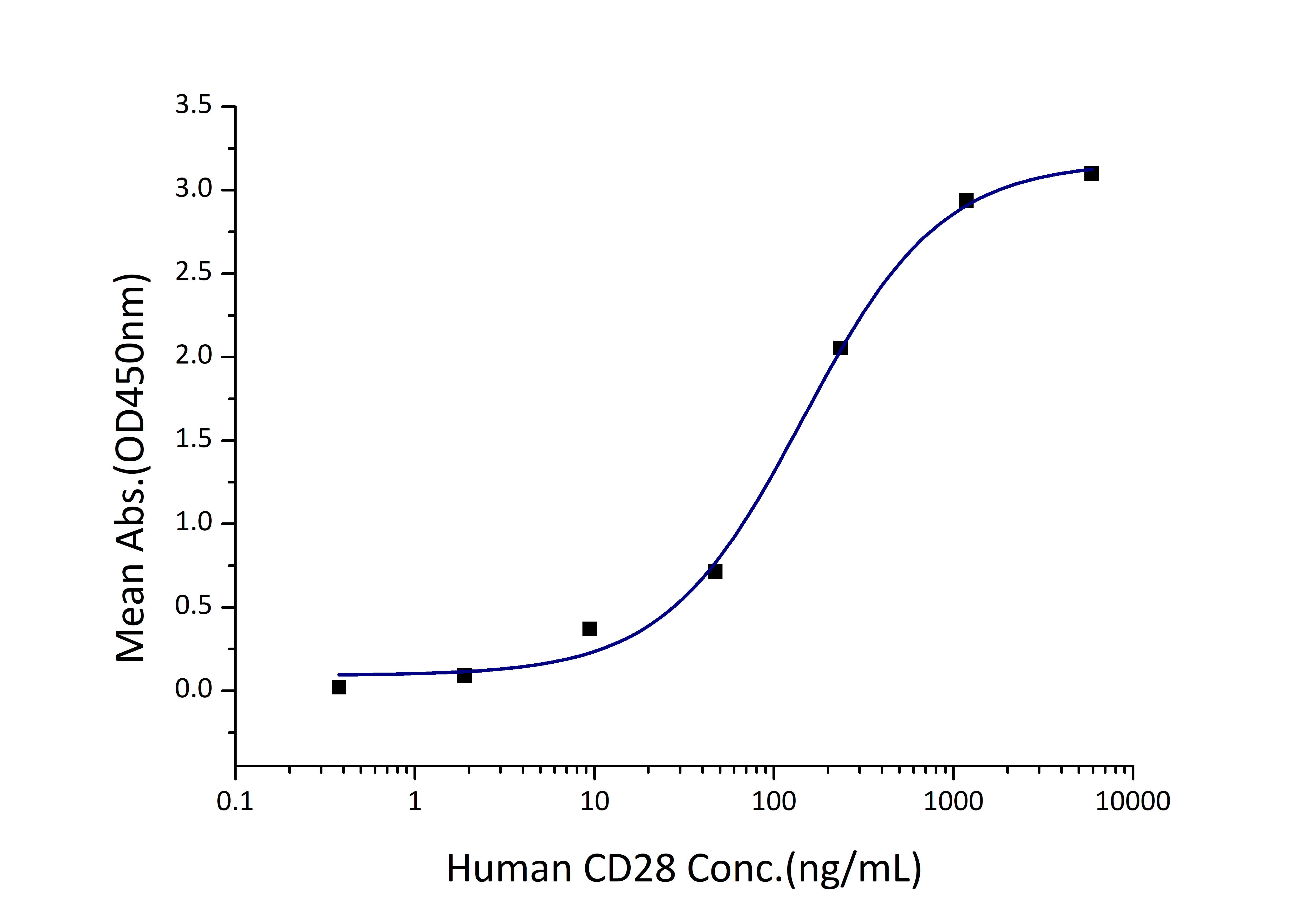
EC50: 72-290 ng/mL
验证数据展示
产品信息
| 纯度 | >95 %, SDS-PAGE |
| 内毒素 | <0.1 EU/μg protein, LAL method |
| 生物活性 | Immobilized Human CD86 (His tag) at 2 μg/mL (100 μL/well) can bind Human CD28 (hFc tag) with a linear range of 72-290 ng/mL. |
| 来源 | HEK293-derived Human CD86 protein Leu26-Pro247 (Accession# P42081-1) with a His tag at the C-terminus. |
| 基因ID | 942 |
| 蛋白编号 | P42081-1 |
| 预测分子量 | 26.2 kDa |
| SDS-PAGE | 37-60 kDa, reducing (R) conditions |
| 组分 | Lyophilized from 0.22 μm filtered solution in PBS, pH 7.4. Normally 5% trehalose and 5% mannitol are added as protectants before lyophilization. |
| 复溶 | Briefly centrifuge the tube before opening. Reconstitute at 0.1-0.5 mg/mL in sterile water. |
| 储存条件 |
It is recommended that the protein be aliquoted for optimal storage. Avoid repeated freeze-thaw cycles.
|
| 运输条件 | The product is shipped at ambient temperature. Upon receipt, store it immediately at the recommended temperature. |
背景信息
CD86 (also known as B7-2) is a costimulatory molecule belonging to the immunoglobulin (Ig) superfamily. CD86 is primarily expressed in antigen-presenting cells (APCs), including B cells, dendritic cells, and macrophages. CD86 has strong structural similarity with another B7 family molecule, CD80 (B7-1). CD86 and CD80 are the ligands for two proteins at the cell surface of T cells, CD28 antigen and cytotoxic T-lymphocyte antigen 4 (CTLA-4). The binding of CD86 or CD80 with CD28 antigen is a costimulatory signal for T cell activation, proliferation, and cytokine production. The binding of CD86 or CD80 with CTLA-4 negatively regulates T-cell activation and diminishes the immune response. However, CD86 and CD80 bind to CTLA-4 with higher affinity than CD28. Defects in CTLA-4-mediated transendocytosis of CD86 are associated with autoimmunity.
参考文献:
1.Bolandi N, et al. (2021). Int J Mol Sci. 22(19):10719
2.Yokozeki H, et al. (1996). J Invest Dermatol.106(1):147-153
3.Baravalle G, et al. (2011). J Immunol. 187(6):2966-2973.
4.Collins M, et al. (2005). Genome Biol. 6(6):223
5.Greaves P, et al. (2013). Blood. 121(5):734-744
6.Kennedy A, et al. (2022). Nat Immunol.23(9):1365-1378

