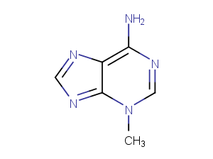
3-Methyladenine

CAS号

5142-23-4

分子式

C6H7N5

主要靶点

Mitophagy|Autophagy|Endogenous Metabolite|PI3K

仅限科研使用

Cat No : CM01277

Print datasheet

Synonyms

Mitophagy|Mitochondrial Autophagy|Inhibitor|Phosphoinositide 3-kinase|PI3K|PI3Kγ|NSC 66389|NSC66389|NSC-66389|Vps34|inhibit|EndogenousMetabolite|Endogenous Metabolite|Autophagy|3 Methyladenine|3Methyladenine|3-Methyladenine|3-MA|3-甲基腺嘌呤


产品信息

CAS号 5142-23-4
分子式 C6H7N5
主要靶点 Mitophagy|Autophagy|Endogenous Metabolite|PI3K
主要通路 自噬|代谢|PI3K/Akt/mTOR 信号通路|自噬
分子量 149.15
纯度 99.51%, 此纯度可做参考,具体纯度与批次有关系,可咨询客服
储存条件 store at low temperature,keep away from direct sunlight,keep away from moisture | Powder: -20°C for 3 years | Shipping with blue ice.
别名 Mitophagy|Mitochondrial Autophagy|Inhibitor|Phosphoinositide 3-kinase|PI3K|PI3Kγ|NSC 66389|NSC66389|NSC-66389|Vps34|inhibit|EndogenousMetabolite|Endogenous Metabolite|Autophagy|3 Methyladenine|3Methyladenine|3-Methyladenine|3-MA|3-甲基腺嘌呤

靶点活性

PI3Kγ:60 μM (in HeLa cells)|VPS34:25 μM (in HeLa cells)

体内活性

方法:为研究 3-Methyladenine 对动脉粥样硬化的影响,将 3-Methyladenine (30 mg/kg) 腹腔注射给 HFD 喂养的 ApoE?/? 小鼠,每周两次,持续八周。 结果:在高脂肪饮食喂养的小鼠中, 3-Methyladenine 治疗显著减少了动脉粥样硬化斑块的大小,并增加了病变的稳定性。3-Methyladenine 具有多种动脉粥样硬化保护作用,包括调节巨噬细胞自噬和泡沫细胞形成以及改变免疫微环境。[4] 方法:为研究自噬的调节作用,将 3-Methyladenine (15 mg/kg?) 单剂量腹腔注射给 LPS 诱导内毒素休克的 C57/BL6 小鼠。 结果: LPS 联合 3-Methyladenine 治疗的动物在内毒素血症后表现出存活率增加,血清炎症介质 TNF-α 和 IL-6 降低。[5]

体外活性

方法:人宫颈癌细胞 HeLa 用 3-Methyladenine (2.5-10 mM) 处理 48 h,使用 Trypan blue dye exclusion assay 检测细胞生长抑制情况。 结果:3-Methyladenine 以时间和剂量依赖的方式降低 HeLa 细胞活力。[1] 方法:脂肪细胞 3T3-L1 在没有血清的情况下用 3-Methyladenine (5 mM) 处理 4 h,使用 Western Blot 方法检测靶点蛋白表达水平。 结果:3-Methyladenine 显著降低了自噬标记物 LC3-II 的细胞内水平,增加了 p62 的表达,表明 3-Methyladenine 有效抑制自噬。[2] 方法:小鼠黑色素瘤细胞 B16 用 2DG (5 mM)、rotenone (1 μM) 和 3-Methyladenine (1.2-5 mM) 处理 24 h,使用 LDH release assay 检测细胞毒性。 结果:3-Methyladenine 剂量依赖性降低 2DG/rotenone 引起的 LDH 释放上调,保护肿瘤细胞免受糖酵解和线粒体呼吸抑制。[3]

溶解度

Ethanol:4 mg/mL (26.81 mM);DMSO:13.75 mg/mL (92.19 mM);H2O:3 mg/mL (20.11 mM)

细胞实验

Cells were seeded in an 8-well coverglass-bottomed chamber for 24 hours (6×10^3 cells per well). Images were acquired automatically at multiple locations on the coverglass using a Nikon TE2000E inverted microscope fitted with a 20× Nikon Plan Apo objective, a linearly-encoded stage, and a Hamamatsu Orca-ER CCD camera. A mercury-arc lamp with two neutral density filters (for a total 128-fold reduction in intensity) was used for fluorescence illumination. The microscope was controlled using NIS-Elements Advanced Research software and housed in a custom-designed 37°C chamber with a secondary internal chamber that delivered humidified 5% CO2. Fluorescence and differential interference contrast images were obtained every 10 min for a period of 48 hours. To analyze live cell imaging movies, the time-lapse records of live cell imaging experiments were exported as an image series and analyzed manually using NIS-Elements Advanced Research software. The criteria for analyses were described previously, and lagging chromosomes in prometaphase were defined as the red fluorescence-positive materials that lingered outside the roughly formed metaphase plate for more than 3 frames (30 min) [2].

动物实验

All rats were fasted for 12 h with free access to water prior to operation. After anesthesia by intraperitoneal (i.p.) injection of 2% sodium pentobarbital (0.25 mL/100 g), they were laid and fixed on the table, routinely shaven, disinfected, and draped. The rat SAP model was induced by 0.1 mL/min speed uniformly retrograde infusion of a freshly prepared 3.5% sodium taurocholate solution (0.1 mL/100 g) into the biliopancreatic duct after laparotomy. Equivalent volume of normal saline solution was substituted for 3.5% sodium taurocholate solution in the sham-operation (SO) control group. The incision was closed with a continuous 3-0-silk suture, and 2 mL/100 g of saline was injected into the back subcutaneously to compensate for the fluid loss. 180 rats were randomly divided into four groups: (1) Acanthopanax treatment group (Aca group, n = 45) where the rats were injected with 0.2% Acanthopanax injection at a dose of 3.5 mg/100 g 3 h after successful modeling via the vena caudalis once, knowing that this dosage was effective as proven in our previous experiment; (2) 3-Methyladenine treatment group (3-methyladenine group, n = 45) where the rats were injected with 100 nmol/μL 3-methyladenine solution at a dose of 1.5 mg/100 g 3 h after successful modeling via the intraperitoneal route once, knowing that this dosage was effective as proven in the literature [6]; (3) SAP model group (SAP group, n = 45) where these rats received an equivalent volume of the normal saline instead of Acanthopanax injection 3 h after successful modeling via the vena caudalis once; (4) SO group (control, n = 45) where these rats received an equivalent volume of the normal saline instead of Acanthopanax injection 3 h after successful sham-operation via the vena caudalis once. The 45 animals in each of the four groups were equally randomized into 3, 12, and 24 h subgroups for postoperative observations [4].

参考文献

1.Hou H, et al. Inhibitors of phosphatidylinositol 3'-kinases promote mitotic cell death in HeLa cells. PLoS One. 2012;7(4):e35665.
2.Wang S, Li F, Qiao R, et al. Arginine-Rich Manganese Silicate Nanobubbles as a Ferroptosis-Inducing Agent for Tumor-Targeted Theranostics[J]. ACS nano. 2018 Dec 26;12(12):12380-12392.
3.Heckmann BL, et al. The autophagic inhibitor 3-methyladenine potently stimulates PKA-dependent lipolysis in adipocytes. Br J Pharmacol. 2013 Jan;168(1):163-71.
4.Kosic M, et al. 3-Methyladenine prevents energy stress-induced necrotic death of melanoma cells through autophagy-independent mechanisms. J Pharmacol Sci. 2021 Sep;147(1):156-167.
5.Dai S, et al. Systemic application of 3-methyladenine markedly inhibited atherosclerotic lesion in ApoE-/- mice by modulating autophagy, foam cell formation and immune-negative molecules. Cell Death Dis. 2016 Dec 1;7(12):e2498.
6.Li Q, et al. Inhibition of autophagy with 3-methyladenine is protective in a lethal model of murine endotoxemia and polymicrobial sepsis. Innate Immun. 2018 May;24(4):231-239.
7.hang C, Liu Z, Zhang Y, et al. Z“Iron free” zinc oxide nanoparticles with ion-leaking properties disrupt intracellular ROS and iron homeostasis to induce ferroptosis[J]. Cell Death & Disease. 2020, 11(3): 1-15.
8.Shang Z, Zhang T, Jiang M, et al. High-carbohydrate, High-fat Diet-induced Hyperlipidemia Hampers the Differentiation Balance of Bone Marrow Mesenchymal Stem Cells by Suppressing Autophagy via the AMPK/mTOR Pathway in Rat Models[J]. 2020.
9.Xia Y, Chen J, Yu Y, et al. Compensatory combination of mTOR and TrxR inhibitors to cause oxidative stress and regression of tumors[J]. Theranostics. 2021, 11(9): 4335.
10.Zhang H, Cui Z, Cheng D, et al. RNF186 regulates EFNB1 (ephrin B1)-EPHB2-induced autophagy in the colonic epithelial cells for the maintenance of intestinal homeostasis[J]. Autophagy . 2020
The molarity calculator equation

Mass (g) = Concentration (mol/L) × Volume (L) × Molecular Weight (g/mol)

质量   浓度   体积   分子量 *
=
×
×

The dilution calculator equation

Concentration (start) × Volume (start) = Concentration (final) × Volume (final)
This equation is commonly abbreviated as: C1V1 = C2V2

浓度 (start) × 体积 (start) = 浓度 (final) × 体积 (final)
×
=
×
C1   V1   C2   V2