验证数据展示
经过测试的应用
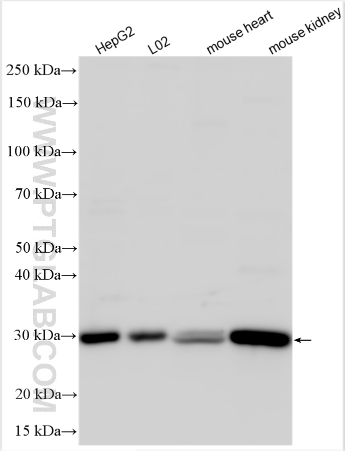
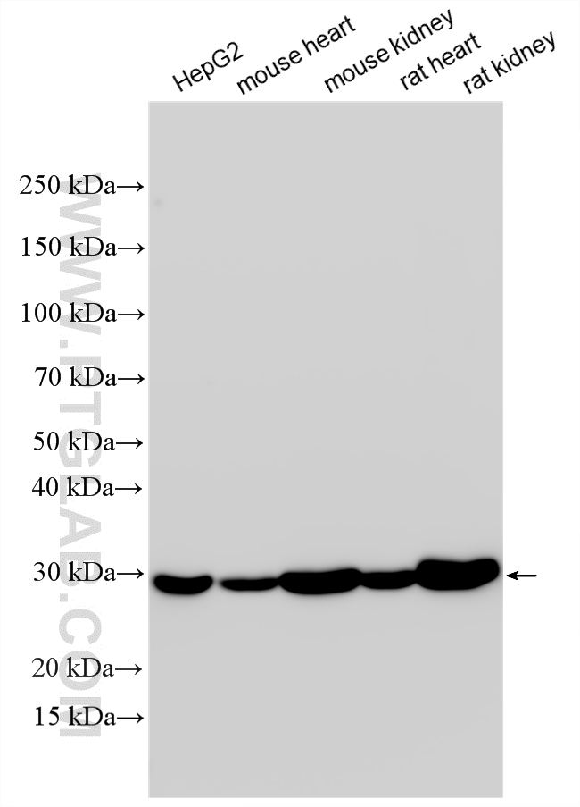
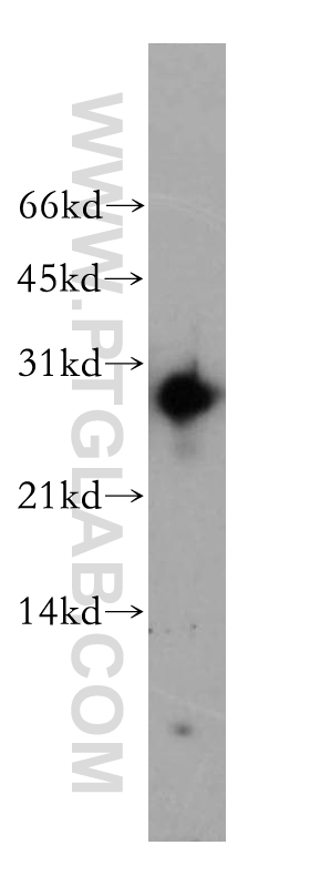
| Positive WB detected in | HepG2 cells, Y79 cells, L02 cells, mouse heart tissue, mouse kidney tissue, rat heart tissue, rat kidney |
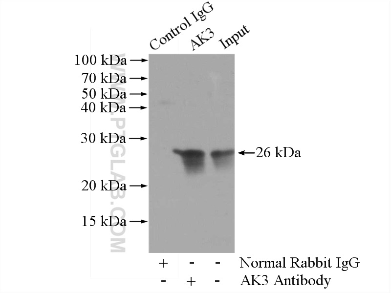
| Positive IP detected in | mouse kidney tissue |
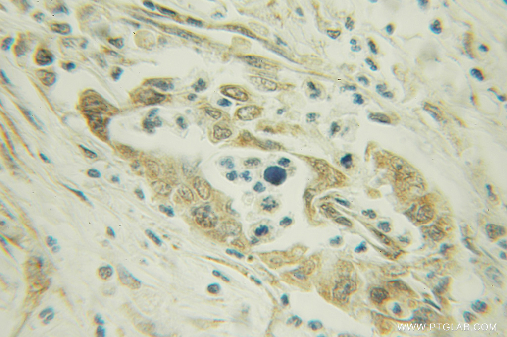
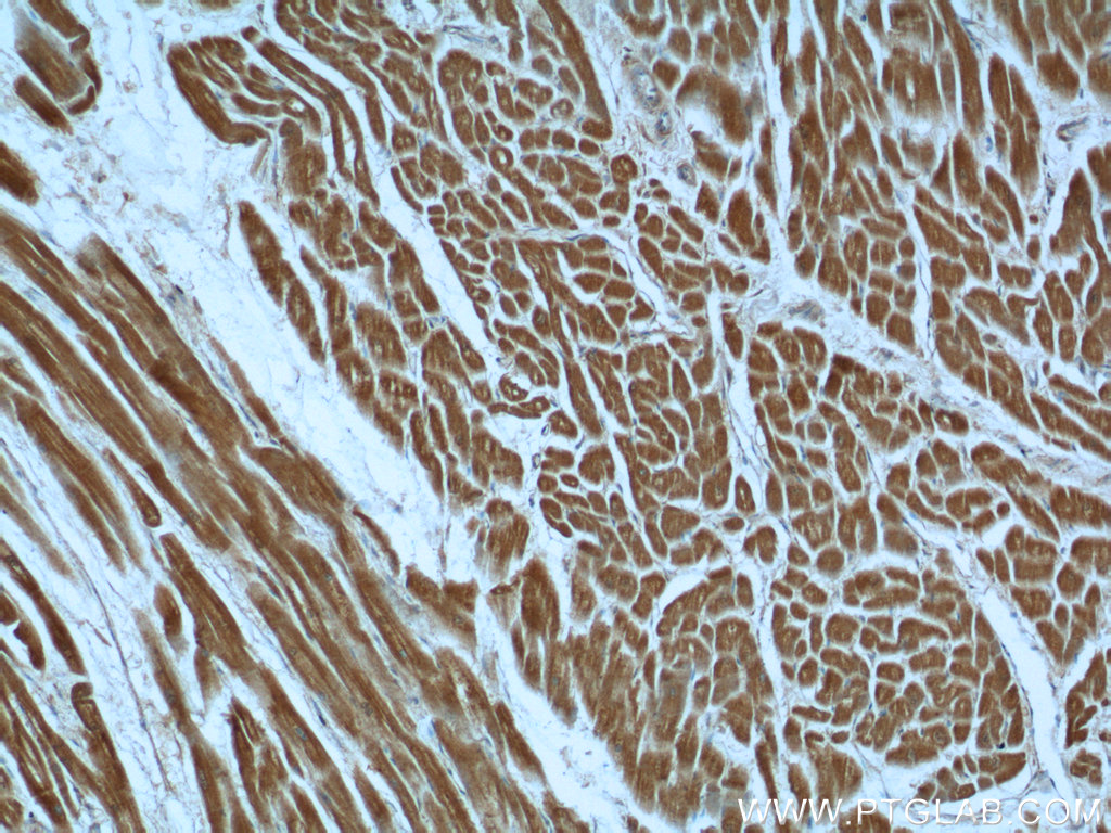
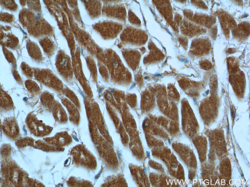
| Positive IHC detected in | human pancreas cancer tissue, human heart tissue Note: suggested antigen retrieval with TE buffer pH 9.0; (*) Alternatively, antigen retrieval may be performed with citrate buffer pH 6.0 |
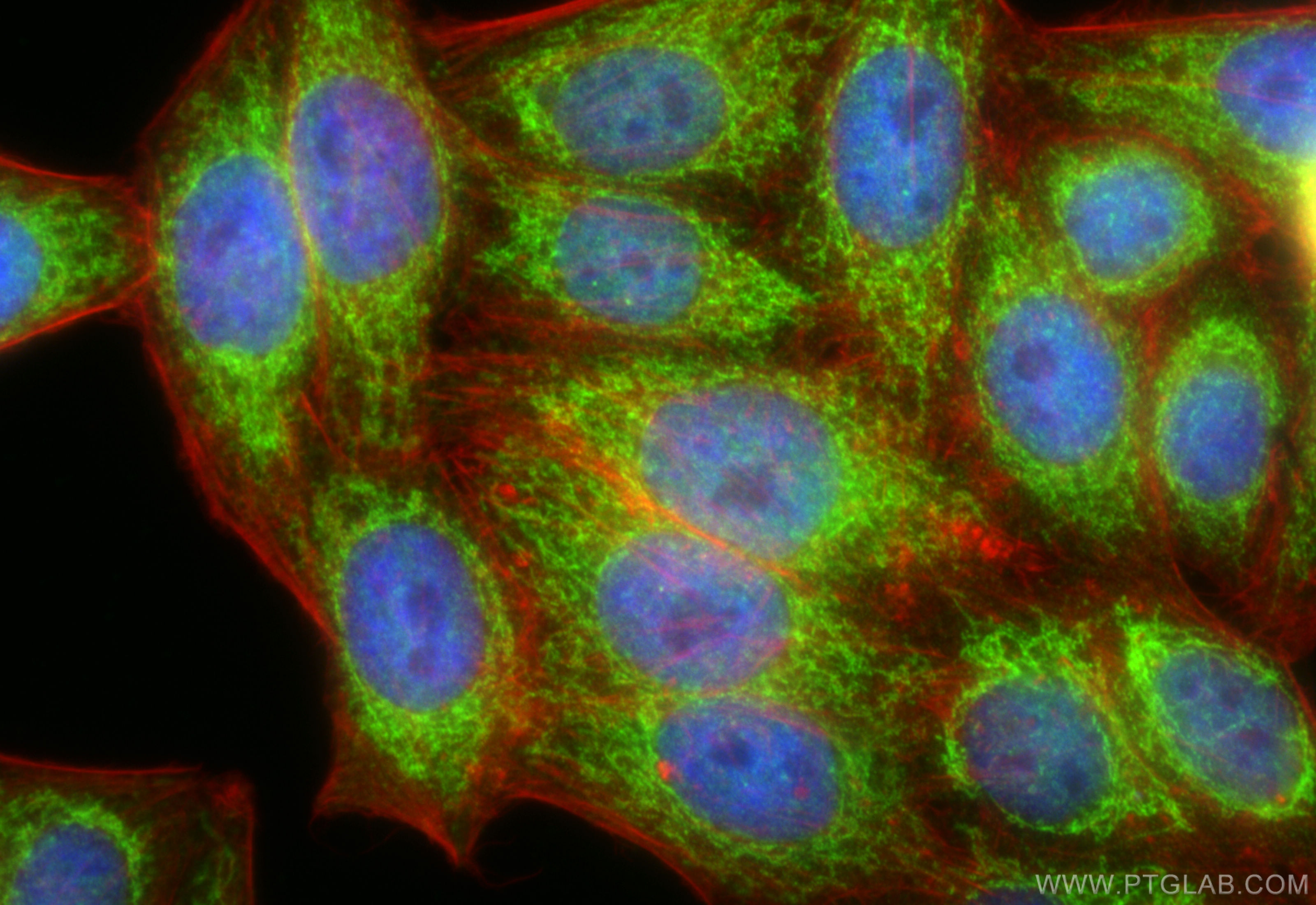
| Positive IF/ICC detected in | HepG2 cells |
推荐稀释比
| 应用 | 推荐稀释比 |
|---|---|
| Western Blot (WB) | WB : 1:5000-1:50000 |
| Immunoprecipitation (IP) | IP : 0.5-4.0 ug for 1.0-3.0 mg of total protein lysate |
| Immunohistochemistry (IHC) | IHC : 1:20-1:200 |
| Immunofluorescence (IF)/ICC | IF/ICC : 1:200-1:800 |
| It is recommended that this reagent should be titrated in each testing system to obtain optimal results. | |
| Sample-dependent, Check data in validation data gallery. | |
发表文章中的应用
| WB | See 2 publications below |
产品信息
12562-1-AP targets AK3 in WB, IHC, IF/ICC, IP, ELISA applications and shows reactivity with human, mouse, rat samples.
| 经测试应用 | WB, IHC, IF/ICC, IP, ELISA Application Description |
| 文献引用应用 | WB |
| 经测试反应性 | human, mouse, rat |
| 文献引用反应性 | human |
| 免疫原 |
CatNo: Ag3146 Product name: Recombinant human AK3 protein Source: e coli.-derived, PGEX-4T Tag: GST Domain: 1-227 aa of BC013771 Sequence: MGASARLLRAVIMGAPGSGKGTVSSRITTHFELKHLSSGDLLRDNMLRGTEIGVLAKAFIDQGKLIPDDVMTRLALHELKNLTQYSWLLDGFPRTLPQAEALDRAYQIDTVINLNVPFEVIKQRLTARWIHPASGRVYNIEFNPPKTVGIDDLTGEPLIQREDDKPETVIKRLKAYEDQTKPVLEYYQKKGVLETFSGTETNKIWPYVYAFLQTKVPQRSQKASVTP 种属同源性预测 |
| 宿主/亚型 | Rabbit / IgG |
| 抗体类别 | Polyclonal |
| 产品类型 | Antibody |
| 全称 | adenylate kinase 3 |
| 别名 | Adenylate kinase 3 alpha-like 1, AK 3, AK3L1, AK6, AKL3L |
| 计算分子量 | 227 aa, 26 kDa |
| 观测分子量 | 26 kDa |
| GenBank蛋白编号 | BC013771 |
| 基因名称 | AK3 |
| Gene ID (NCBI) | 50808 |
| RRID | AB_2305373 |
| 偶联类型 | Unconjugated |
| 形式 | Liquid |
| 纯化方式 | Antigen affinity purification |
| UNIPROT ID | Q9UIJ7 |
| 储存缓冲液 | PBS with 0.02% sodium azide and 50% glycerol, pH 7.3. |
| 储存条件 | Store at -20°C. Stable for one year after shipment. Aliquoting is unnecessary for -20oC storage. |
背景介绍
AK3(Adenylate kinase 3) is also named as AK3L1, AK6, AKL3L and belongs to the adenylate kinase family. It is located in the mitochondrial matrix and is a GTP:AMP phosphotransferase(PMID:2546792). AK3 can be detected AK3 at an apparent molecular mass of 28 kD by western blot analysis and subcellular fractionation of mouse liver and kidney detected Ak3 in the mitochondrial fraction(PMID:11485571). This antibody may also recognize AK4 due to the high homology.
实验方案
| Product Specific Protocols | |
|---|---|
| IF protocol for AK3 antibody 12562-1-AP | Download protocol |
| IHC protocol for AK3 antibody 12562-1-AP | Download protocol |
| IP protocol for AK3 antibody 12562-1-AP | Download protocol |
| WB protocol for AK3 antibody 12562-1-AP | Download protocol |
| Standard Protocols | |
|---|---|
| Click here to view our Standard Protocols |
发表文章
| Species | Application | Title |
|---|---|---|
Mol Med Rep MicroRNA‑214 upregulates HIF‑1α and VEGF by targeting ING4 in lung cancer cells. | ||

