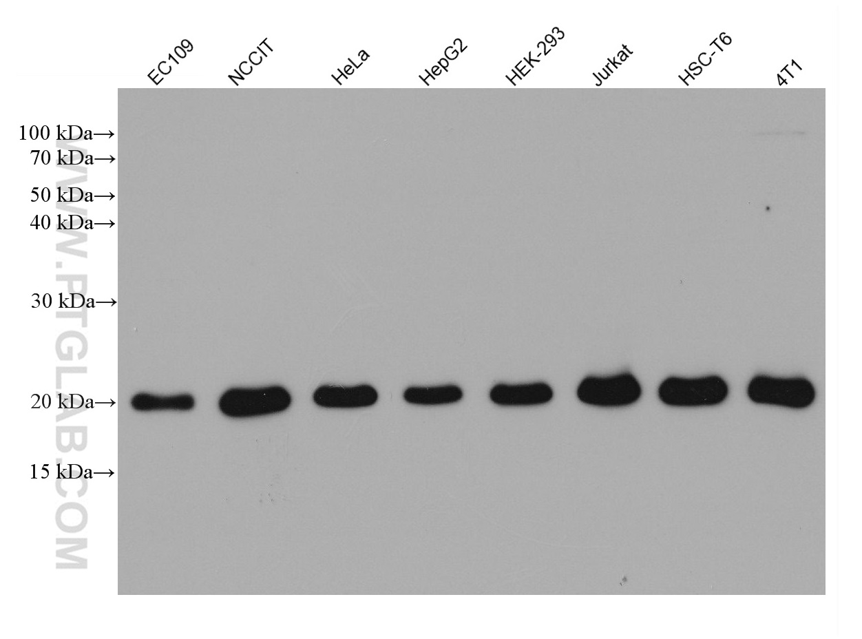
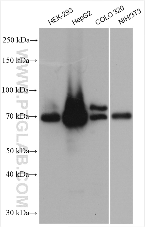
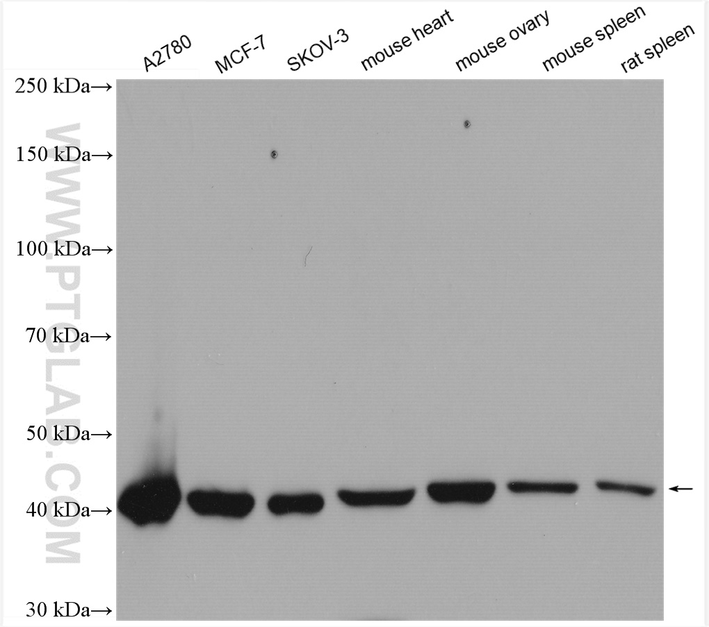
Various lysates were subjected to SDS PAGE followed by western blot with 67763-1-Ig (GPX4 antibody) at dilution of 1:5000 incubated at room temperature for 1.5 hours.
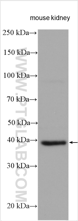
mouse kidney tissue were subjected to SDS PAGE followed by western blot with 20886-1-AP (FSP1 antibody) at dilution of 1:1500 incubated at room temperature for 1.5 hours.
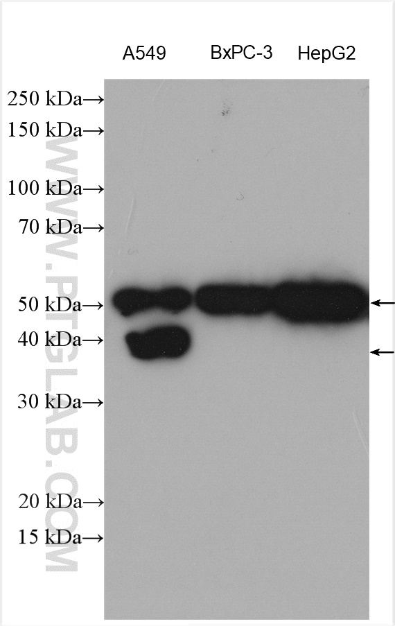
Various lysates were subjected to SDS PAGE followed by western blot with 26864-1-AP (SLC7A11/xCT antibody) at dilution of 1:1000 incubated at room temperature for 1.5 hours.
HeLa cells were subjected to SDS PAGE followed by western blot with 15193-1-AP (CD98 antibody at dilution of 1:20000 incubated at room temperature for 1.5 hours.
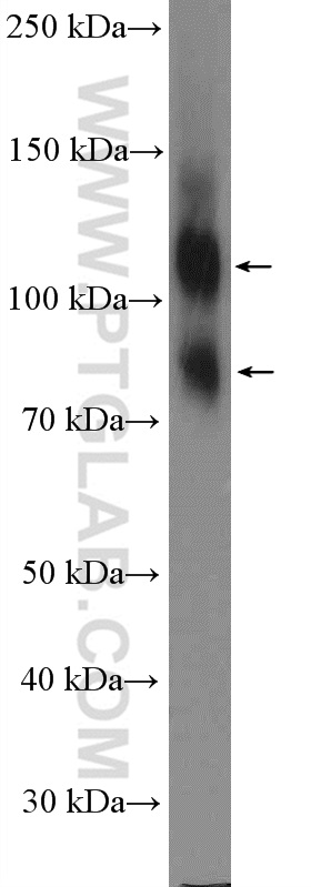
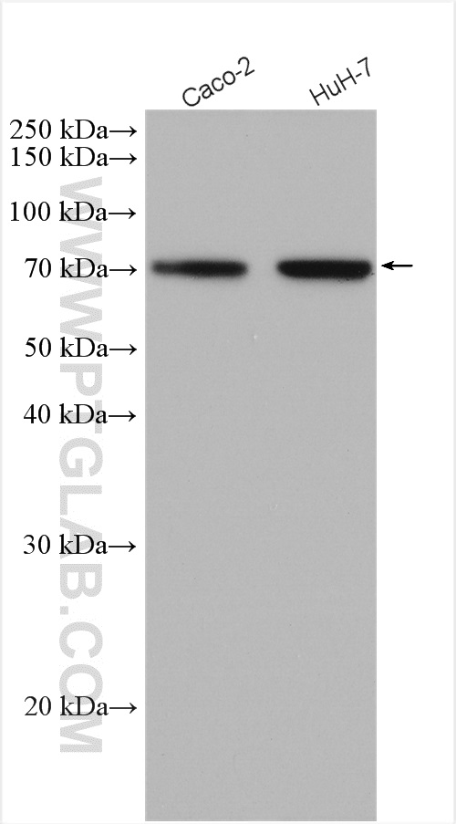
Various cell lysates were subjected to SDS PAGE followed by western blot with 20507-1-AP (DMT1 antibody) at dilution of 1:800 incubated at room temperature for 1.5 hours.
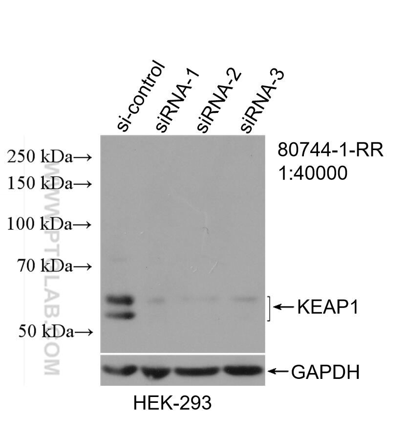
WB result of KEAP1 antibody (80744-1-RR; 1:40000; incubated at room temperature for 1.5 hours) with sh-Control and sh-KEAP1 transfected HEK-293 cells.
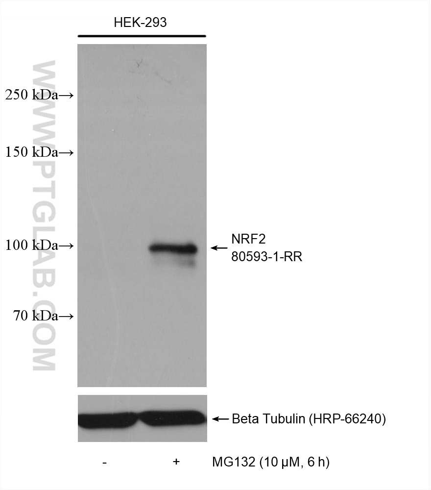
Non-treated and MG 132 treated HEK-293 cells were subjected to SDS PAGE followed by western blot with 80593-1-RR (NRF2, NFE2L2 antibody) at dilution of 1:2500 incubated at room temperature for 1.5 hours.
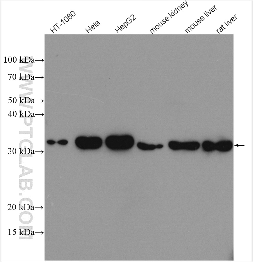
Various lysates were subjected to SDS PAGE followed by western blot with 10701-1-AP (HO-1 antibody) at dilution of 1:3000 incubated at room temperature for 1.5 hours.
Various lysates were subjected to SDS PAGE followed by western blot with 22401-1-AP (ACSL4 antibody) at dilution of 1:6000 incubated at room temperature for 1.5 hours.
Various lysates were subjected to SDS PAGE followed by western blot with 14877-1-AP (DHODH antibody) at dilution of 1:8000 incubated at room temperature for 1.5 hours.
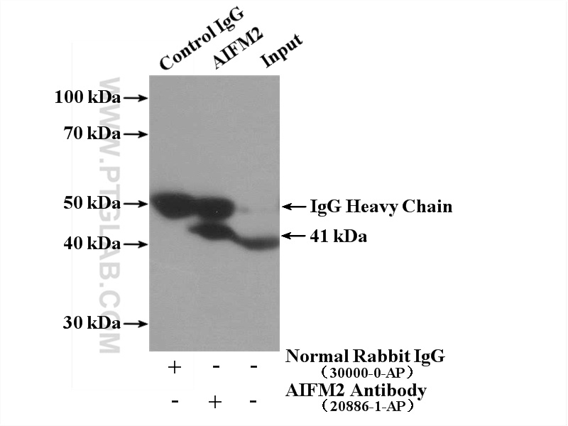
IP Result of anti-FSP1 (IP:20886-1-AP, 4ug; Detection:20886-1-AP 1:300) with L02 cells lysate 1800ug.
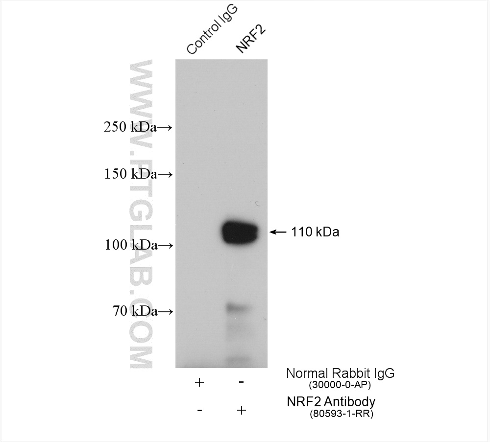
IP result of anti-NRF2, NFE2L2(IP:80593-1-RR, 4ug; Detection:80593-1-RR 1:800) with HeLa cells lysate 2520 ug.
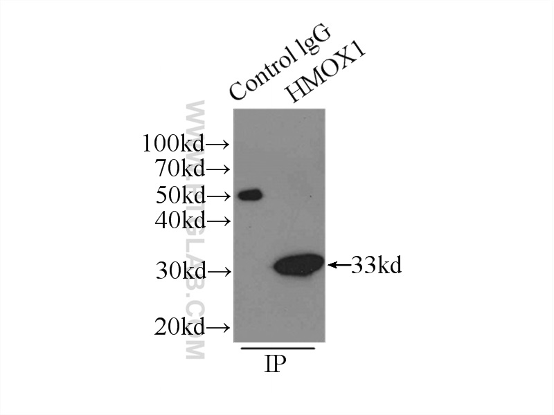
IP Result of anti-HMOX1 (IP:10701-1-AP, 3ug; Detection:10701-1-AP 1:1000) with HeLa cells lysate 3000ug.
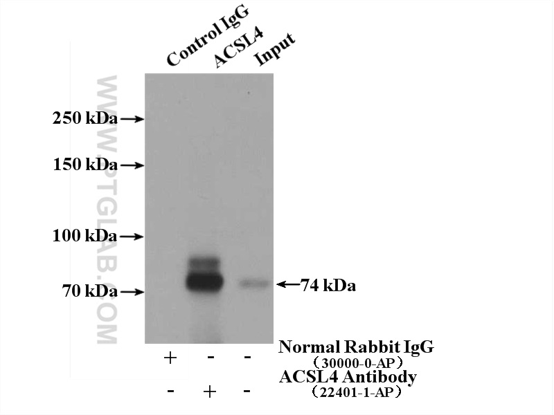
IP Result of anti-ACSL4 (IP:22401-1-AP, 4ug; Detection:22401-1-AP 1:1000) with COLO 320 cells lysate 2000ug.
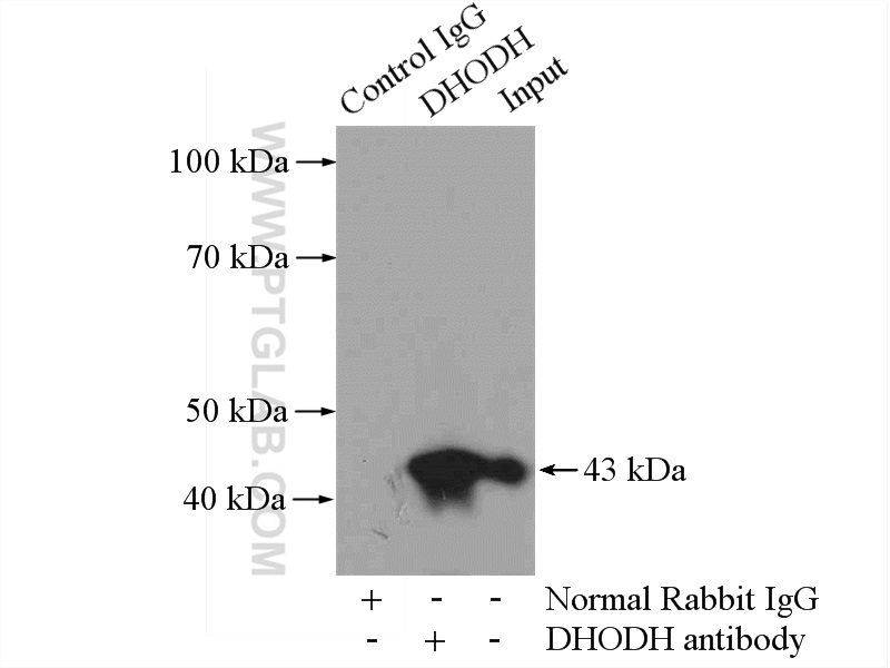
IP Result of anti-DHODH (IP:14877-1-AP, 4ug; Detection:14877-1-AP 1:1000) with mouse spleen tissue lysate 4000ug.
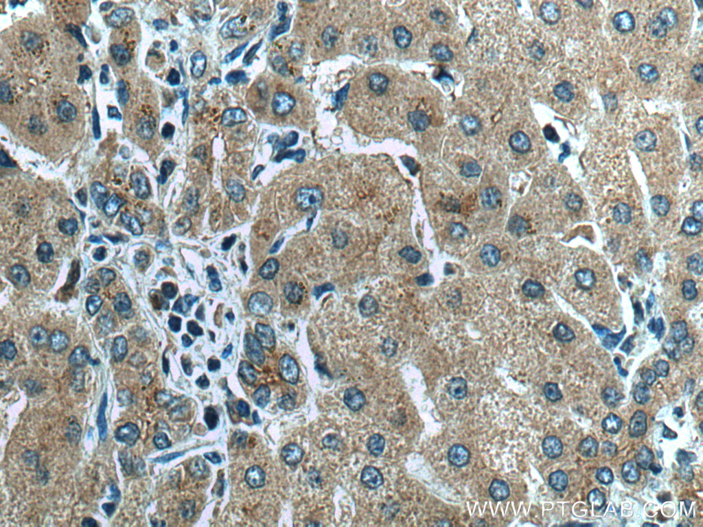
Immunohistochemical analysis of paraffin-embedded human liver cancer tissue slide using 67763-1-Ig (GPX4 antibody) at dilution of 1:2000 (under 40x lens). Heat mediated antigen retrieval with Tris-EDTA buffer (pH 9.0).
Immunohistochemical analysis of paraffin-embedded human liver tissue slide using 20886-1-AP (AIFM2/ FSP1 antibody) at dilution of 1:200 (under 40x lens). Heat mediated antigen retrieval with Tris-EDTA buffer (pH 9.0).
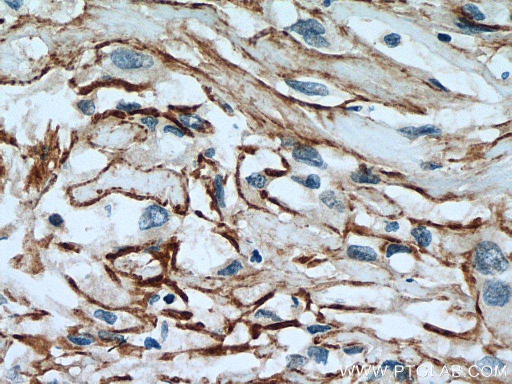
Immunohistochemical analysis of paraffin-embedded human renal cell carcinoma tissue slide using 26864-1-AP (SLC7A11/xCT antibody) at dilution of 1:200 (under 40x lens). Heat mediated antigen retrieval with Tris-EDTA buffer (pH 9.0).
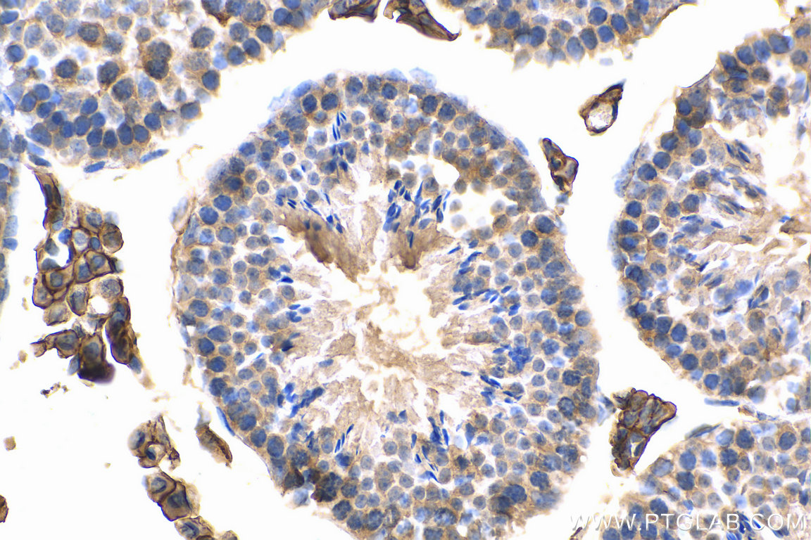
Immunohistochemical analysis of paraffin-embedded mouse testis tissue slide using 15193-1-AP (CD98/SLC3A2 antibody) at dilution of 1:200 (under 40x lens). Heat mediated antigen retrieval with Tris-EDTA buffer (pH 9.0).
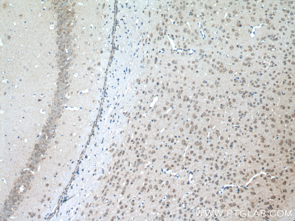
Immunohistochemical analysis of paraffin-embedded mouse brain tissue slide using 20507-1-AP (SLC11A2 antibody) at dilution of 1:200 (under 10x lens. Heat mediated antigen retrieval with Tris-EDTA buffer (pH 9.0).
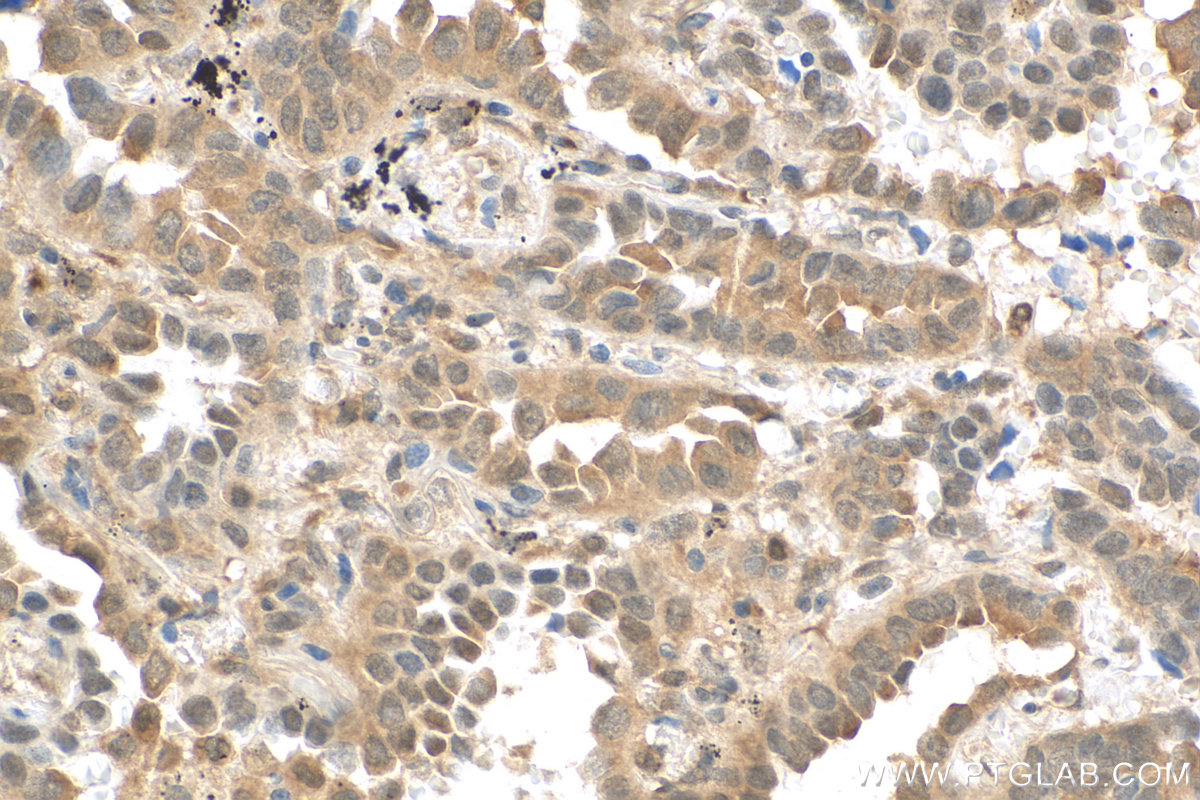
Immunohistochemical analysis of paraffin-embedded human lung cancer tissue slide using 80744-1-RR (KEAP1 antibody) at dilution of 1:1000 (under 40x lens). Heat mediated antigen retrieval with Tris-EDTA buffer (pH 9.0).
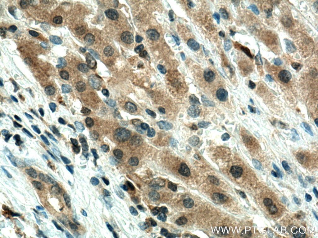
Immunohistochemical analysis of paraffin-embedded human liver cancer tissue slide using 10701-1-AP (HMOX1 antibody at dilution of 1:200 (under 40x lens). Heat mediated antigen retrieval with Tris-EDTA buffer (pH 9.0).
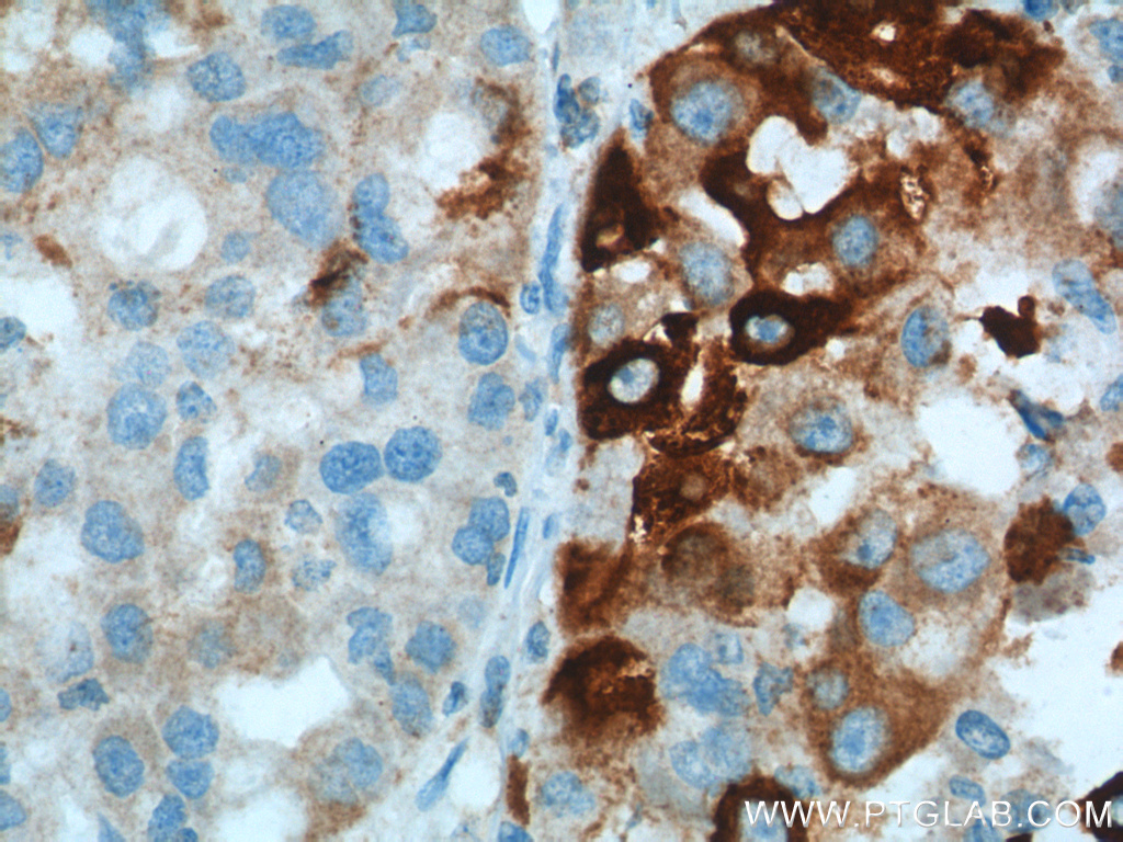
Immunohistochemical analysis of paraffin-embedded human placenta slide using 22401-1-AP (ACSL4 antibody at dilution of 1:50.
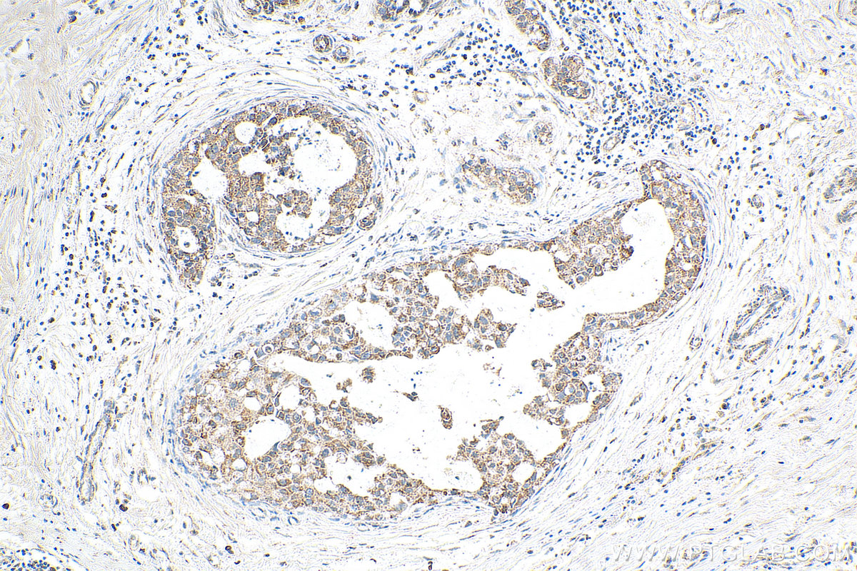
Immunohistochemical analysis of paraffin-embedded human breast cancer tissue slide using 14877-1-AP (DHODH antibody) at dilution of 1:200 (under 10x lens). Heat mediated antigen retrieval with Tris-EDTA buffer (pH 9.0).
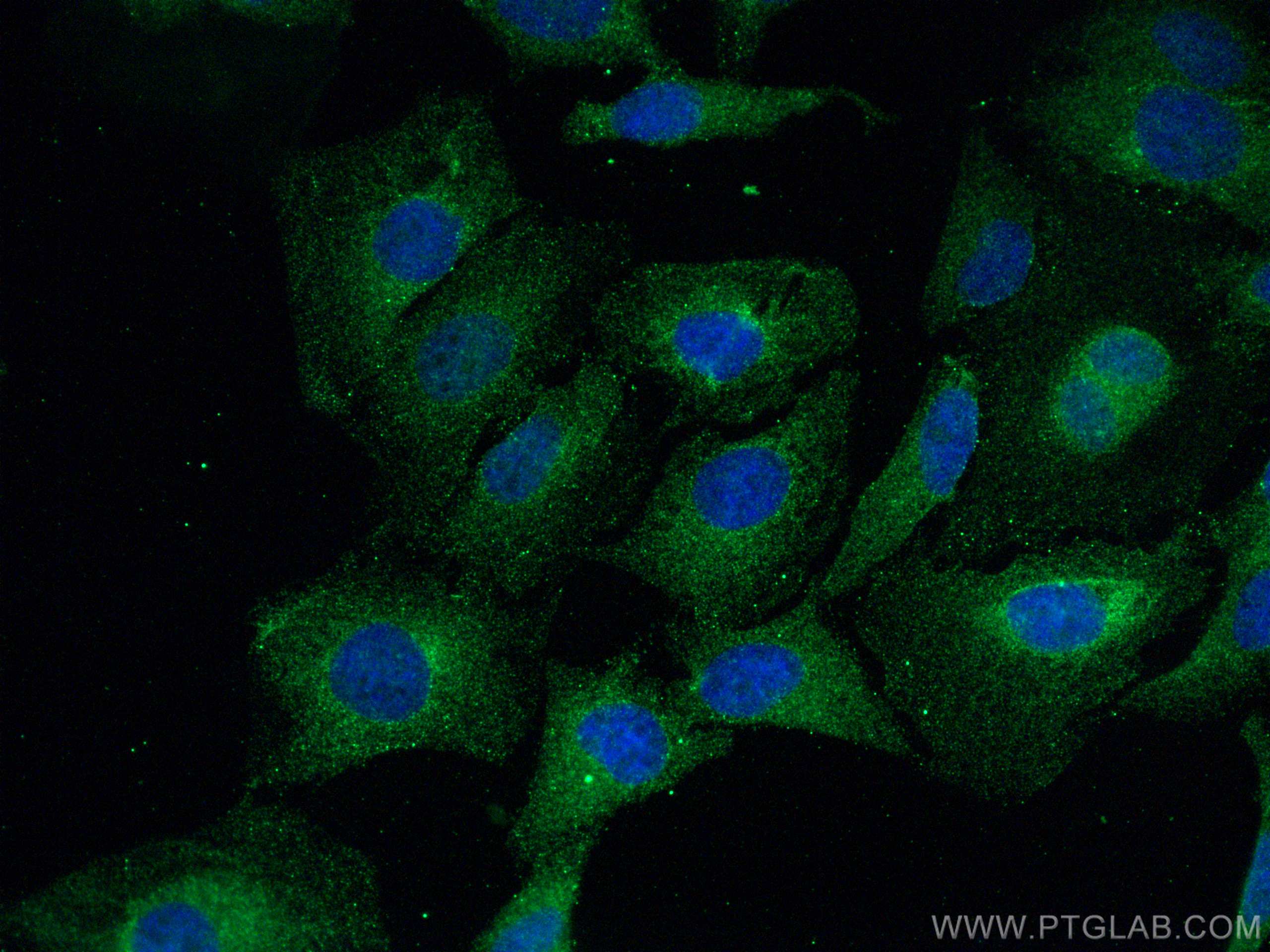
Immunofluorescent analysis of (4% PFA) fixed HeLa cells using GPX4 antibody (67763-1-Ig, Clone: 3F5G5 ) at dilution of 1:800 and CoraLite®488-Conjugated AffiniPure Goat Anti-Mouse IgG(H+L).
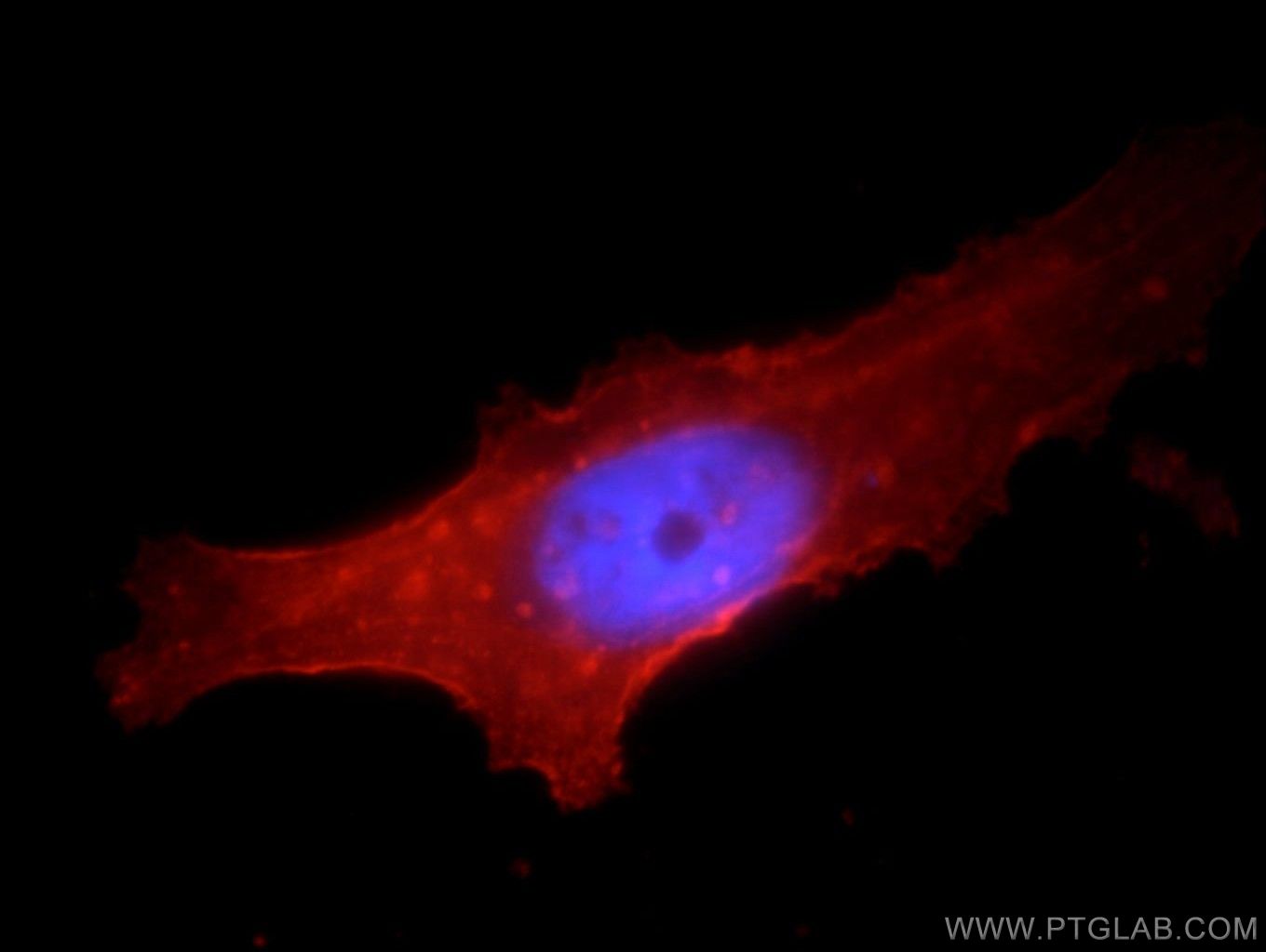
Immunofluorescent analysis of HepG2 cells, using SLC3A2 antibody 15193-1-AP at 1:25 dilution and Rhodamine-labeled goat anti-rabbit IgG (red). Blue pseudocolor = DAPI (fluorescent DNA dye).
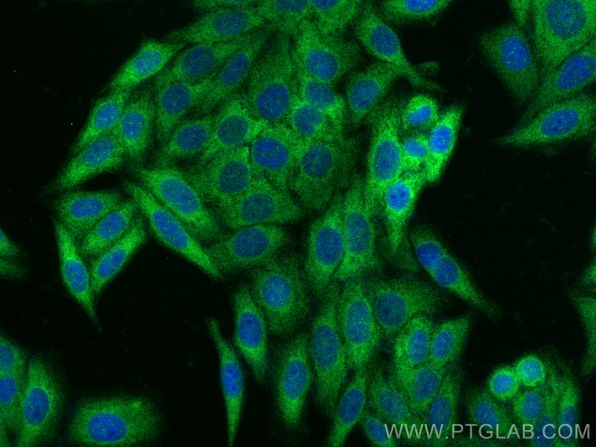
Immunofluorescent analysis of (-20°C Ethanol) fixed HepG2 cells using KEAP1 antibody (80744-1-RR, Clone: 5O18 ) at dilution of 1:1000 and CoraLite®488-Conjugated AffiniPure Goat Anti-Rabbit IgG(H+L).
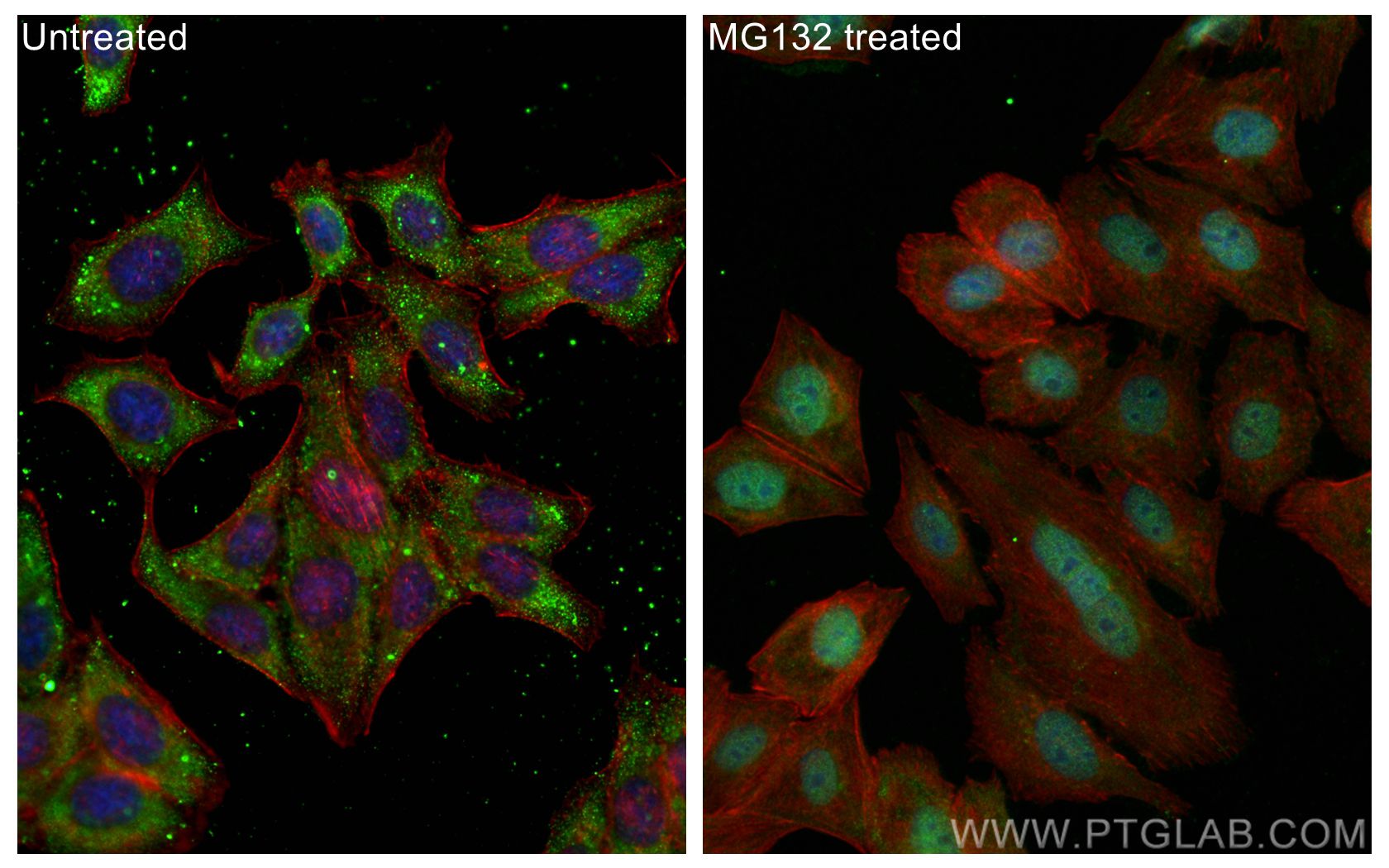
Immunofluorescent analysis of (-20°C Ethanol) fixed MG132 treated HepG2 cells using NRF2, NFE2L2 antibody (80593-1-RR, Clone: 1I21 ) at dilution of 1:600 and CoraLite®488-Conjugated AffiniPure Goat Anti-Rabbit IgG(H+L), CL594-Phalloidin (red).
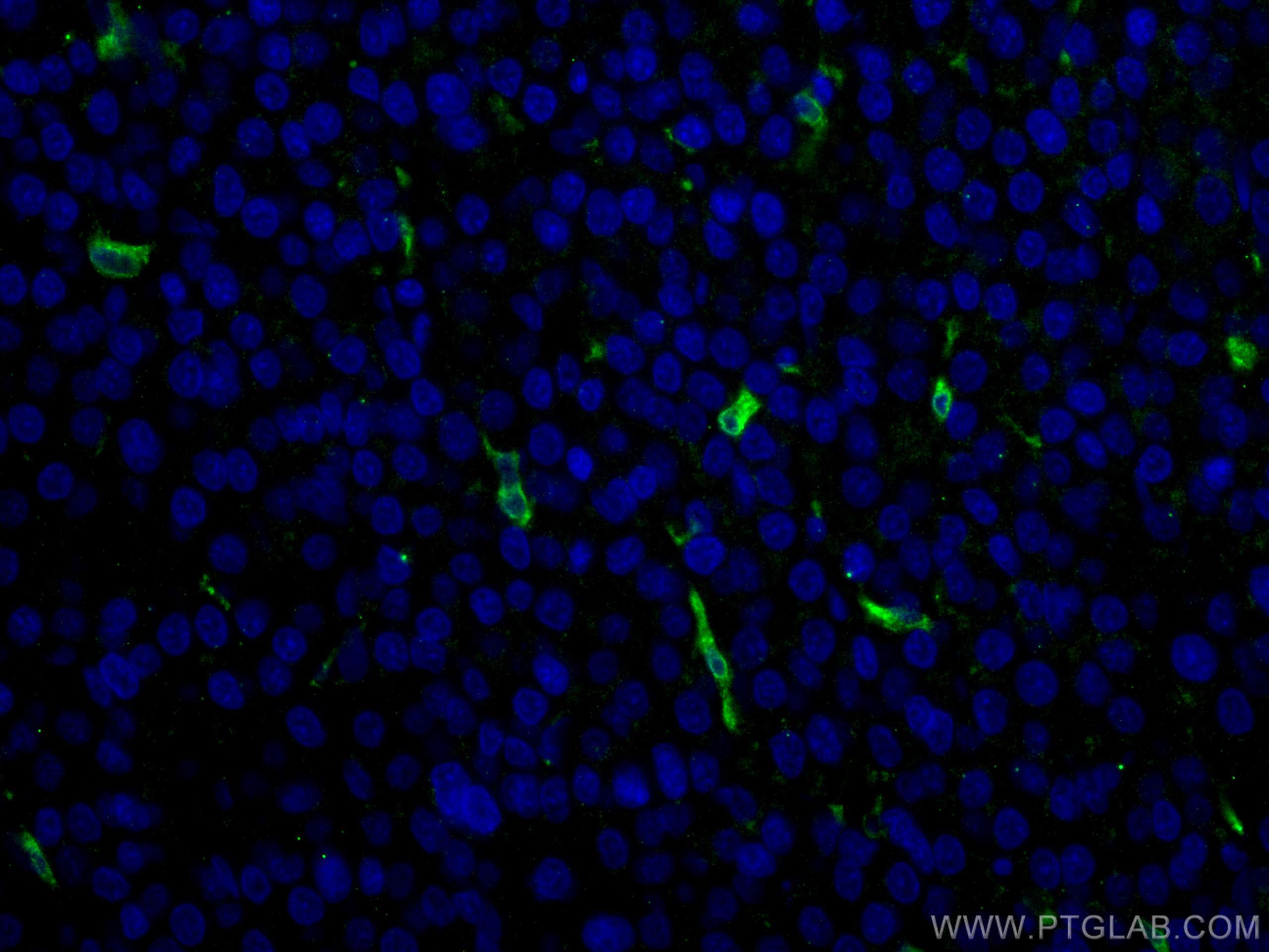
Immunofluorescent analysis of (4% PFA) fixed human liver cancer tissue using HO-1/HMOX1 antibody (10701-1-AP) at dilution of 1:400 and CoraLite®488-Conjugated AffiniPure Goat Anti-Rabbit IgG(H+L).
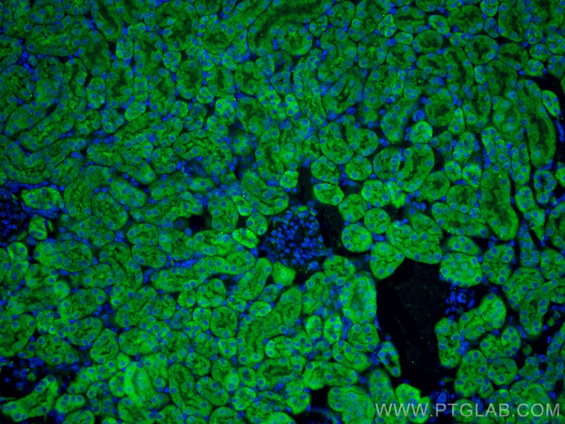
Immunofluorescent analysis of (4% PFA) fixed mouse kidney tissue using DHODH antibody (14877-1-AP) at dilution of 1:200 and CoraLite®488-Conjugated AffiniPure Goat Anti-Rabbit IgG(H+L).
