Recombinant Human Fas/CD95 protein (His Tag)
种属
Human
纯度
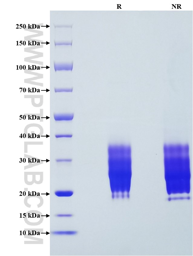
>95 %, SDS-PAGE
标签
His Tag
生物活性
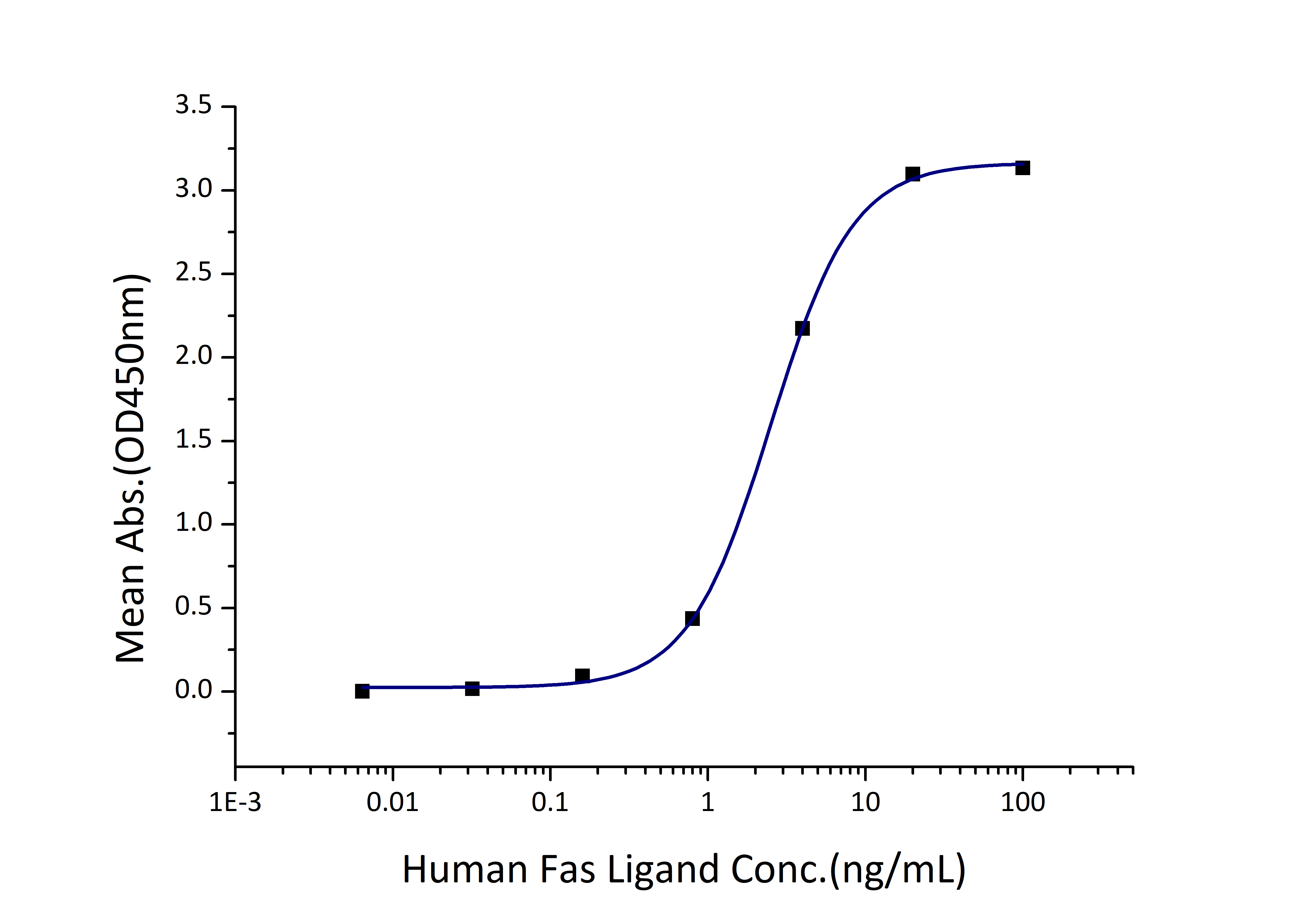
EC50: 1-5 ng/mL
验证数据展示
产品信息
| 纯度 | >95 %, SDS-PAGE |
| 内毒素 | <0.1 EU/μg protein, LAL method |
| 生物活性 | Immobilized Human Fas (His tag) at 2 μg/mL (100 μL/well) can bind Human Fas Ligand (hFc tag) with a linear range of 1-5 ng/mL. |
| 来源 | HEK293-derived Human Fas protein Gln26-Asn173 (Accession# P25445-1) with His tag at the C-terminus. |
| 基因ID | 355 |
| 蛋白编号 | P25445-1 |
| 预测分子量 | 17.4 kDa |
| SDS-PAGE | 20-36 kDa, reducing (R) conditions |
| 组分 | Lyophilized from 0.22 μm filtered solution in PBS, pH 7.4. Normally 5% trehalose and 5% mannitol are added as protectants before lyophilization. |
| 复溶 | Briefly centrifuge the tube before opening. Reconstitute at 0.1-0.5 mg/mL in sterile water. |
| 储存条件 |
It is recommended that the protein be aliquoted for optimal storage. Avoid repeated freeze-thaw cycles.
|
| 运输条件 | The product is shipped at ambient temperature. Upon receipt, store it immediately at the recommended temperature. |
背景信息
Fas, also known as TNFRSF6, CD95, and APO-1, is a transmembrane glycoprotein belonging to the tumor necrosis factor (TNF) receptor superfamily. It can mediate apoptosis by ligation with an agonistic anti-Fas antibody or Fas ligand. Stimulation of Fas results in the aggregation of its intracellular death domains, leading to the formation of the death-inducing signaling complex (DISC). FAS-mediated apoptosis plays a role in the maintenance of cell homeostasis and in the deletion of useless or autoreactive T cells. Alterations in the CD95/CD95L pathway have been involved in several disease conditions, including autoimmune diseases, chronic inflammation and cancer.
参考文献:
1.N Itoh. et al. (1991). Cell. 66(2):233-43.
2.M E Peter. et al. (2003). Cell Death Differ. 10(1):26-35.
3.M E Peter. et al. (2015). Cell Death Differ. 22(4):549-59.
4.Vesna Risso. et al. (2022). Cell Death Dis. 13(3):248.

