验证数据展示
 at dilution of 1:2000 (under 40x lens). Heat mediated antigen retrieval with Tris-EDTA buffer (pH 9.0).")
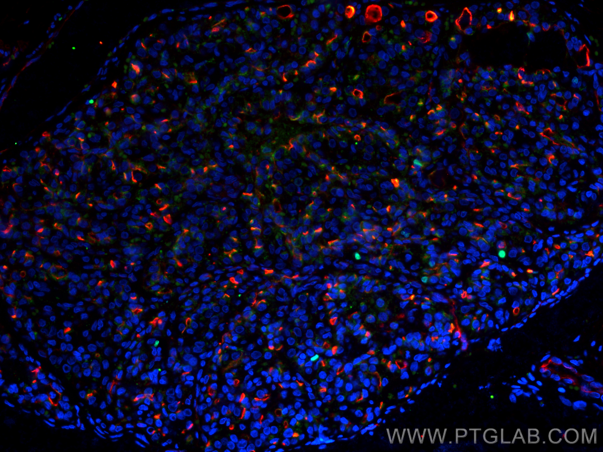
 fixed paraffin-embedded human lung cancer tissue using KI67 antibody (84192-3-RR, Clone: 241499B1 ) at dilution of 1:200 and Multi-rAb CoraLite ® Plus 488-Goat Anti-Rabbit Recombinant Secondary Antibody (H+L) (<a class='green' href='/productredirect?CatalogNo=RGAR002' target='_blank'>RGAR002</a>), ICAM-1 antibody (<a class='green' href='/productredirect?CatalogNo=60299-1-Ig' target='_blank'>60299-1-Ig</a>, Clone: 2F9A8, red). Heat mediated antigen retrieval with Tris-EDTA buffer (pH 9.0).")
 fixed paraffin-embedded human lung cancer tissue using KI67 antibody (84192-3-RR, Clone: 241499B1 ) at dilution of 1:200 and Multi-rAb CoraLite ® Plus 488-Goat Anti-Rabbit Recombinant Secondary Antibody (H+L) (<a class='green' href='/productredirect?CatalogNo=RGAR002' target='_blank'>RGAR002</a>), ICAM-1 antibody (<a class='green' href='/productredirect?CatalogNo=60299-1-Ig' target='_blank'>60299-1-Ig</a>, Clone: 2F9A8, red). Heat mediated antigen retrieval with Tris-EDTA buffer (pH 9.0).")
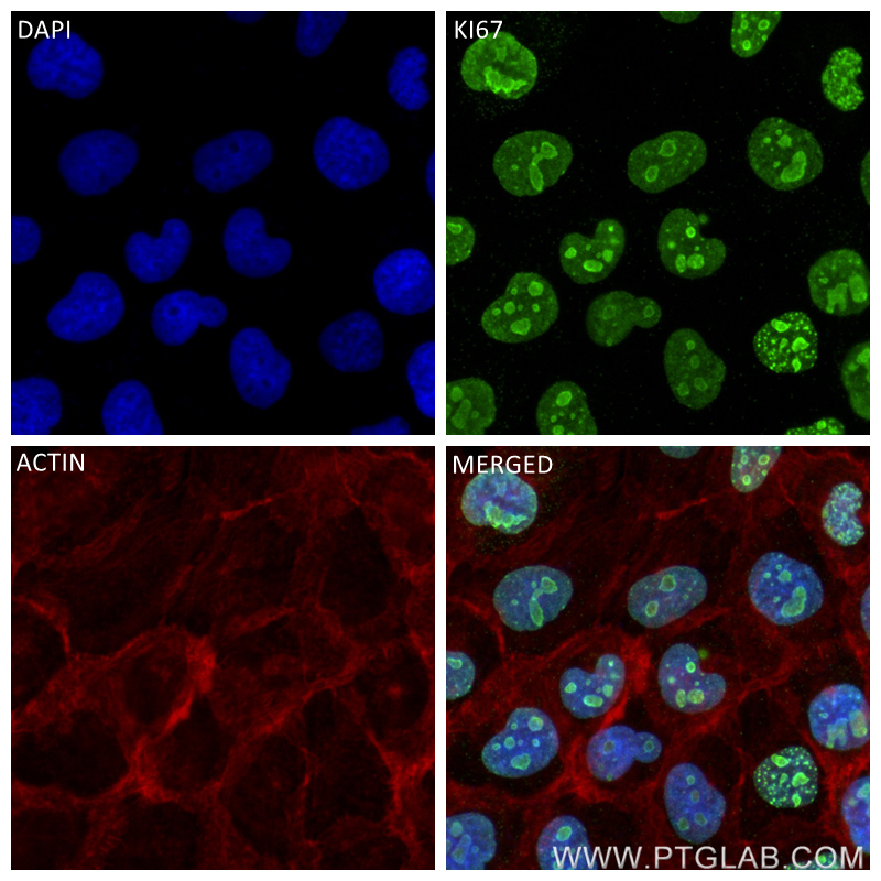
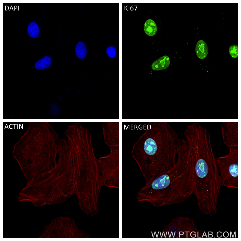
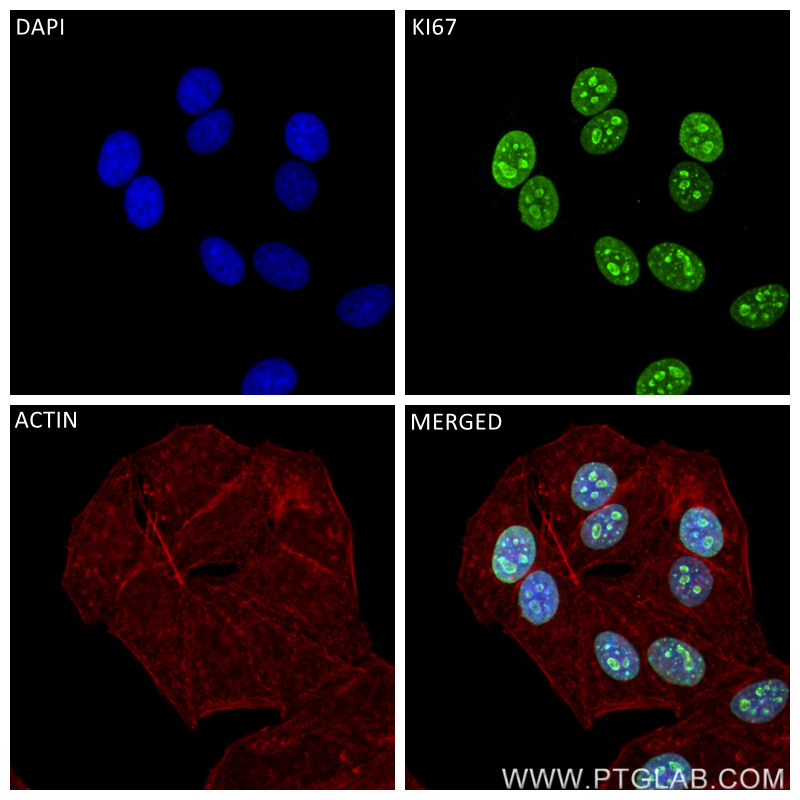
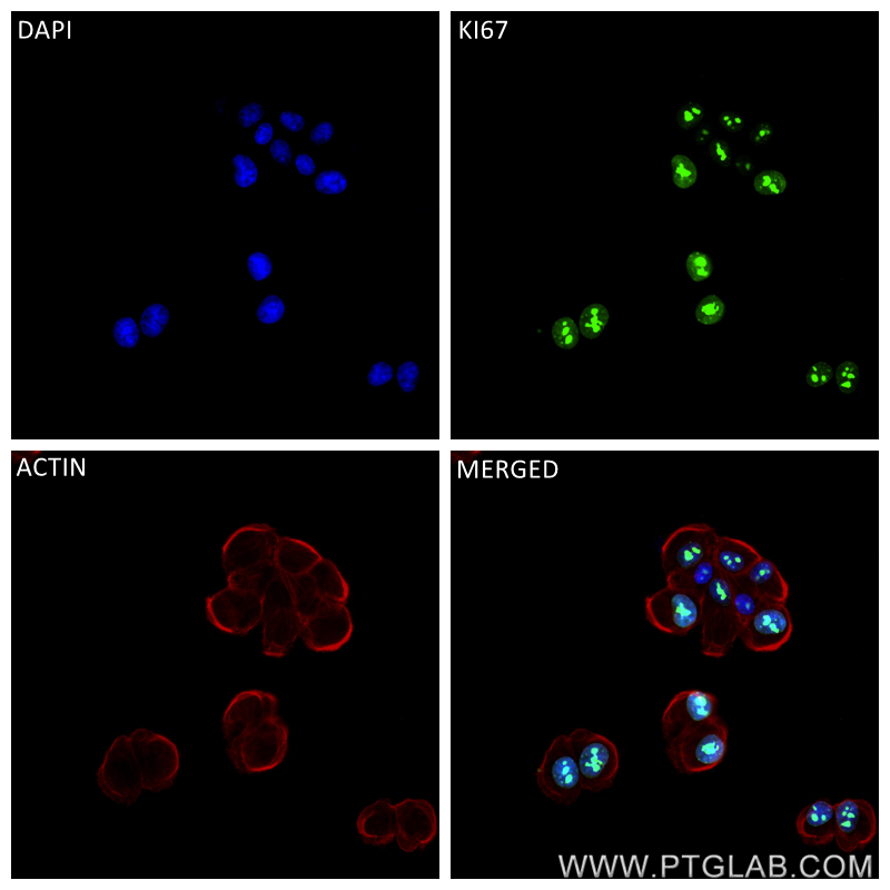
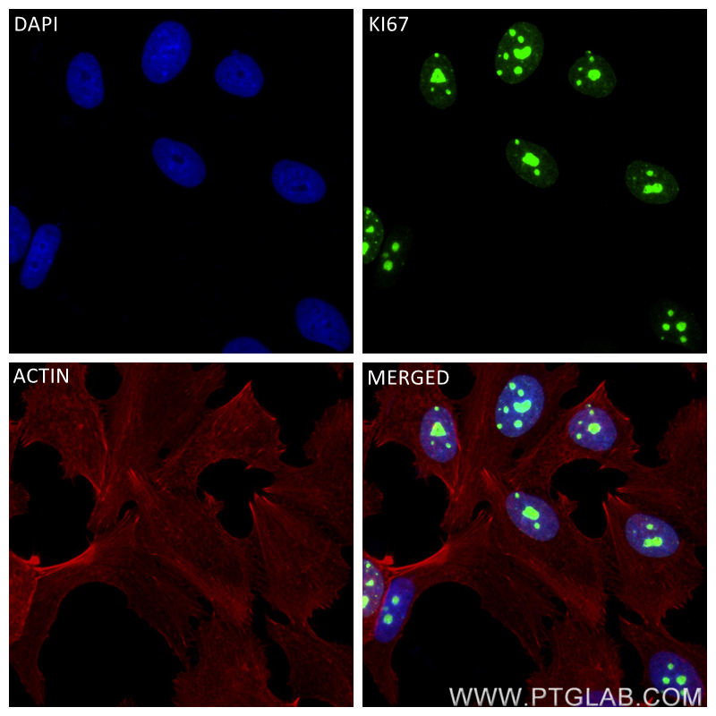
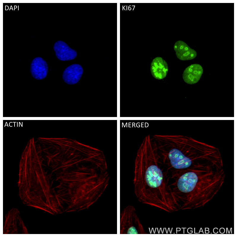
 fixed HeLa cells using KI67 antibody (84192-3-RR, Clone: 241499B1 ) at dilution of 1:250 and CoraLite®488-Conjugated Goat Anti-Rabbit IgG(H+L) (<a class='green' href='/productredirect?CatalogNo=SA00013-2' target='_blank'>SA00013-2</a>), CL594-Phalloidin (red).")
 at dilution of 1:16000 incubated at room temperature for 1.5 hours.")
经过测试的应用
| Positive WB detected in | MCF-7 cells, HeLa cells |
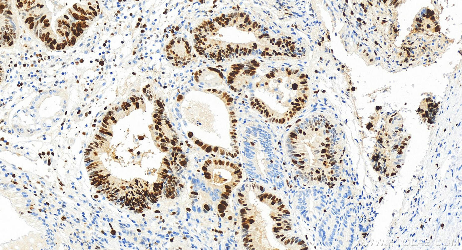
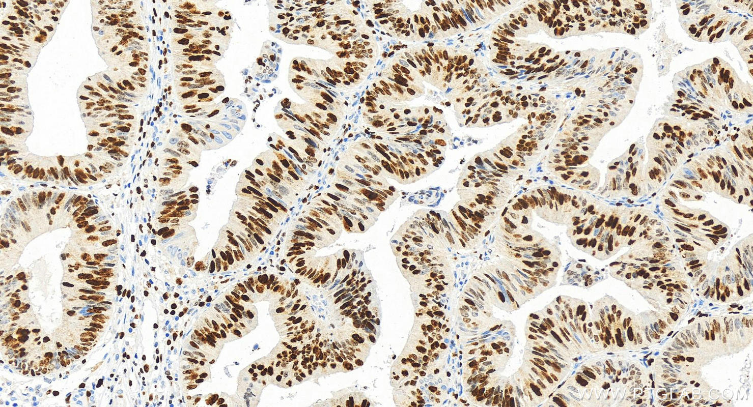
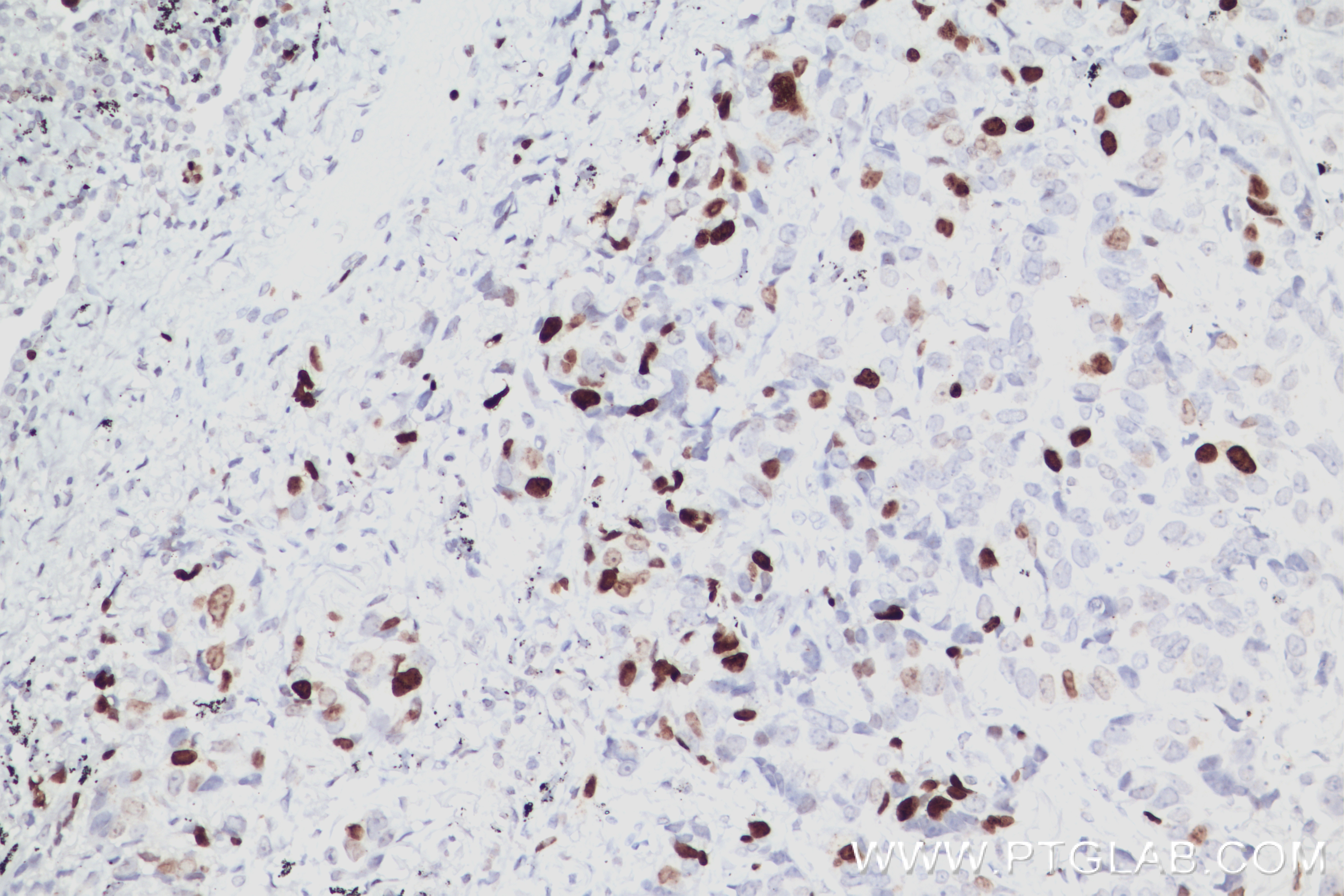
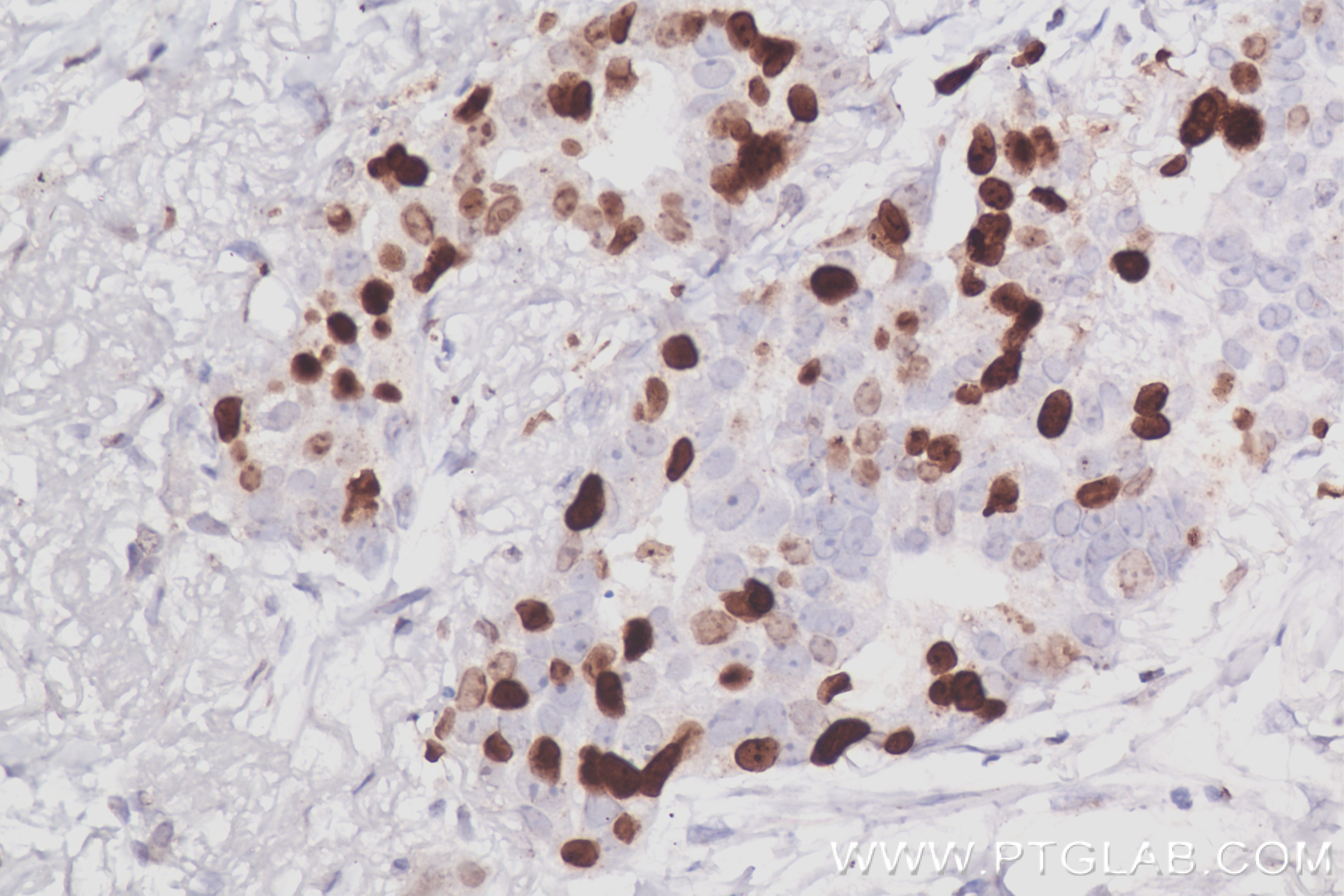
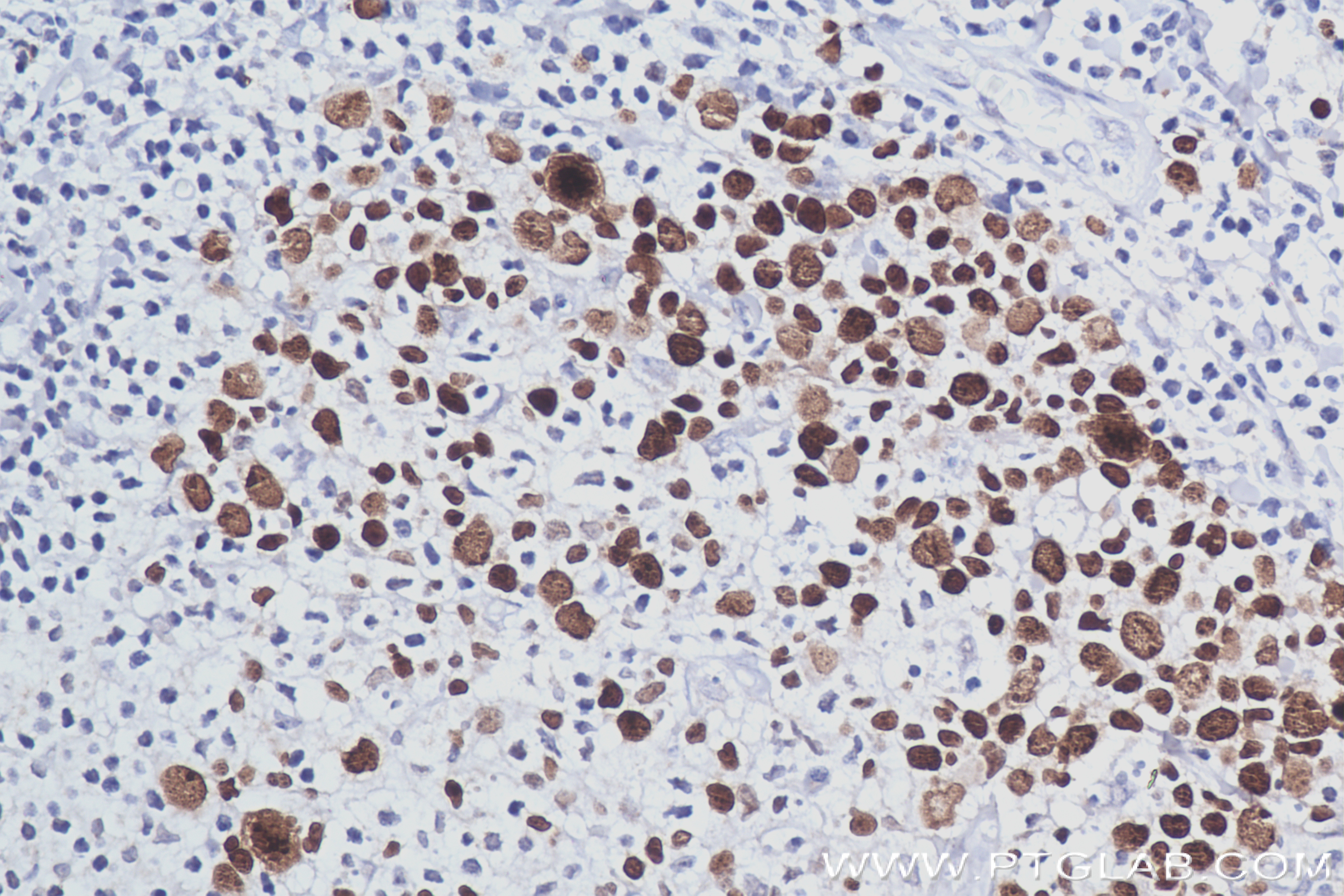
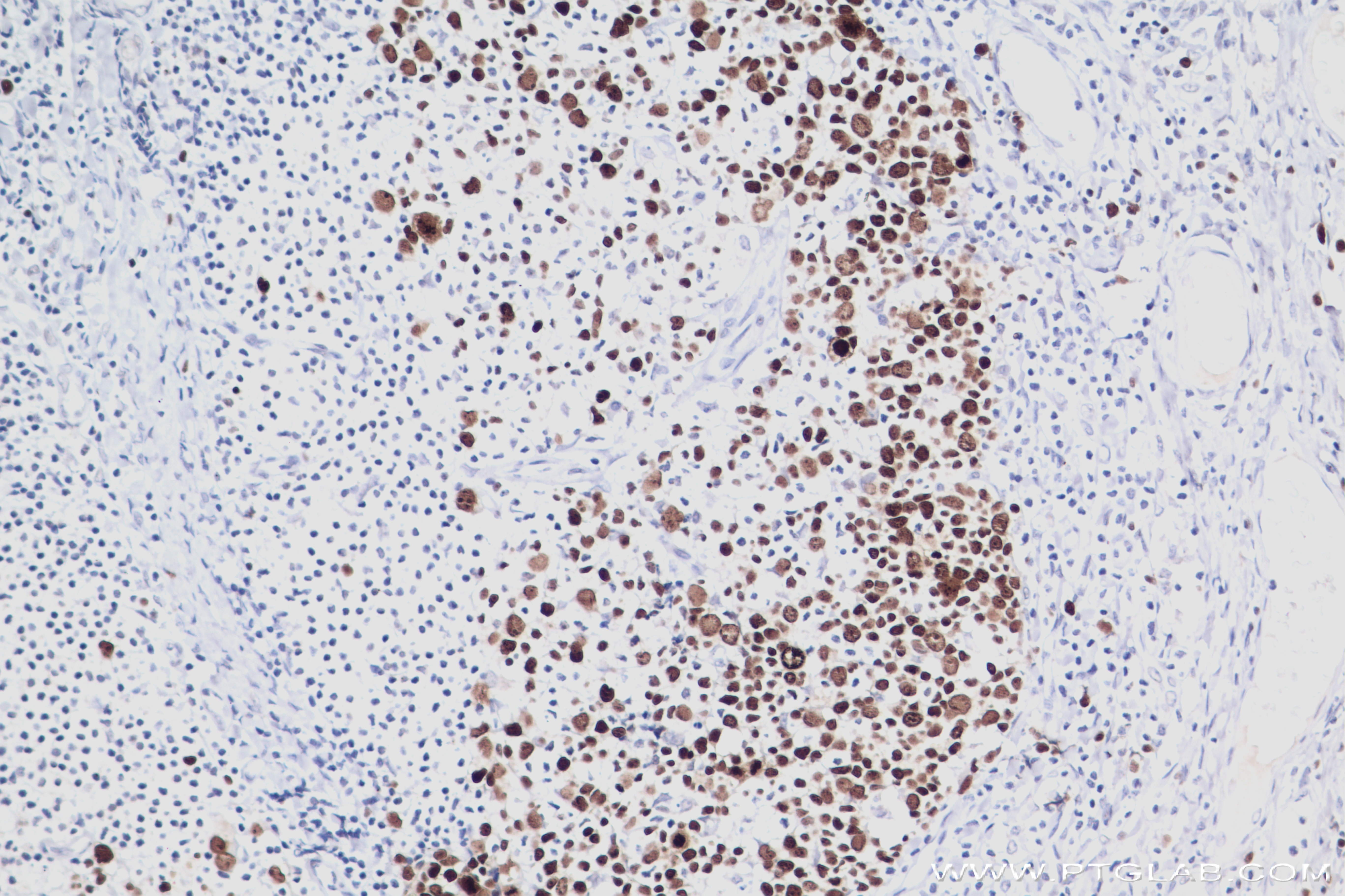
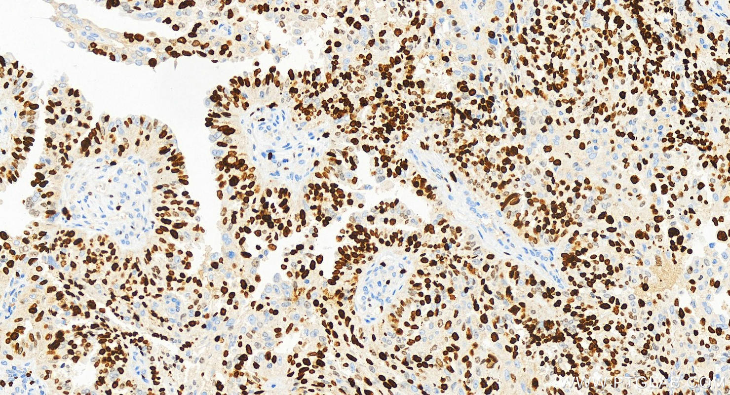
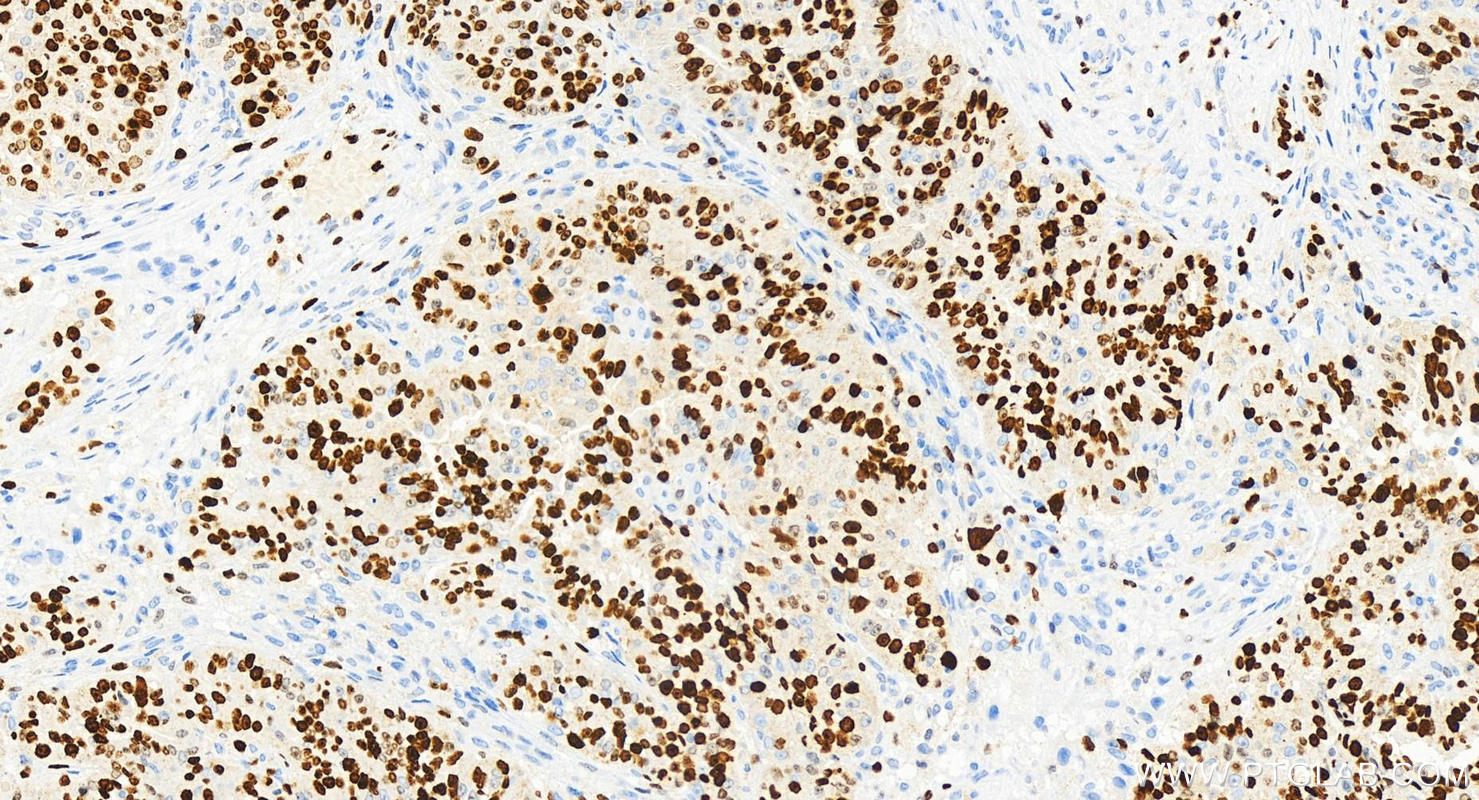
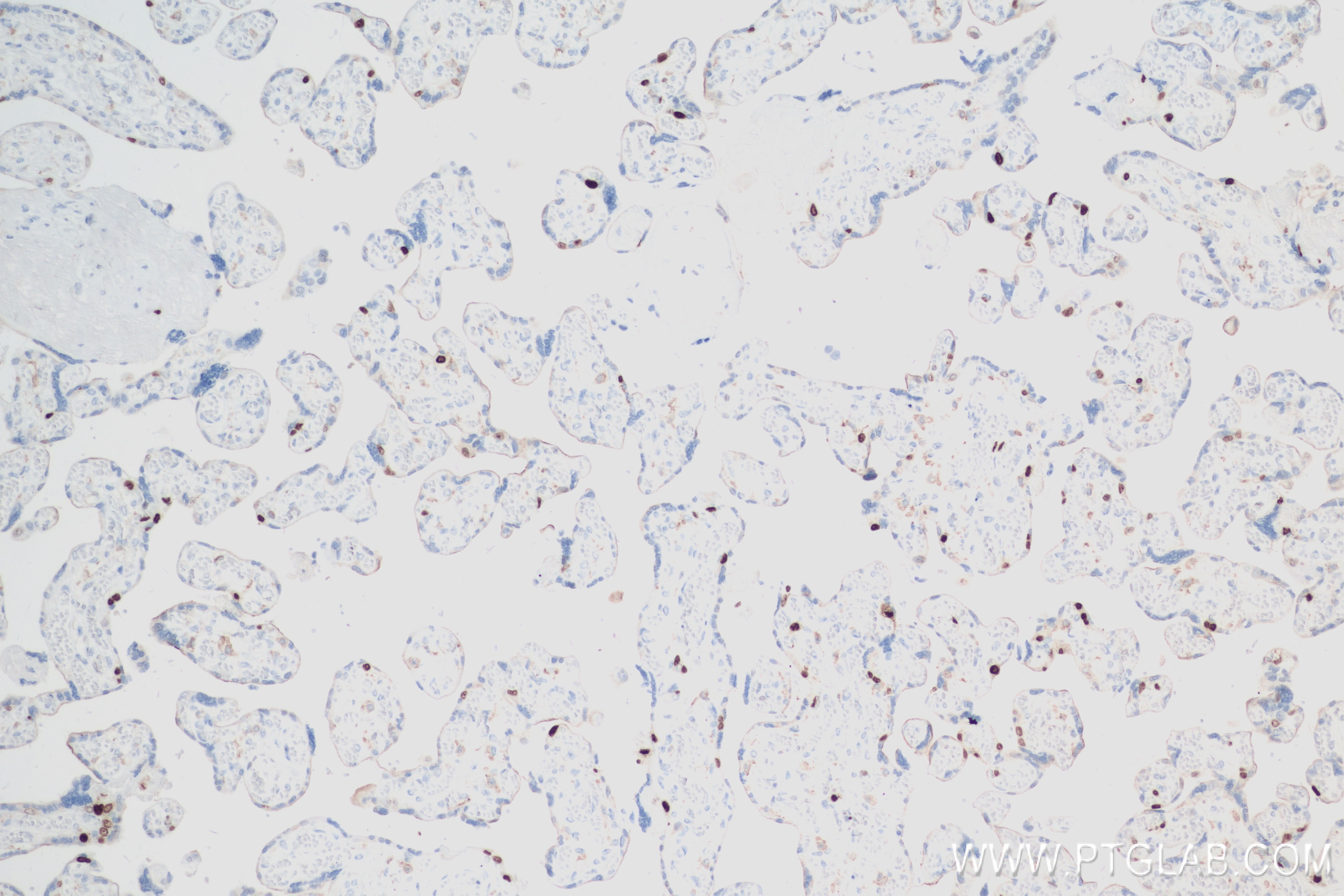
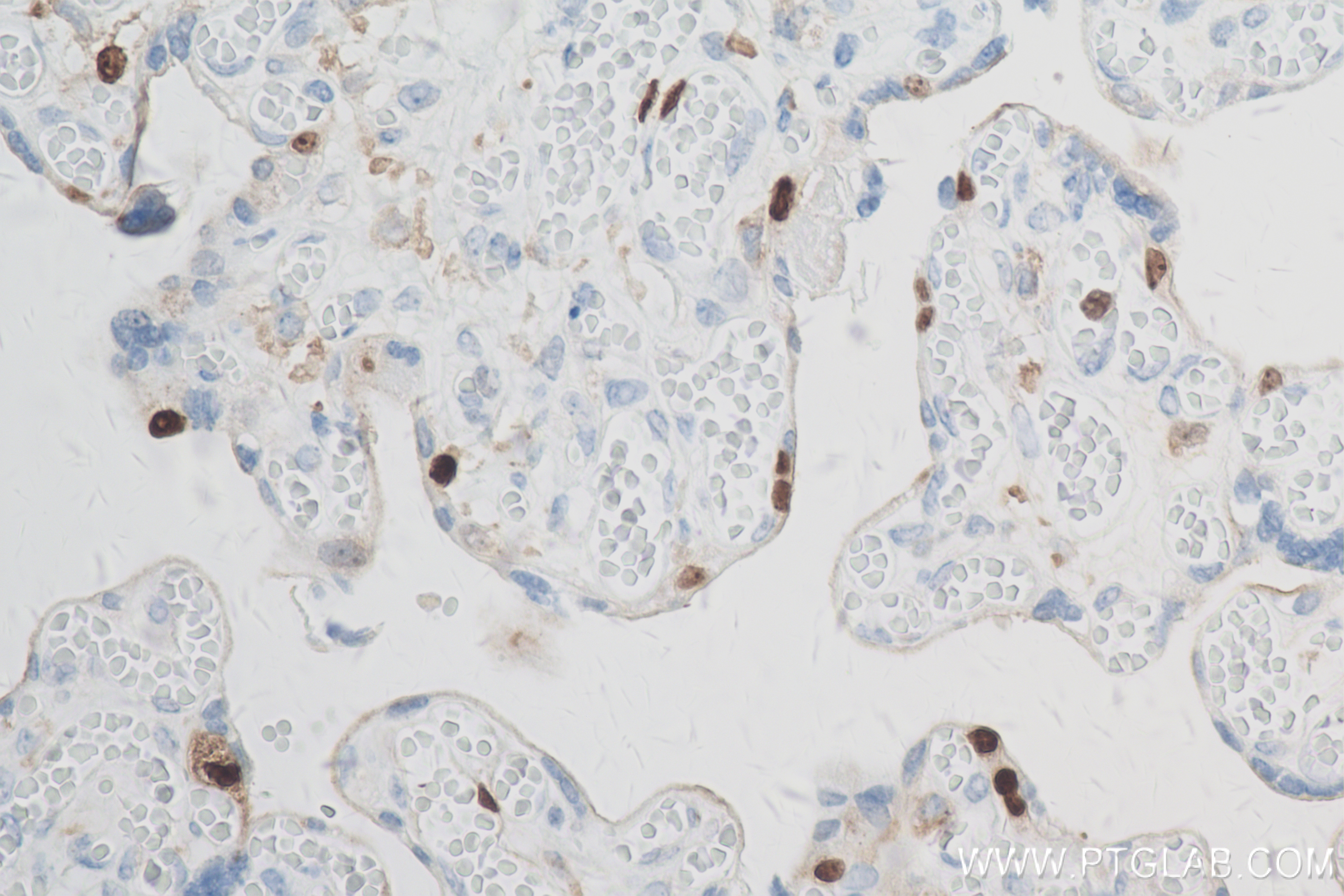
| Positive IHC detected in | human tonsillitis tissue, human colon cancer tissue, human lung cancer tissue, human malignant melanoma tissue, human ovarian cancer, human placenta tissue Note: suggested antigen retrieval with TE buffer pH 9.0; (*) Alternatively, antigen retrieval may be performed with citrate buffer pH 6.0 |
| Positive IF-P detected in | human lung cancer tissue, human thyroid cancer tissue |
| Positive IF/ICC detected in | HeLa cells, U2OS cells, MCF-7 cells, hTERT-RPE1 cells, HepG2 cells, A549 cells, A431 cells |
推荐稀释比
| Application | Dilution |
|---|---|
| Western Blot (WB) | WB : 1:5000-1:50000 |
| Immunohistochemistry (IHC) | IHC : 1:1000-1:4000 |
| Immunofluorescence (IF)-P | IF-P : 1:50-1:500 |
| Immunofluorescence (IF)/ICC | IF/ICC : 1:100-1:400 |
| It is recommended that this reagent should be titrated in each testing system to obtain optimal results. | |
| Sample-dependent, Check data in validation data gallery. | |
产品信息
84192-3-RR targets Ki-67 in WB, IHC, IF/ICC, IF-P, ELISA applications and shows reactivity with human samples.
| Tested Applications | WB, IHC, IF/ICC, IF-P, ELISA Application Description |
| Tested Reactivity | human |
| Immunogen | Peptide 种属同源性预测 |
| Host / Isotype | Rabbit / IgG |
| Class | Recombinant |
| Type | Antibody |
| Full Name | antigen identified by monoclonal antibody Ki-67 |
| Synonyms | MKI67, KI67, Ki 67, Antigen KI-67, Antigen identified by monoclonal antibody Ki-67 |
| Calculated Molecular Weight | 359 kDa |
| GenBank Accession Number | NM_002417 |
| Gene Symbol | KI67 |
| Gene ID (NCBI) | 4288 |
| Conjugate | Unconjugated |
| Form | Liquid |
| Purification Method | Protein A purification |
| UNIPROT ID | P46013 |
| Storage Buffer | PBS with 0.02% sodium azide and 50% glycerol pH 7.3. |
| Storage Conditions | Store at -20°C. Stable for one year after shipment. Aliquoting is unnecessary for -20oC storage. |
背景介绍
The Ki-67 protein (also known as MKI67) is a cellular marker for proliferation. Ki67 is present during all active phases of the cell cycle (G1, S, G2 and M), but is absent in resting cells (G0). Cellular content of Ki-67 protein markedly increases during cell progression through S phase of the cell cycle. Therefore, the nuclear expression of Ki67 can be evaluated to assess tumor proliferation by immunohistochemistry. It has been demonstrated to be of prognostic value in breast cancer. In head and neck cancer, several studies have reported an association between high proliferative activity and poorer prognosis.
实验方案
| Product Specific Protocols | |
|---|---|
| WB protocol for Ki-67 antibody 84192-3-RR | Download protocol |
| IHC protocol for Ki-67 antibody 84192-3-RR | Download protocol |
| IF protocol for Ki-67 antibody 84192-3-RR | Download protocol |
| Standard Protocols | |
|---|---|
| Click here to view our Standard Protocols |