验证数据展示
 at dilution of 1:2000 incubated at room temperature for 1.5 hours. This data was developed using the same antibody clone with 82719-15-PBS in a different storage buffer formulation.")
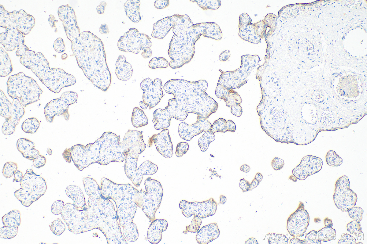
 at dilution of 1:400 (under 20x lens). Heat mediated antigen retrieval with Tris-EDTA buffer (pH 9.0). This data was developed using the same antibody clone with 82719-15-PBS in a different storage buffer formulation.")
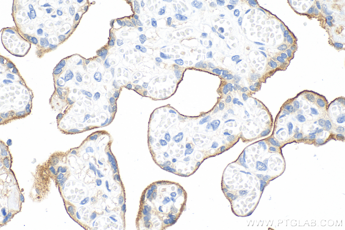
 at dilution of 1:400 (under 20x lens). Heat mediated antigen retrieval with Tris-EDTA buffer (pH 9.0). This data was developed using the same antibody clone with 82719-15-PBS in a different storage buffer formulation.")
 fixed paraffin-embedded human tonsillitis tissue using PD-L1/CD274 antibody (<a class='green' href='/productredirect?CatalogNo=82719-15-RR' target='_blank'>82719-15-RR</a>, Clone: 2H4 ) at dilution of 1:400 and CoraLite®488-Conjugated Goat Anti-Rabbit IgG(H+L) (<a class='green' href='/productredirect?CatalogNo=SA00013-2' target='_blank'>SA00013-2</a>). Heat mediated antigen retrieval with Tris-EDTA buffer (pH 9.0). This data was developed using the same antibody clone with 82719-15-PBS in a different storage buffer formulation.")
 kinetic assays of <a class='green' href='/productredirect?CatalogNo=82719-15-RR' target='_blank'>82719-15-RR</a> against Human PD-L1/CD274 were performed. The affinity constant is below 1 pM.")
产品信息
82719-15-PBS targets PD-L1/CD274 in WB, IHC, IF-P, Indirect ELISA applications and shows reactivity with human samples.
| 经测试应用 | WB, IHC, IF-P, Indirect ELISA Application Description |
| 经测试反应性 | human |
| 免疫原 | PD-L1/CD274 fusion protein Ag12432 种属同源性预测 |
| 宿主/亚型 | Rabbit / IgG |
| 抗体类别 | Recombinant |
| 产品类型 | Antibody |
| 全称 | CD274 molecule |
| 别名 | CD274, PD-L1, PD L1, hPD-L1, B7-H1 |
| 计算分子量 | 290 aa, 33 kDa |
| 观测分子量 | 50 kDa |
| GenBank蛋白编号 | BC074984 |
| 基因名称 | PD-L1 |
| Gene ID (NCBI) | 29126 |
| 偶联类型 | Unconjugated |
| 形式 | Liquid |
| 纯化方式 | Protein A purification |
| UNIPROT ID | Q9NZQ7 |
| 储存缓冲液 | PBS only , pH 7.3 |
| 储存条件 | Store at -80°C. The product is shipped with ice packs. Upon receipt, store it immediately at -80°C |
背景介绍
PD-L1, also known as CD274 or B7H1, stands for programmed cell death ligand 1. It is a type I transmembrane protein that is thought to repress immune responses by binding to its receptor (PD1), thus inhibiting T-cell activation, proliferation, and cytokine production. It contains V-like and C-like immunoglobulin domains. PD-L1 expression is regulated by various cytokines, such as TNF-α or LPS (ISSN: 1848-7718). Increased expression of this protein in certain types of cancers, e.g., renal cell carcinoma or colon cancer, correlates with poor prognosis.
What is the molecular weight of PD-L1?
Depending on the isoform, the calculated molecular weight of the protein varies between 20 and 33 kDa (176-290 aa).
What are the isoforms of PD-L1?
According to NCBI, three different isoforms have been identified. There are significant differences in the untranslated and protein coding regions.
What is the subcellular localization and tissue specificity of PD-L1?
It is predicted to localize in the plasma membrane of various cell types, with a particularly high expression in placental trophoblast and subsets of immune cells. High levels of PD-L1 protein have also been detected in lung and colon tissues.
What is the function of PD-L1 in immune responses?
PD-L1 is critical for the induction and maintenance of immune self-tolerance during infection or inflammation in normal tissues. The interaction of PD-L1 and its receptors is responsible for preventing auto-immune phenotypes and balancing the overall immune response in situations such as pregnancy or tissue allografts. The interaction between PD-L1 and PD-1 or B7.1 starts an inhibitory signaling cascade, which results in the decreased proliferation of antigen-specific T-cells and increased survival of regulatory T-cells (PMID: 15240681).
How can PD-L1's implication in cancer be used as a drug target?
In certain tumors, high expression of PD-L1 serves as a stop-sign to inhibit the recognition of cancer cells by T-cells (PMID: 23087408). The interaction between PD-L1 and its receptors (PD1 and B7.1) is a mechanism for the tumor to evade the host immune response (PMID: 29357948). Several mAbs have been developed to target that interaction and thus prevent the inactivation of cytotoxic T-cells by the tumor (PMIDs: 23890059, 18173375).