验证数据展示
 at dilution of 1:800 incubated at room temperature for 1.5 hours.")
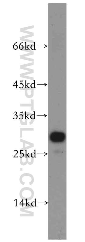
 with mouse skeletal muscle tissue lysate 4000ug.")
经过测试的应用
| Positive WB detected in | mouse skeletal muscle tissue |
| Positive IP detected in | mouse skeletal muscle tissue |
推荐稀释比
| 应用 | 推荐稀释比 |
|---|---|
| Western Blot (WB) | WB : 1:500-1:1000 |
| Immunoprecipitation (IP) | IP : 0.5-4.0 ug for 1.0-3.0 mg of total protein lysate |
| It is recommended that this reagent should be titrated in each testing system to obtain optimal results. | |
| Sample-dependent, Check data in validation data gallery. | |
发表文章中的应用
| WB | See 3 publications below |
产品信息
20635-1-AP targets PPAPDC3 in WB, IP, ELISA applications and shows reactivity with human, mouse, rat samples.
| 经测试应用 | WB, IP, ELISA Application Description |
| 文献引用应用 | WB |
| 经测试反应性 | human, mouse, rat |
| 文献引用反应性 | human |
| 免疫原 | PPAPDC3 fusion protein Ag14676 种属同源性预测 |
| 宿主/亚型 | Rabbit / IgG |
| 抗体类别 | Polyclonal |
| 产品类型 | Antibody |
| 全称 | phosphatidic acid phosphatase type 2 domain containing 3 |
| 别名 | C9orf67, KIAA0515, NET39, PPAPDC3 |
| 计算分子量 | 271 aa, 29 kDa |
| 观测分子量 | 29 kDa |
| GenBank蛋白编号 | BC006362 |
| 基因名称 | PPAPDC3 |
| Gene ID (NCBI) | 84814 |
| RRID | AB_10696180 |
| 偶联类型 | Unconjugated |
| 形式 | Liquid |
| 纯化方式 | Antigen affinity purification |
| UNIPROT ID | Q8NBV4 |
| 储存缓冲液 | PBS with 0.02% sodium azide and 50% glycerol , pH 7.3 |
| 储存条件 | Store at -20°C. Stable for one year after shipment. Aliquoting is unnecessary for -20oC storage. |
背景介绍
PPAPDC3(Phosphatidic acid phosphatase type 2 domain-containing protein 3) is also named as C9orf67 and belongs to the PA-phosphatase related phosphoesterase family. It plays a role as negative regulator of myoblast differentiation, in part through effects on MTOR signaling.
实验方案
| Product Specific Protocols | |
|---|---|
| WB protocol for PPAPDC3 antibody 20635-1-AP | Download protocol |
| IP protocol for PPAPDC3 antibody 20635-1-AP | Download protocol |
| Standard Protocols | |
|---|---|
| Click here to view our Standard Protocols |
发表文章
| Species | Application | Title |
|---|---|---|
Genome Biol Specific nuclear envelope transmembrane proteins can promote the location of chromosomes to and from the nuclear periphery. | ||
Cells Lamin A/C Assembly Defects in LMNA-Congenital Muscular Dystrophy Is Responsible for the Increased Severity of the Disease Compared with Emery-Dreifuss Muscular Dystrophy. | ||
Front Cell Dev Biol Nuclear envelope transmembrane proteins involved in genome organization are misregulated in myotonic dystrophy type 1 muscle |