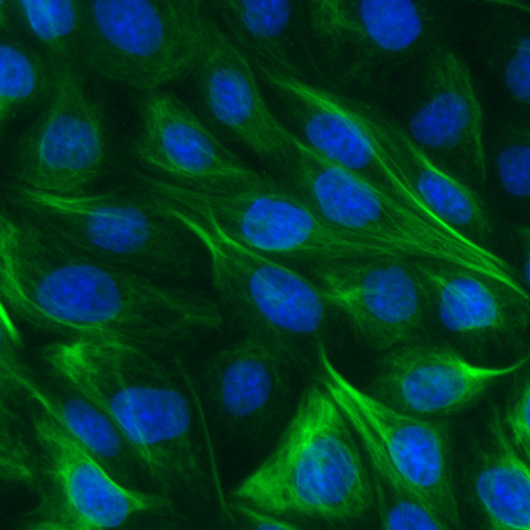
Validation Data Gallery
Product Information
anti-vimentin VHH/ Nanobody conjugated to the fluorophore ATTO488 for IF, & microscopy
| Description | Vimentin-Label specifically binds to the Vimentin intermediate filament protein. Due to its small size, the Vimentin-Labelenables higher image quality in epifluorescence, confocal, and super-resolution microscopy:
• Specific probe for direct immunostaining of Vimentin filaments • Higher image resolution • Less than 4 nm epitope-label displacement minimizes linkage error • Superior accessibility and labeling of epitopes in crowded cellular/organelle environments • Improved target binding due to an avidity effect from the bivalent form of Vimentin-Label |
| Applications | Monoclonal |
| Host | Alpaca |
| Specificity/Target | Vimentin-Label specifically binds to the vimentin intermediate filament protein. Validated on canine MDCK cells, rodent BHK cells and on human A549 cells upon stimulation with TGFβ. |
| Conjugate | ATTO 488 |
| Physical State | Liquid |
| Suggested Dilution | IF/ICC: 1:200 – 1:400 |
| Type | Nanobody |
| Class | Recombinant |
| RRID | AB_2631392 |
| Excitation/ Emission | Excitation range 480 - 510 nm (λabs= 501 nm) Emission range 520 - 560 nm (λfl= 523 nm) |
| Storage Buffer | 1x PBS, 0.09% sodium azide. |
| Storage Condition | Shipped at ambient temperature. Upon receipt store at 4°C. Do not freeze. Protect from light. |
| Size | 10 μL; 100 μL |
| Note | This product is for research use only, not for diagnostic or therapeutic use |
Documentation
| SDS |
|---|
| vba488_SDS_Vimentin-Label ATTO 488 (EN) |
| Datasheet |
|---|
| Vimentin-Label Atto488 Datasheet |
| Brochure |
|---|
| Chromotek nanobodies brochure (PDF) |
Publications
| Application | Title |
|---|---|
Nat Neurosci Panoptic imaging of transparent mice reveals whole-body neuronal projections and skull-meninges connections. | |
Light Sci Appl Repeated photoporation with graphene quantum dots enables homogeneous labeling of live cells with extrinsic markers for fluorescence microscopy. | |
bioRxiv Genetic removal of Nlrp3 protects against sporadic and R345W Efemp1-induced basal laminar deposit formation |


