
Ovarian Cancer
Proteintech offers a wide range of markers and related antibodies for ovarian cancer.
|
Tumor Suppressor Genes and Oncogenes |
Introduction
Epithelial ovarian cancer (EOC) is the most aggressive form of the gynecological cancer types and is the fourth most common cause of female cancer death in developed countries (1). The ovary is a complex tissue, and although ovarian cancers can originate from germ cells or granulosa-theca cells, most ovarian cancers have an epithelial histology.
EOC can be found in women at any age. However, it is most common in women over the age of 50. Unfortunately, the disease is mainly diagnosed at an advanced stage due to a lack of early biomarkers and confusing clinical pathology. Treatment of EOC is mainly based on a combination of surgery with chemotherapy. The survival rate, however, is poor, at <5 years (2). Early diagnosis is therefore fundamental to achieving a higher cure rate.
Histopathological Types
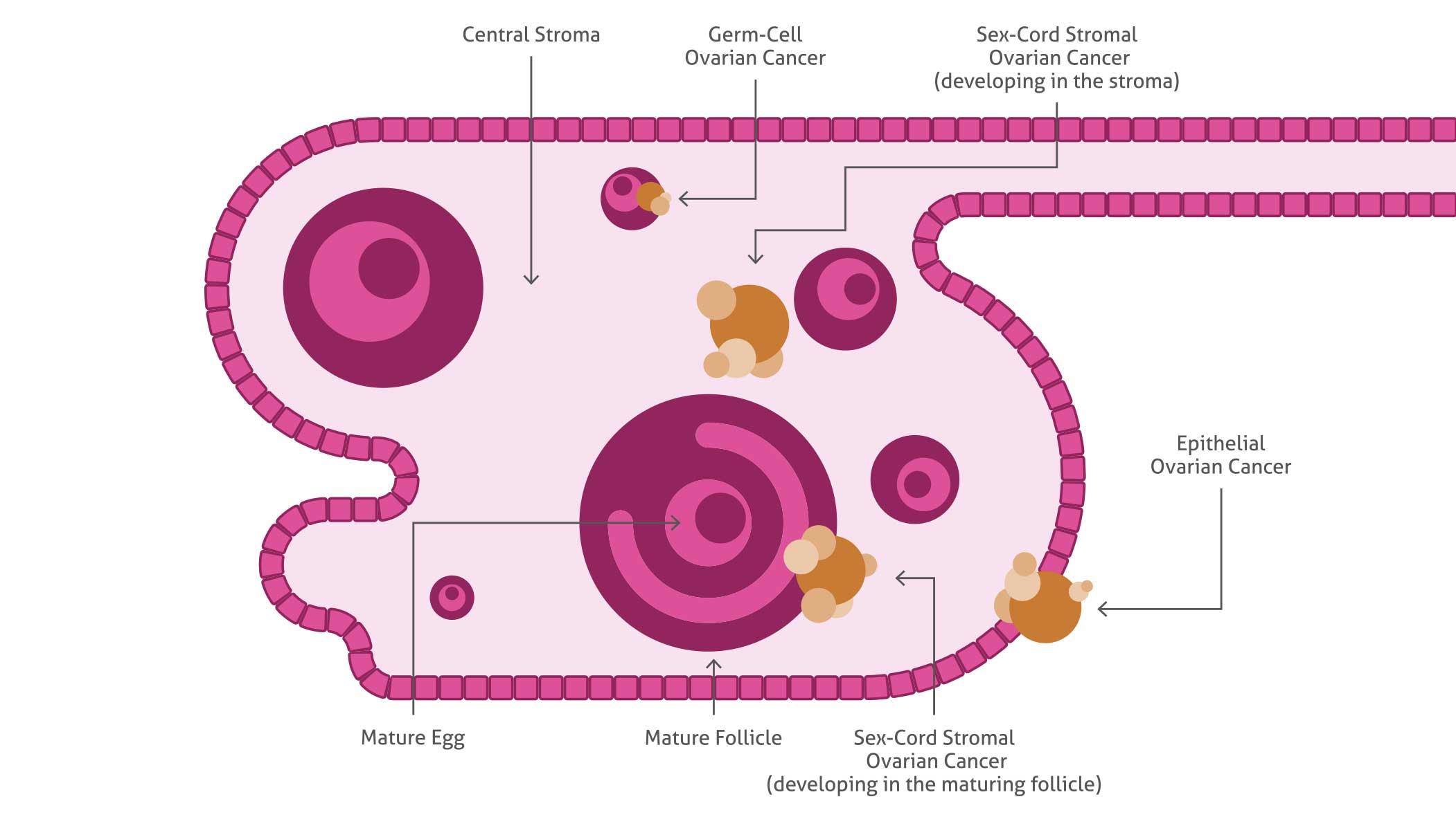 |
| Schema of the ovary showing the histopathological types of ovarian cancer. Epithelial ovarian cancer is the most common cause. |
Symptoms of Ovarian Cancer
| Weight loss | Discomfort in the pelvic area |
| Abdominal swelling/bloating | Constant tiredness |
| Frequent need to urinate | Constipation |
| Quickly feeling full when eating |
Tumor Suppressor Genes and Oncogenes
Several putative tumor suppressor genes and well-established oncogenes have been identified in the development of ovarian cancer.
Broadly, all EOC patients have mutations in the p53 (10442-1-AP) gene (3), (4) and loss of functional p53 protein is crucial for a cell. The TP53 (21891-1-AP) gene encodes a transcription factor that plays a critical role in regulating cell cycle progression, DNA repair, or cell death.(5), (6).
Besides the large number of already known potential targets in ovarian cancer, the identification of new targets is essential for understanding EOC mechanisms and identifying the disease at an early stage. Recent data demonstrate, for instance, that WT1 (12609-1-AP) is expressed at a high frequency in patients with EOC. (7)
WT1 is normally required for formation of the genitourinary system and mesothelial tissues. Recently, it was also demonstrated that the well-known heatshock protein 90 (Hsp90) is a novel target protein in ovarian cancer. Hsp90 (13171-1-AP) provides chaperoning activity for different proteins; many of these are members of oncogenic pathways. In ovarian cancer, inhibition of Hsp90 has been shown to have anti-proliferative effects (8), (9), (10); however, the exact molecular mechanism remains unclear.
Due to the heterogeneity of ovarian cancer, it is of high importance to characterize and identify the different abnormalities present in ovarian cancer.
Related Products
| Antibody Name | Catalog number |
| HSP90 | 13171-1-AP |
| K-Ras | 12063-1-AP |
| p53 | 10442-1-AP |
| p53 | 21891-1-AP |
| WT1 | 12609-1-AP |
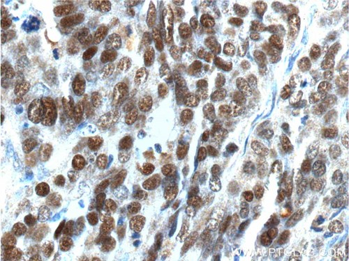 |
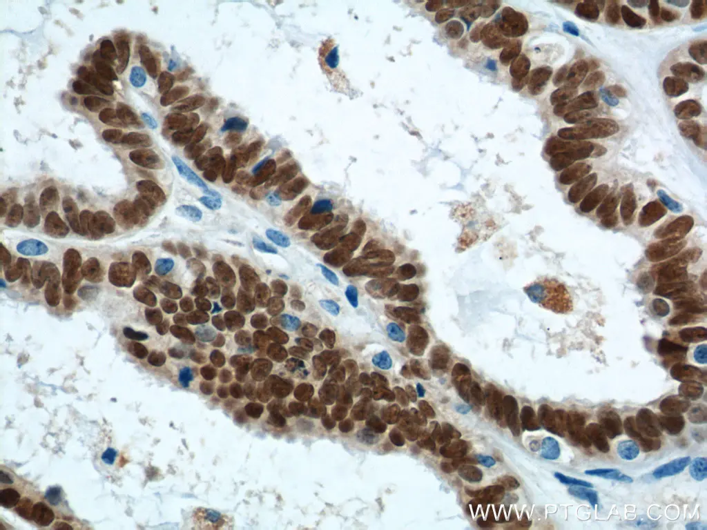
| IHC of paraffin-embedded human ovary tissue slide using 21891-1-AP (p53 Antibody) at dilution of 1:200 (40 x lens) |
Product FocusHER2/ErbB2 Antibody |
|
| Catalog Number | HER2, also known as ErbB2 and Neu, is a 185-kDa transmembrane glycoprotein that is a member of the epidermal growth factor (EGF) receptor family of receptor tyrosine kinases. It has no ligand-binding domain of its own and therefore cannot bind growth factors. Amplification and/or overexpression of HER2 has been reported in numerous cancers, including breast and ovarian tumors |
| 60311-1-Ig | |
| Type | |
| Mouse Monoclonal | |
| Applications | |
| ELISA, FC, IHC | |
Biomarkers
Despite the intensive use of conventional methods to detect ovarian cancer progression, ovarian cancer remains a commonly fatal gynecological disease. The current challenge is to detect ovarian cancer at an early stage. Effective screening methods have so far failed due to a lack of specificity and sensitivity. Biomarkers for precise detection of ovarian cancer are greatly needed to monitor and understand details of ovarian cancer tumorigenesis.
The latest studies report that HE4 (14406-1-AP) can be referred as a biomarker for ovarian carcinoma.11 HE4 is expressed in a number of normal tissues, and it is also highly expressed in a number of tumor cell lines.
Metastasis to the peritoneal surface requires the adhesion of ovarian cancer cells to the mesothelial cells. Epithelial cell-adhesion molecule (EpCAM, CD326, 21050-1- AP) functions as an epithelial-specific intercellular cell-adhesion molecule.(12), (13) It is of special interest as a potential diagnostic and prognostic marker for ovarian cancer.
Osteopontin has been shown in multiple studies, including, among others, a microarray analysis of ovarian cancer cell lines and healthy human ovarian surface epithelial cell cultures, to be a potential biomarker.(14) However, to date, the functional mechanisms of osteopotin in ovarian cancer remain poorly understood.(15)
Related Products
| Antibody Name | Catalog Number | Antibody Name | Catalog Number |
| CD138/Syndecan-1 | 10593-1-AP | HE4 | 14406-1-AP |
| CD146/MCAM | 17564-1-AP | HNF1B | 12525-1-AP |
| CD147 | 11989-1-AP | HOXA7, NCOA6 | 25241-1-AP |
| CD34 | 14486-1-AP | MUC1/CA15-3 | 19976-1-AP |
| CX3CR1 | 13885-1-AP | Osteopontin | 22952-1-AP |
| EPCAM, CD326 | 21050-1-AP | PD-1 | 18106-1-lg |
| PR | 66300-1-lg | SALL4 | 24500-1-AP |
Product Focus |
||
| Catalog Number | Type | Applications |
| 60261-1-lg | Mouse Monoclonal | ELISA, IHC |
|
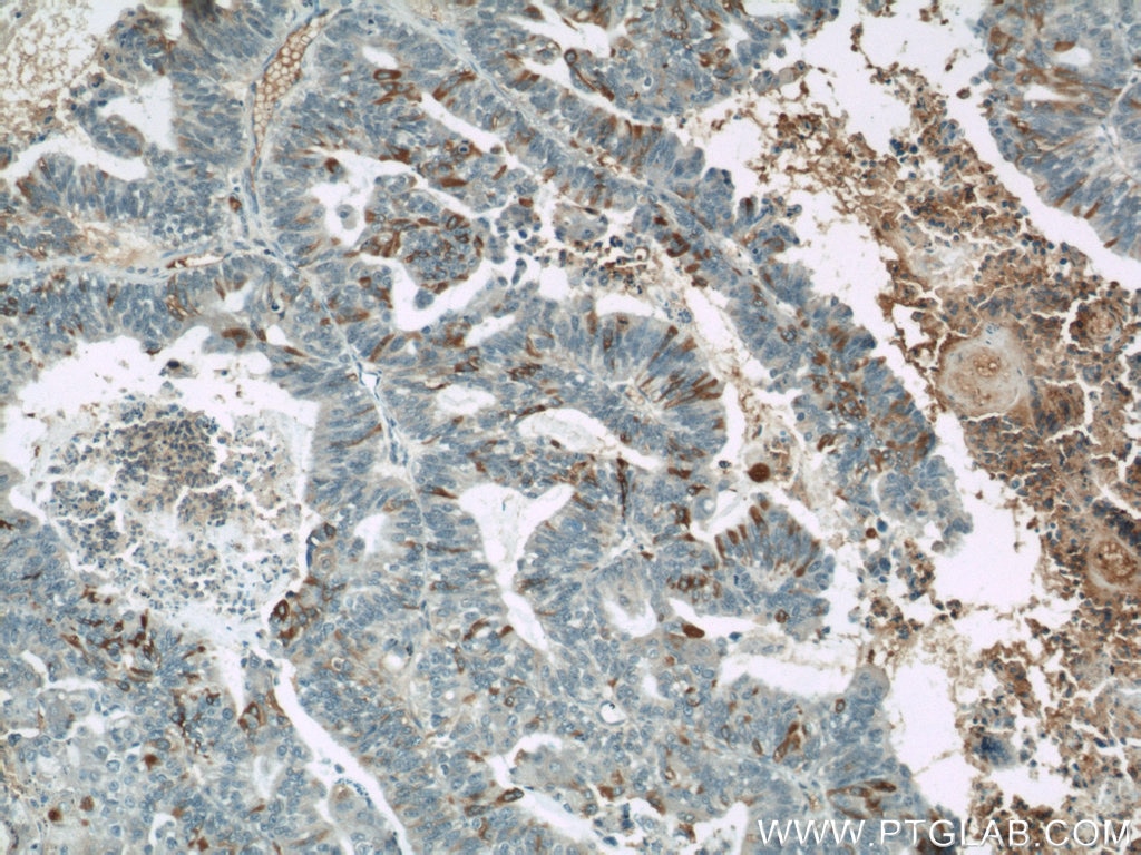
IHC of paraffin-embedded human ovary tumor using 60261-1-Ig (MUC16,CA125 antibody) at dilution of 1:50 (10x lens). |
| MUC16/CA125 is a glycosylated transmembrane mucin that is overexpressed in the majority of ovarian cancers. It is an important factor involved in cell adhesion, motility, and invasion of ovarian cancer. On the reverse side, knockdown experiments of CA125 showed a decrease in invasion of OCÈ |
Product Focus |
||
| Catalog Number | Type | Applications |
| 10256-1-AP | Rabbit Polyclonal | ELISA, FC, IHC, IP, WB |
|
The paired box 8 (PAX8) gene is a transcription factor expressed during organogenesis of the thyroid gland, kidney, and Müllerian ducts. PAX8 belongs to a family of 9 proteins (PAX1-PAX9) where each member is directly implicated in the transcription of various genes, PAX members typically containing a paired box domain and a paired-type homeodomain. PAX8 is expressed in a high percentage of ovarian serous, endometrioid, and clearcell carcinomas, but only rarely in primary ovarian mucinous adenocarcinomas. In this setting, PAX8 is also a useful marker for distinguishing ovarian carcinomas from those of mammary origin, and is shown to be significant in many more forms of cancer. It is thought to regulate the expression of Wilms tumour suppressor (WT1) gene and mutations in PAX8 have been associated with Wilms tumour cells, thyroid and ovarian carcinomas. Research continues to shed light on its diagnostic value. Differentiating metastatic female genital tract tumours from primary ovarian tumours is one of the most important issues in this field. Recent evidence supports the utility of PAX8 factor, especially in determining Müllerian tumours. Differentiation of ovarian metastatic tumours (especially with breast origin) from primary ovarian tumours can also be performed based on this IHC staining. The molecular mechanism of PAX8 in the carcinogenesis up to now remains unclear and requires further studies. |
||
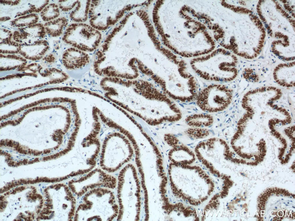 |
| Immunohistochemical analysis of paraffin-embedded human ovary tumor tissue slide using 10336-1-AP (PAX8 Antibody) at dilution of 1:1200 (under 10x lens). Heat mediated antigen retrieval with Tris-EDTA buffer (pH 9.0). |
 |
| Immunohistochemical analysis of paraffin-embedded human ovary tumor tissue slide using 10336-1-AP (PAX8 Antibody) at dilution of 1:1200 (under 40x lens). Heat mediated antigen retrieval with Tris-EDTA buffer (pH 9.0). |
Signaling
Many different signaling pathways are activated in ovarian cancer and current studies focus on the exact mechanism and molecules involved in signaling pathways that are frequently activated in ovarian cancer. The PI3K (20584-1-AP) pathway has been shown to play a significant role in the majority of ovarian cancer cases. The activation can occur either by autocrine/paracrine signaling or by different targets like PTEN (22034-1-AP) or Akt (10176-2-AP). The inhibition of PI3K has been shown to prevent ovarian cancer growth in xenografts models, for instance.(16), (17) Another important player is IL-6 (21865-1-AP). Once the IL-6 receptor is activated, activation of the JAK-STAT cascade follows (17670-1-AP, 10253-2-AP). STAT has been reported to be crucial for the regulation of different biological functions like proliferation or apoptosis.(18)
The number of cytokines involved in ovarian cancer is numerous, as shown by the activation of NF-κB. NF-κB shows activation in the majority of EOC cases (19), (20) and it is well known that multiple cytokines or growth factors can lead to NF-κB activation. Following the activation of signaling pathways, many different downstream mechanisms get switched on. Ovarian cancer cells show increased resistance to apoptosis and, at a more advanced stage, cancer cells metastasize. For instance, matrix metalloproteinases (MMP2, 10373-2-AP; MMP7, 10374-2-AP) have been found in EOC, thereby increasing invasion.(21) More effector proteinases, such as Kallikreins (Kallikrein 8, 14232-1-AP), have been found in EOC.(22)
| Antibody Name | Catalog Number |
| Akt | 10176-2-AP |
| Cathepsin B | 12216-1-AP |
| IGFBP2 | 11065-3-AP |
| IL-6 | 21865-1-AP |
| Jak2 | 17670-1-AP |
| Kallikrein 8 | 14232-1-AP |
| MMP2 | 10373-2-AP |
| MMP7 | 10374-2-AP |
| NF-KB | 14220-1-AP |
References
1 Jemal A, et al., Global cancer statistics. CA Cancer J Clin. 2011.
2 Jayson GC, et al., Ovarian cancer. Lancet. 2014; 384: 1376–1388.
3 Curtin JC, et al., p53 in human embryonal carcinoma: identification of a transferable, transcriptional repression domain in the N-terminal region of p53. Oncogene. 2005;24:1481–1490
4. D’Souza S, et al., The gene encoding p202, an interferon-inducible negative regulator of the p53 tumour suppressor, is a target of p53-mediated transcriptional repression. J Biol Chem. 2001;276:298–305.
5 Berchuck A, et al. Overexpression of p53 is not a feature of benign and early-stage borderline epithelial ovarian tumors. Gynecol. Oncol 1994;52:232–236.
6 Havrilesky L, et al. Prognostic significance of p53 mutation and p53 overexpression in advanced epithelial ovarian cancer: a Gynecologic Oncology Group study. J. Clin. Oncol 2003;21:3814–3825
7 Hylander B, et al., Expression of Wilms tumor gene (WT1) in epithelial ovarian cancer, Gynecol Oncol. 2006 Apr;101(1):12-7.
8 Banerji U, et al., Pharmacokineticpharmacodynamic relationships for the heat shock protein 90 molecular chaperone inhibitor 17-allylamino, 17-demethoxygeldanamycin in human ovarian cancer xenograft models. Clin Cancer Res. 2005;11:7023–7032.
9 Sain N, et al., Potentiation of paclitaxel activity by the HSP90 inhibitor 17-allylamino-17-demethoxygeldanamycin in human ovarian carcinoma cell lines with high levels of activated AKT. Mol Cancer Ther. 2006;5:1197–1208.
10 Maloney A, et al., Gene and protein expression profiling of human ovarian cancer cells treated with the heat shock protein 90 inhibitor 17-allylamino- 17-demethoxygeldanamycin. Cancer Res. 2007;67:3239–3253.
11 Molina R., et al., HE4 a novel tumour marker for ovarian cancer: comparison with CA 125 and ROMA algorithm in patients with gynaecological diseases. Tumour Biol. 2011 Dec;32(6):1087-95.
12 Montagnana M., et al., HE4 in ovarian cancer: from discovery to clinical application. Adv Clin Chem. 2011;55:1-20.
13 Lee M., et al. Prognostic impact of epithelial cell adhesion molecule in ovarian cancer patients, J Gynecol Oncol. 2014 Oct; 25(4): 352–354.
14 Jae-Hoon K., et al., Osteopontin as a Potential Diagnostic Biomarker for Ovarian Cancer. JAMA. 2002;287(13):1671-1679.
15 Song G., et al., Osteopontin promotes ovarian cancer progression and cell survival and increases HIF-1alpha expression through the PI3-K/Akt pathway. Cancer Sci. 2008 Oct;99(10):1901-7.
16 Raynaud FL, et al. Pharmacologic characterization of a potent inhibitor of class I phophatidylinositide 3-kinases. Cancer Res 2007; 67:5840–5850.
17 Hu L, Hofmann J, Lu Y, Mills GB, Jaffe RB. Inhibition of phophatidylinositol 3’ kinase increases efficacy of paclitaxel in in vitro and in vivo ovarian cancer models. Cancer Res 2002; 62:1087–1092
18 Burke WM, et al. Inhibition of constitutively active Stat3 suppresses growth of human ovarian and breast cancer cells. Oncogene 2001; 20:7925–7934.
19 YG, et al. Curcumin inhibits tumor growth and angiogenesis in ovarian carcinoma by targeting the nuclear factor-κB pathway. Clin. Cancer Res 2007;13:3423–3430.
20 Samanta AK, et al., Overexpression of MEKK3 confers resistance to apoptosis through activation of NF κB. J. Biol. Chem 2004;279:7576–7583.
21 Sood AK, et al. Stress hormone-mediated invasion of ovarian cancer cells. Clin. Cancer Res 2006;15:369–375.
22 Paliouras M, et al. Human tissue kallikreins: the cancer biomarker family. Cancer Lett 2007;28:61–79.


