Recombinant Human CEACAM-1/CD66a protein (His Tag)
种属
Human
纯度
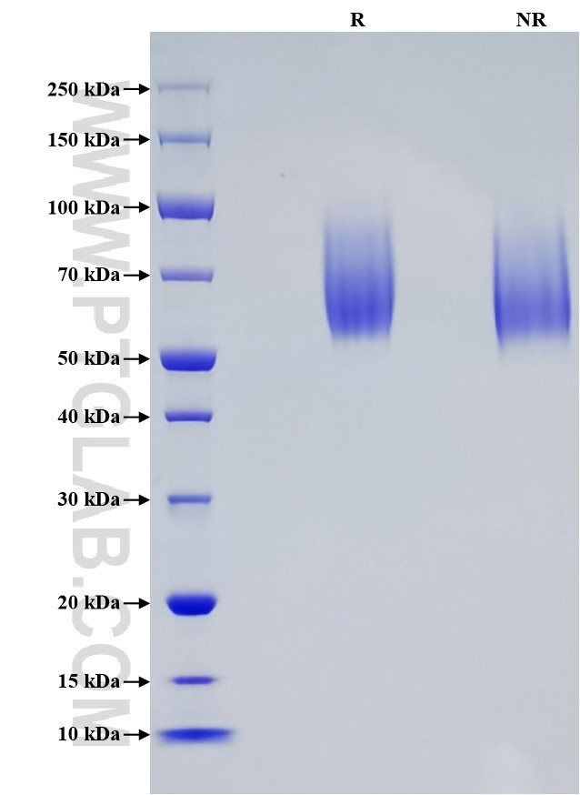
>95 %, SDS-PAGE
标签
His Tag
生物活性
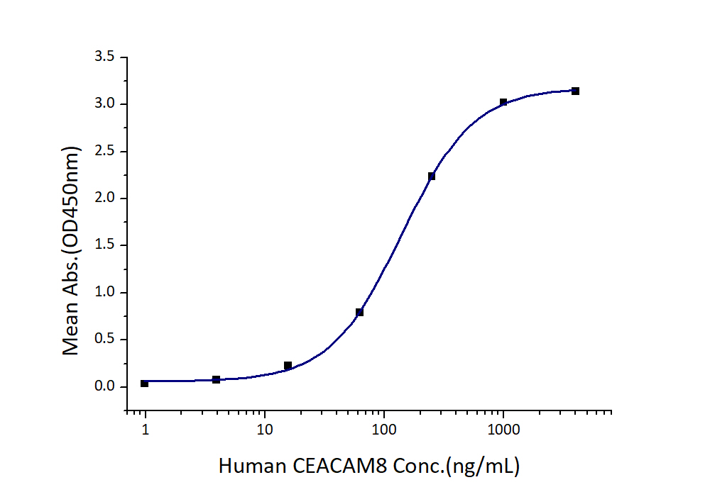
EC50: 70-280 ng/mL
验证数据展示
产品信息
| 纯度 | >95 %, SDS-PAGE |
| 内毒素 | <0.1 EU/μg protein, LAL method |
| 生物活性 | Immobilized Human CEACAM-1 (His tag) at 2 μg/mL (100 μL/well) can bind Human CEACAM-8 (hFc tag) with a linear range of 70-280 ng/mL. |
| 来源 | HEK293-derived Human CEACAM-1 protein Gln35-Gly428 (Accession# P13688-1) with a His tag at the C-terminus. |
| 基因ID | 634 |
| 蛋白编号 | P13688-1 |
| 预测分子量 | 47.5 kDa |
| SDS-PAGE | 55-90 kDa, reducing (R) conditions |
| 组分 | Lyophilized from 0.22 μm filtered solution in PBS, pH 7.4. Normally 5% trehalose and 5% mannitol are added as protectants before lyophilization. |
| 复溶 | Briefly centrifuge the tube before opening. Reconstitute at 0.1-0.5 mg/mL in sterile water. |
| 储存条件 |
It is recommended that the protein be aliquoted for optimal storage. Avoid repeated freeze-thaw cycles.
|
| 运输条件 | The product is shipped at ambient temperature. Upon receipt, store it immediately at the recommended temperature. |
背景信息
Carcinoembryonic antigen-related cell adhesion molecule 1 (CEACAM1), belonging to the immunoglobulin superfamily. It is cell adhesion protein that mediates homophilic cell adhesion in a calcium-independent manner. CEACAM1 is expressed incolumnar epithelial cells of the colon, T cells, granulocytes and lymphocytes. It plays a role as coinhibitory receptor in immune response, insulin action and functions also as an activator during angiogenesis. Its coinhibitory receptor function is phosphorylation- and PTPN6 -dependent, which in turn, suppress signal transduction of associated receptors by dephosphorylation of their downstream effectors. Plays a role in immune response, of T cells, natural killer (NK) and neutrophils. It inhibits cell migration and cell scattering through interaction with FLNA and interferes with the interaction of FLNA with RALA.
参考文献:
1.Huang YH, et al. (2015). Nature. 517(7534):386–390
2.Hosomi S, et al. (2013). Eur J Immunol. 43(9):2473-2483
3.Chen Z, et al. (2008). J Immunol.180(9):6085-6093
4.Klaile E, et al. (2005). J Cell Sci. 118(Pt 23):5513–5524

