Recombinant Human CEACAM6 protein (Myc Tag, His Tag)
ED50
12-46 ng/mL
Species
Human
Purity
>95 %, SDS-PAGE
GeneID
4680
Accession
P40199
验证数据展示
Technical Specifications
| Purity | >95 %, SDS-PAGE |
| Endotoxin Level | <1.0 EU/μg protein, LAL method |
| Biological Activity |
Immobilized Human CEACAM6 (Myc tag, His tag) at 2 μg/mL (100 μL/well) can bind Human CEACAM8 (hFc tag) with a linear range of 12-46 ng/mL. |
| Source | HEK293-derived Human CEACAM6 protein Lys35 -Gly320 (Accession# P40199) with a Myc tag and a His tag at the C-terminus. |
| Predicted Molecular Mass | 33.8 kDa |
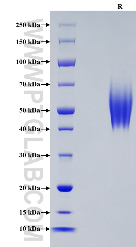
| SDS-PAGE | 42-65 kDa, reducing (R) conditions |
| Formulation | Lyophilized from sterile PBS, pH 7.4. Normally 5% trehalose and 5% mannitol are added as protectants before lyophilization. |
| Reconstitution | Briefly centrifuge the tube before opening. Reconstitute at 0.1-0.5 mg/mL in sterile water. |
| Storage |
It is recommended that the protein be aliquoted for optimal storage. Avoid repeated freeze-thaw cycles.
|
| Shipping | The product is shipped at ambient temperature. Upon receipt, store it immediately at the recommended temperature. |
Background
Carcinoembryonic antigen-related cell adhesion molecule 6 (CEACAM6), belonging to the immunoglobulin superfamily, is cell-adhesion protein on neutrophils. CEACAM6 is expressed in neutrophils and numerous tumor cell lines. CEACAM6 mediates homophilic and heterophilic cell adhesion with other carcinoembryonic antigen-related cell adhesion molecules, such as CEACAM5 and CEACAM8. It plays a role in neutrophil adhesion to cytokine-activated endothelial cells and plays a role as an oncogene by promoting tumor progression; positively regulates cell migration, cell adhesion to endothelial cells and cell invasion. CEACAM6 is also involved in the metastatic cascade process by inducing gain resistance to anoikis of pancreatic adenocarcinoma and colorectal carcinoma cells.
References:
1.Kuroki M, et al. (2001). Journal of leukocyte biology. 70(4): 543-50
2.Kuijpers TW, et al. (1992). J Cell Biol. 118(2):457-66
3.Blumenthal RD, et al. (2005). Cancer research. 65(19):8809–8817
4.Ordoñez C, et al. (2000). Cancer Res. 60(13):3419-24
5.Duxbury MS, et al. (2004). Oncogene. 23(2):465-73

