Recombinant Human Galectin-3 protein (His Tag)
种属
Human
纯度
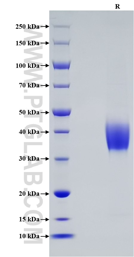
>95 %, SDS-PAGE
标签
His Tag
生物活性
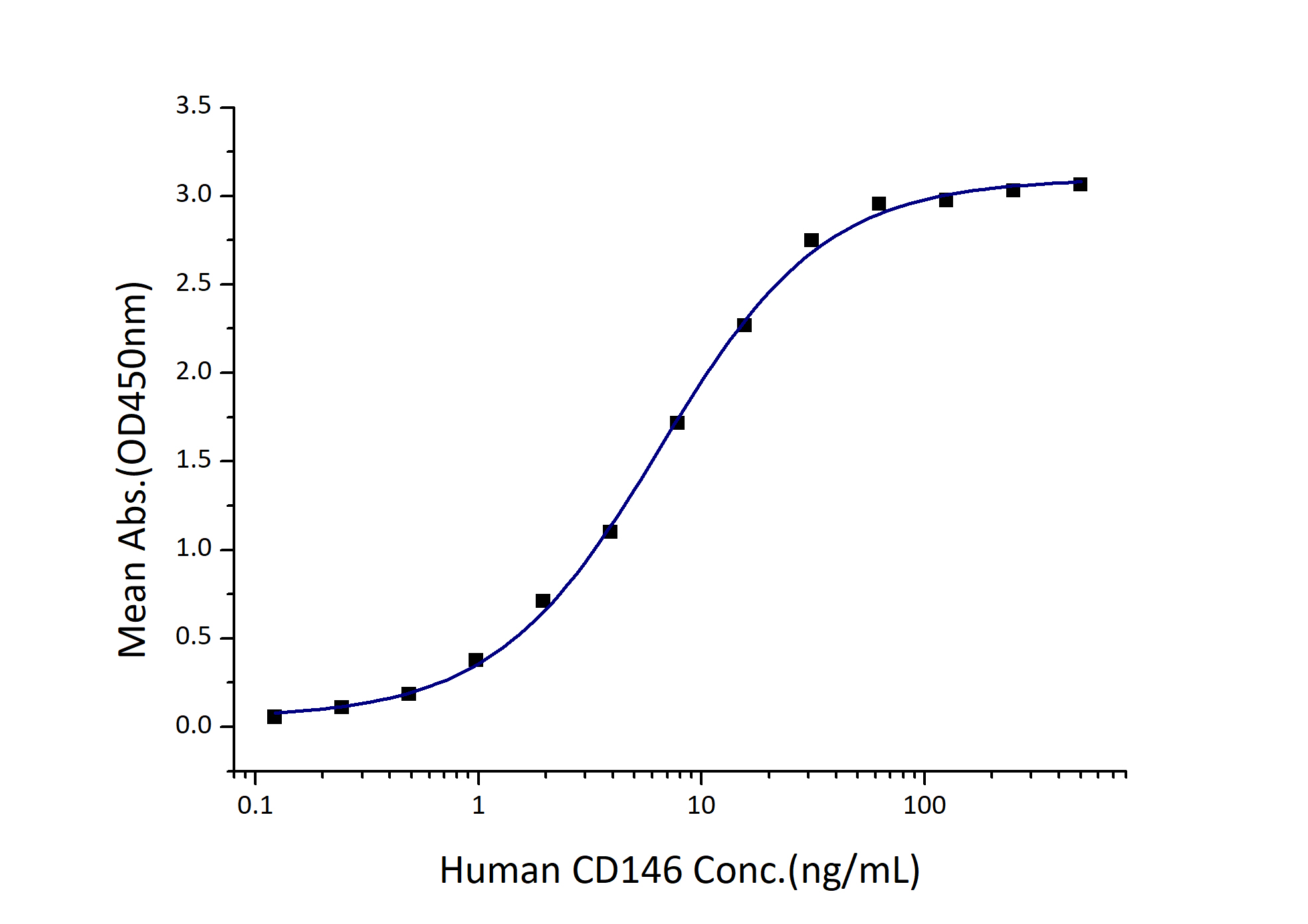
EC50: 3-12 ng/mL
验证数据展示
产品信息
| 纯度 | >95 %, SDS-PAGE |
| 内毒素 | <0.1 EU/μg protein, LAL method |
| 生物活性 |
Immobilized Human Galectin-3 (His tag) at 0.5 μg/mL (100 μL/well) can bind Human CD146 (GST tag) with a linear range of 3-12 ng/mL. |
| 来源 | HEK293-derived Human Galectin-3 protein Ala2-Ile250 (Accession# P17931) with a His tag at the C-terminus. |
| 基因ID | 3958 |
| 蛋白编号 | P17931 |
| 预测分子量 | 26.8 kDa |
| SDS-PAGE | 32-45 kDa, reducing (R) conditions |
| 组分 | Lyophilized from 0.22 μm filtered solution in PBS, pH 7.4. Normally 5% trehalose and 5% mannitol are added as protectants before lyophilization. |
| 复溶 | Briefly centrifuge the tube before opening. Reconstitute at 0.1-0.5 mg/mL in sterile water. |
| 储存条件 |
It is recommended that the protein be aliquoted for optimal storage. Avoid repeated freeze-thaw cycles.
|
| 运输条件 | The product is shipped at ambient temperature. Upon receipt, store it immediately at the recommended temperature. |
背景信息
Galectins are a family of animal lectins defined by shared characteristic amino-acid sequences and affinity for β-galactose-containing oligosac-charides. Galectin-3, a 31-kDa member of the β-galactoside-binding proteins, contains one carbohydrate recognition domain (CRD) and a proline- and glycine-rich N-terminal domain through which is able to form oligomers. Galectin-3 is widely expressed in many normal tissues and a variety of tumors. It is found intracellularly in nucleus and cytoplasm or secreted outside of cell, being present on the cell surface or in the extracellular space. Galectin-3 is involved in various biological processes including cell growth, adhesion, differentiation, apoptosis, angiogenesis, immune response, neoplastic transformation and metastasis. Elevated serum galectin-3 levels have been reported in patients with breast, gastrointestinal, lung, or ovarian cancer, melanoma, and non-Hodgkin's lymphoma.
参考文献:
1. Barondes SH. et al. (1994) J Biol Chem. 269(33):20807-10.
2. Iurisci I. et al. (2000)  Clin Cancer Res. 6(4):1389-93.
3. Takenaka Y. et al. (2002) Glycoconj J. 19(7-9):543-9.
4. Dumic J. et al.(2006) Biochim Biophys Acta. 1760(4):616-35.


