- Featured Product
- KD/KO Validated
Beclin 1 Polyclonal antibody
Beclin 1 Polyclonal Antibody for WB, IHC, IF/ICC, IF-P, IF-Fro, IP, ELISA
Host / Isotype
Rabbit / IgG
Reactivity
human, mouse, rat and More (7)
Applications
WB, IHC, IF/ICC, IF-P, IF-Fro, IP, ELISA and More (2)
Conjugate
Unconjugated
989
验证数据展示
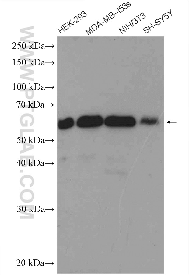
 with sh-Control and sh-Beclin 1 transfected HEK-293 cells.")
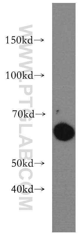
 at dilution of 1:6000 incubated at room temperature for 1.5 hours.")
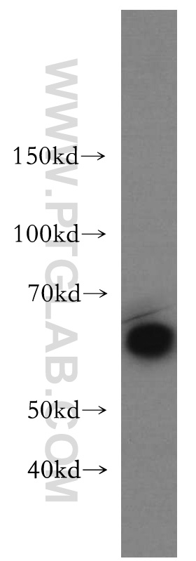
 with MDA-MB-453s cells lysate 2800 ug.")
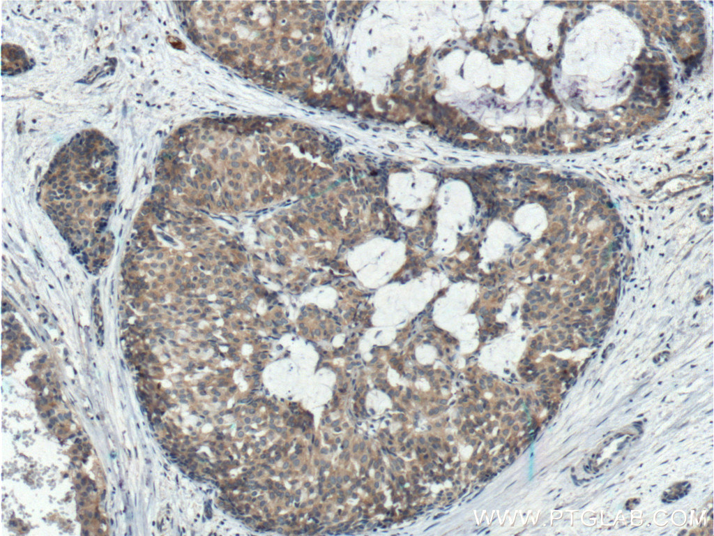
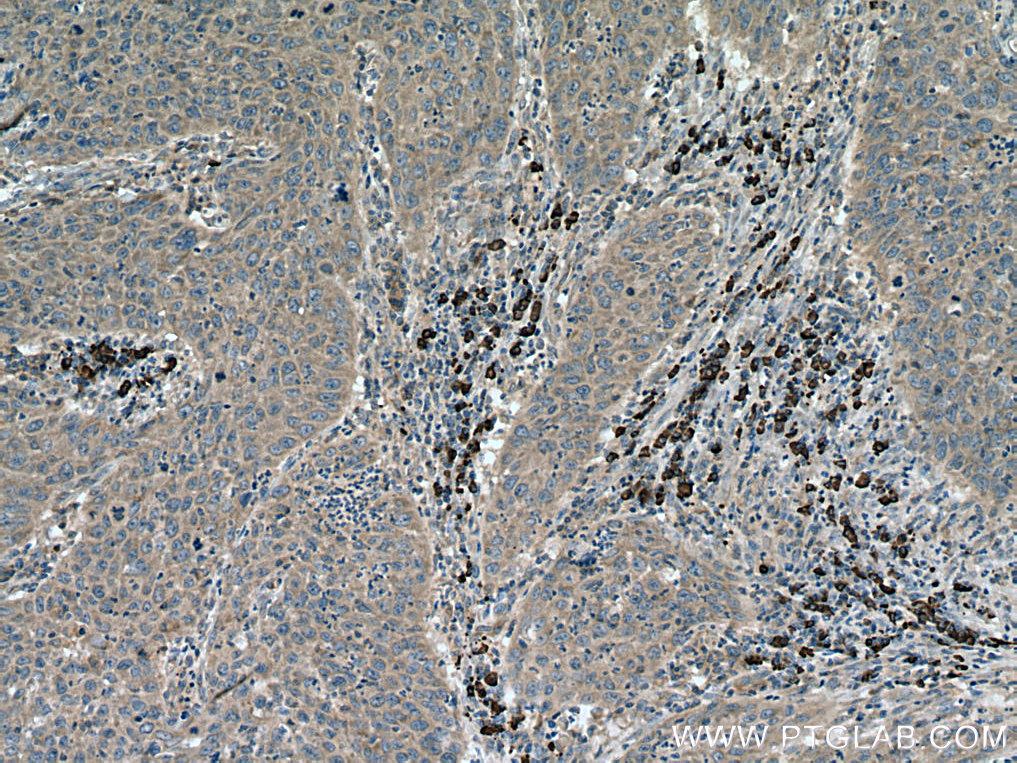
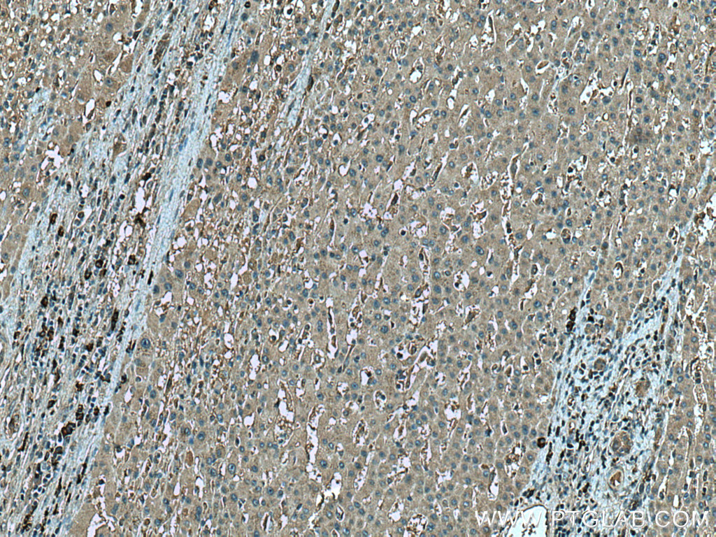
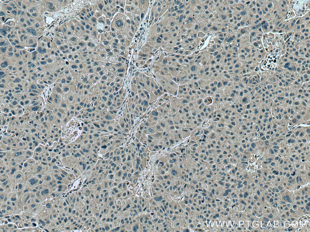
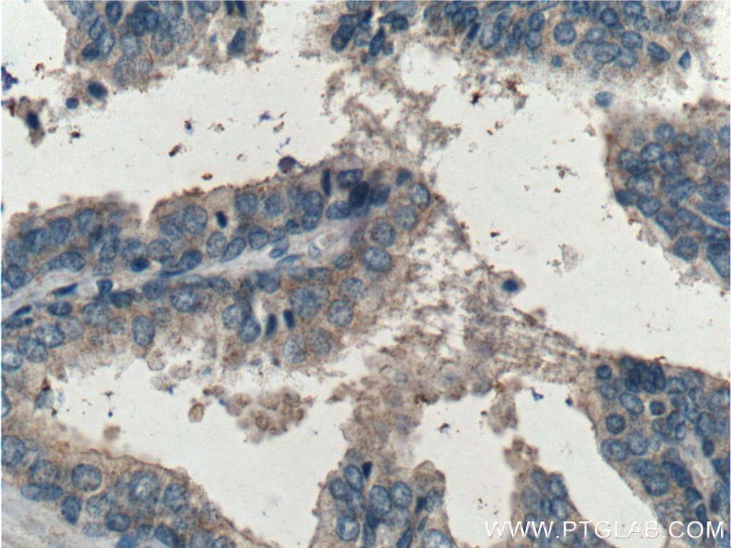
 at dilution of 1:200 (under 10x lens). Heat mediated antigen retrieval with Tris-EDTA buffer (pH 9.0).")
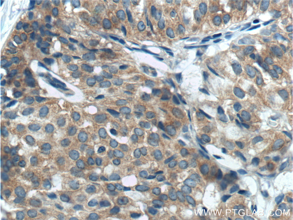
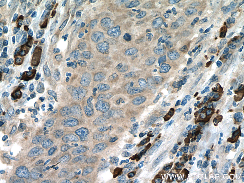
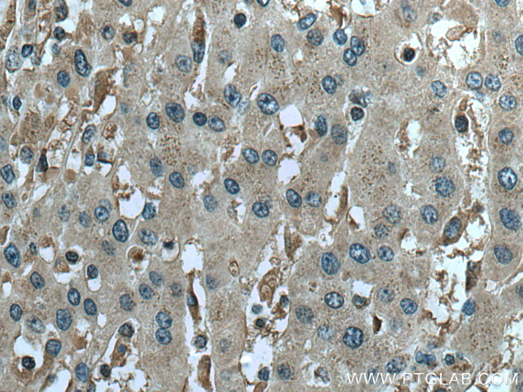
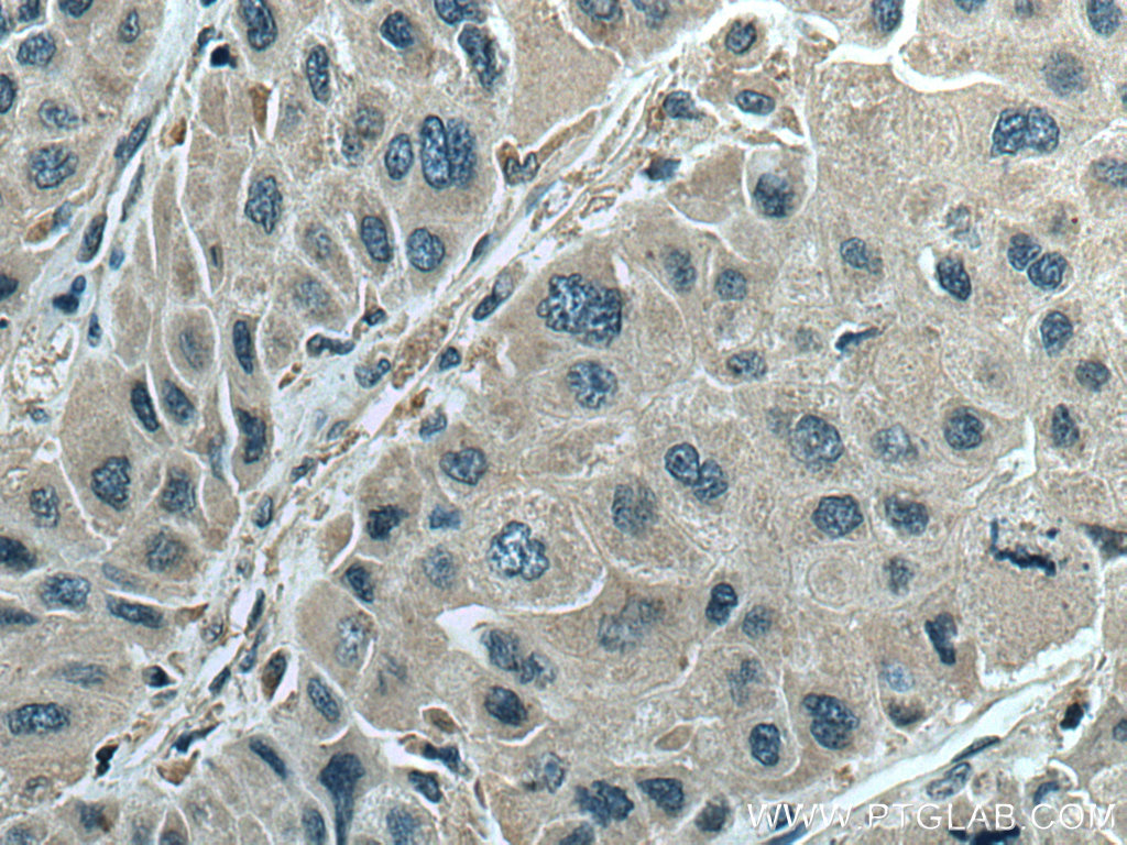
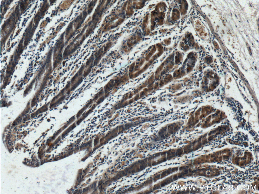
 at dilution of 1:200 (under 40x lens). Heat mediated antigen retrieval with Tris-EDTA buffer (pH 9.0).")
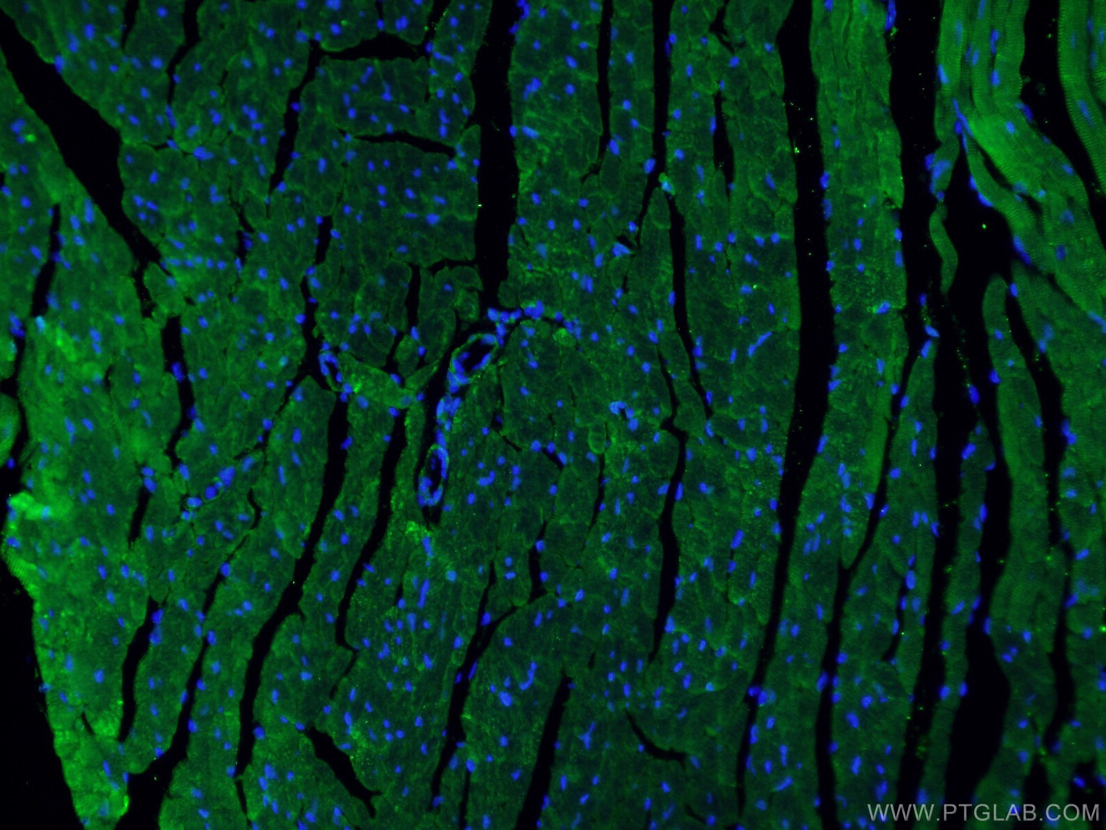
 fixed mouse heart tissue using 11306-1-AP (Beclin 1 antibody) at dilution of 1:50 and Alexa Fluor 488-Conjugated AffiniPure Goat Anti-Rabbit IgG(H+L).")
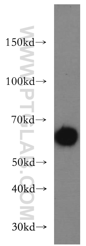
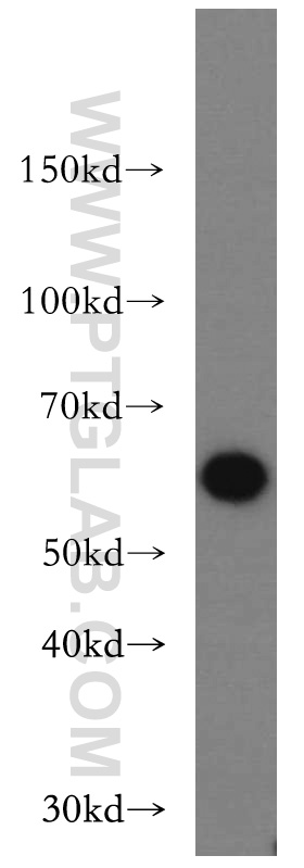
 at dilution of 1:8000 incubated at room temperature for 1.5 hours.")
 at dilution of 1:8000 incubated at room temperature for 1.5 hours.")
 at dilution of 1:8000 incubated at room temperature for 1.5 hours.")
 at dilution of 1:10000 incubated at room temperature for 1.5 hours.")
 fixed mouse heart tissue using Beclin 1 antibody (11306-1-AP) at dilution of 1:400 and CoraLite®488-Conjugated AffiniPure Goat Anti-Rabbit IgG(H+L).")
 fixed paraffin-embedded mouse heart tissue using Beclin 1 antibody (11306-1-AP) at dilution of 1:200 and CoraLite®488-Conjugated AffiniPure Goat Anti-Rabbit IgG(H+L) (<a class='green' href='/productredirect?CatalogNo=SA00013-2' target='_blank'>SA00013-2</a>). Heat mediated antigen retrieval with Tris-EDTA buffer (pH 9.0).")
 at dilution of 1:200 and CoraLite®488-Conjugated AffiniPure Goat Anti-Rabbit IgG(H+L) (<a class='green' href='/productredirect?CatalogNo=SA00013-2' target='_blank'>SA00013-2</a>).")
 at dilution of 1:200 and CoraLite®488-Conjugated AffiniPure Goat Anti-Rabbit IgG(H+L) (<a class='green' href='/productredirect?CatalogNo=SA00013-2' target='_blank'>SA00013-2</a>).")
 fixed NIH/3T3 cells using Beclin 1 antibody (11306-1-AP) at dilution of 1:400 and CoraLite®488-Conjugated AffiniPure Goat Anti-Rabbit IgG(H+L).")
经过测试的应用
| Positive WB detected in | C6 cells, HEK-293 cells, Raji cells, L02 cells, PC-3 cells, A431 cells, HT-1080 cells, human placenta tissue, mouse brain tissue, rat brain tissue, HeLa cells, NIH/3T3 cells, MDA-MB-453s cells, SH-SY5Y cells |
| Positive IP detected in | MDA-MB-453s cells |
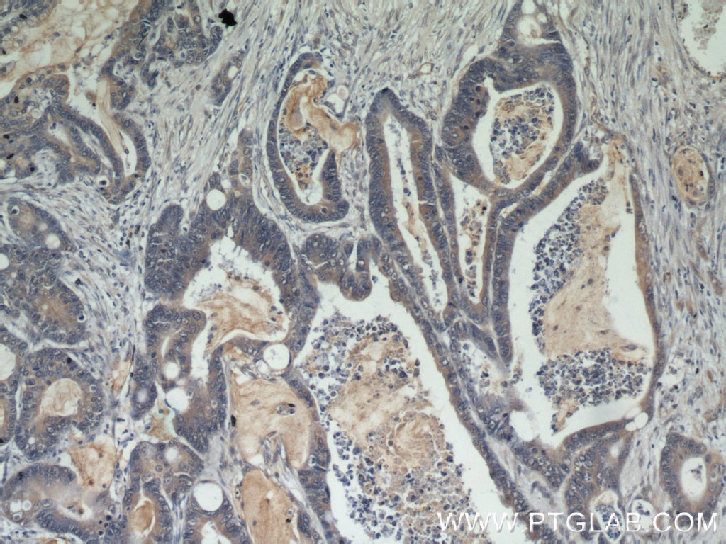
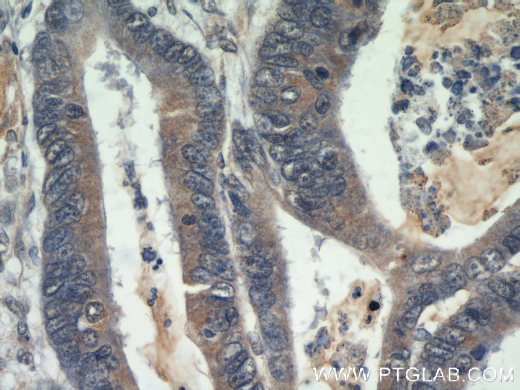
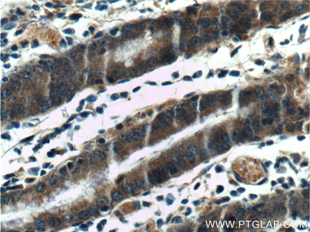
| Positive IHC detected in | human breast cancer tissue, human breast hyperplasia tissue, human prostate hyperplasia tissue, human cervical cancer tissue, human colon cancer tissue, human stomach tissue, human liver cancer tissue Note: suggested antigen retrieval with TE buffer pH 9.0; (*) Alternatively, antigen retrieval may be performed with citrate buffer pH 6.0 |
| Positive IF-P detected in | mouse heart tissue |
| Positive IF-Fro detected in | mouse heart tissue |
| Positive IF/ICC detected in | NIH/3T3 cells |
Planning an IHC experiment? We recommend our IHCeasy BECN1 Ready-To-Use IHC Kit. BECN1 primary antibody included.
推荐稀释比
| Application | Dilution |
|---|---|
| Western Blot (WB) | WB : 1:1000-1:10000 |
| Immunoprecipitation (IP) | IP : 0.5-4.0 ug for 1.0-3.0 mg of total protein lysate |
| Immunohistochemistry (IHC) | IHC : 1:50-1:500 |
| Immunofluorescence (IF)-P | IF-P : 1:50-1:500 |
| Immunofluorescence (IF)-FRO | IF-FRO : 1:50-1:500 |
| Immunofluorescence (IF)/ICC | IF/ICC : 1:200-1:800 |
| It is recommended that this reagent should be titrated in each testing system to obtain optimal results. | |
| Sample-dependent, Check data in validation data gallery. | |
产品信息
11306-1-AP targets Beclin 1 in WB, IHC, IF/ICC, IF-P, IF-Fro, IP, CoIP, ELISA applications and shows reactivity with human, mouse, rat samples.
| Tested Applications | WB, IHC, IF/ICC, IF-P, IF-Fro, IP, ELISA Application Description |
| Cited Applications | WB, IHC, IF, IP, CoIP, ELISA |
| Tested Reactivity | human, mouse, rat |
| Cited Reactivity | human, mouse, rat, pig, chicken, zebrafish, hamster, goat, fish, duck |
| Immunogen | Beclin 1 fusion protein Ag1843 种属同源性预测 |
| Host / Isotype | Rabbit / IgG |
| Class | Polyclonal |
| Type | Antibody |
| Full Name | beclin 1, autophagy related |
| Synonyms | BECN1, Beclin-1-C 37 kDa, Beclin-1-C 35 kDa, Beclin-1, Beclin1 |
| Calculated Molecular Weight | 52 kDa |
| Observed Molecular Weight | 52-60 kDa |
| GenBank Accession Number | BC010276 |
| Gene Symbol | Beclin 1 |
| Gene ID (NCBI) | 8678 |
| RRID | AB_2259061 |
| Conjugate | Unconjugated |
| Form | Liquid |
| Purification Method | Antigen affinity purification |
| UNIPROT ID | Q14457 |
| Storage Buffer | PBS with 0.02% sodium azide and 50% glycerol pH 7.3. |
| Storage Conditions | Store at -20°C. Stable for one year after shipment. Aliquoting is unnecessary for -20oC storage. |
背景介绍
Beclin 1, also known as ATG6 or VPS30, interacts with various cofactors (e.g. Ambra1, Barkor (Atg14), Rubicon, or UVRAG) to regulate the lipid kinase Vps34 and promote the formation of the BECLIN1-Vps34-Vps15 complex, hence inducing autophagy. Its function (via the BH3 domain) is inhibited by Bcl-2 or Bcl-XL. Beclin 1 (BECN1) is a crucial molecule in the control of the autophagic activity, and its activity is regulated by multiple mechanisms, including the post-translational modification, protein-protein interaction, and subcellular localization. It plays a role in crosstalk between apoptosis and autophagy. It has been reported that Beclin 1 can be cleaved into fragments of 50, 37 and 35 kDa during apoptosis. It is involved in many disorders, including neurodegeneration and cancer (tumorigenesis). Beclin 1 is a mammalian tumor suppressor, and its gene is monoallelically deleted in 75% of ovarian, 50% of breast, and 40% of prostate cancers. Decreased expression of Beclin 1 has also been observed in human brain and lung tumors. The level of Beclin 1 was decreased in the affected brain regions of patients with Alzheimer's disease early in the disease process. Recent studies have also shown that gain and loss of Beclin 1 function affects the death of heart cells.
实验方案
| Product Specific Protocols | |
|---|---|
| WB protocol for Beclin 1 antibody 11306-1-AP | Download protocol |
| IHC protocol for Beclin 1 antibody 11306-1-AP | Download protocol |
| IF protocol for Beclin 1 antibody 11306-1-AP | Download protocol |
| IP protocol for Beclin 1 antibody 11306-1-AP | Download protocol |
| Standard Protocols | |
|---|---|
| Click here to view our Standard Protocols |
发表文章
| Species | Application | Title |
|---|---|---|
Cell TEX264 drives selective autophagy of DNA lesions to promote DNA repair and cell survival | ||
Signal Transduct Target Ther Targeting CRL4 suppresses chemoresistant ovarian cancer growth by inducing mitophagy | ||
Mol Cancer Overcoming multi-drug resistance in SCLC: a synergistic approach with venetoclax and hydroxychloroquine targeting the lncRNA LYPLAL1-DT/BCL2/BECN1 pathway | ||
ACS Nano Neutrophil Nanovesicle Protects against Experimental Autoimmune Encephalomyelitis through Enhancing Myelin Clearance by Microglia | ||
Cancer Commun (Lond) Targeting autophagy overcomes cancer-intrinsic resistance to CAR-T immunotherapy in B-cell malignancies | ||