WB Figures
WB analysis using 14479-1-AP
Various lysates were subjected to SDS PAGE followed by western blot with 14479-1-AP (Calbindin-D28k antibody) at dilution of 1:30000 incubated at room temperature for 1.5 hours.
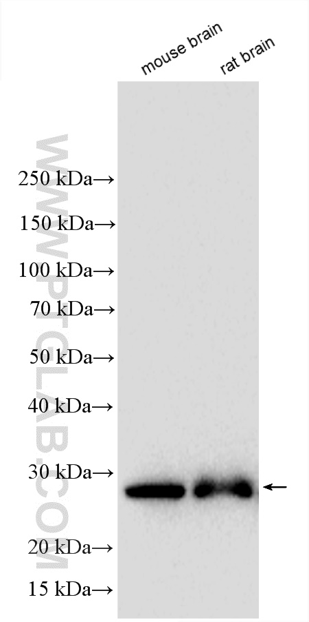
WB analysis using 14479-1-AP
Various lysates were subjected to SDS PAGE followed by western blot with 14479-1-AP (Calbindin-D28k antibody) at dilution of 1:4000 incubated at room temperature for 1.5 hours.
WB analysis using 14479-1-AP
Various lysates were subjected to SDS PAGE followed by western blot with 14479-1-AP (Calbindin-D28k antibody) at dilution of 1:15000 incubated at room temperature for 1.5 hours. (Negative control: NIH/3T3, C6)
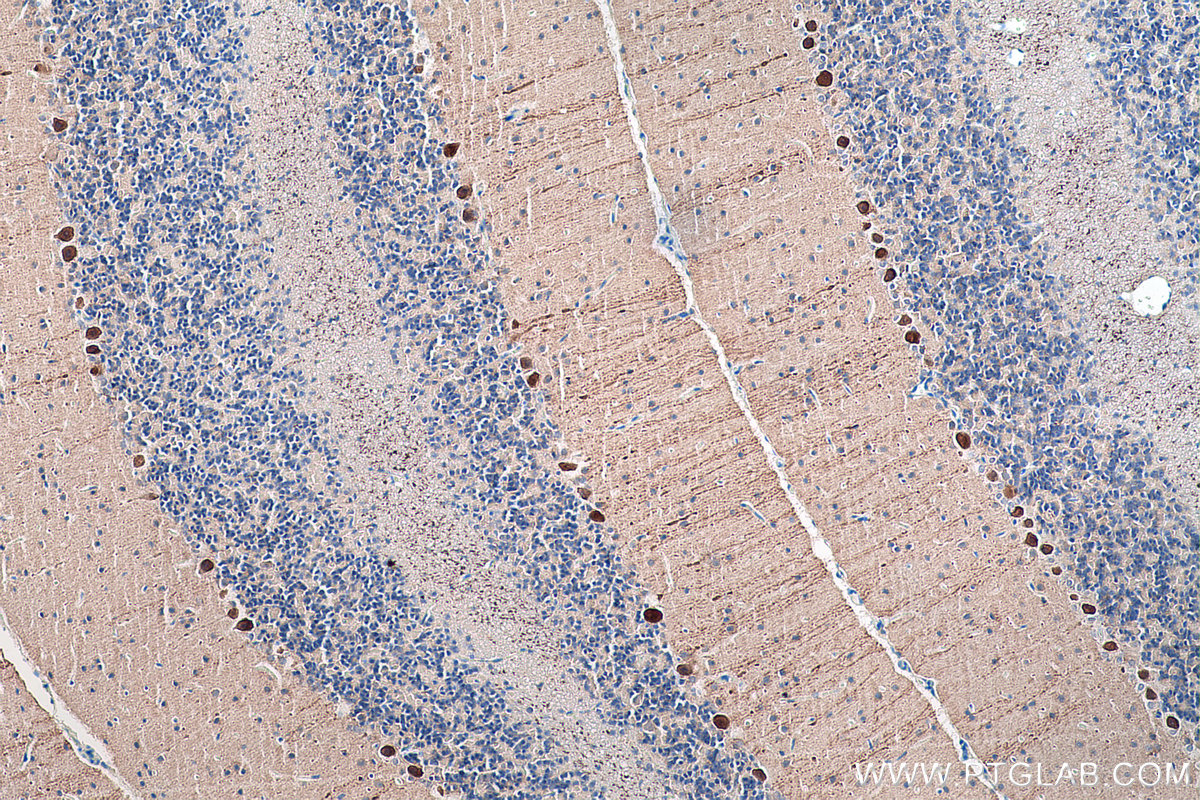
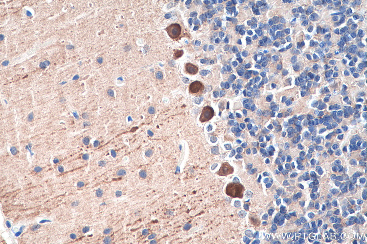
IHC staining of hippocampus using 14479-1-AP
IHC result of Calbindin antibody (14479-1-AP, 1:5000 in 1% BSA 0.3%Tx-100 Tris buffer incubated at 4℃ overnight) with freshly formalin fixed human hippocampus (floating section). Secondary antibody biotinylated anti-rabbit lgG 1:200 in 1% BSA 0.3%Tx-100 Tris buffer for 2 hours at room temperature. .
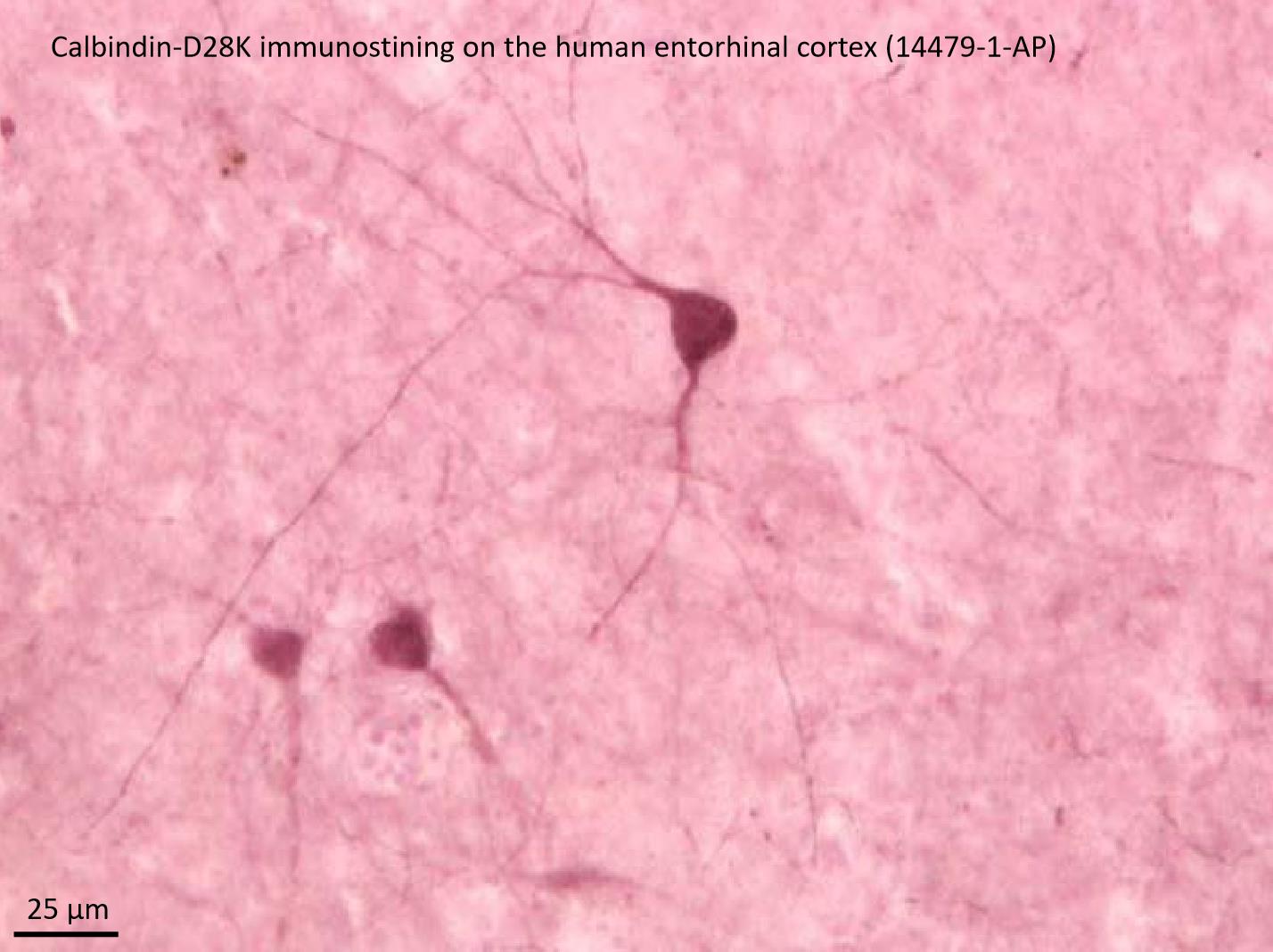
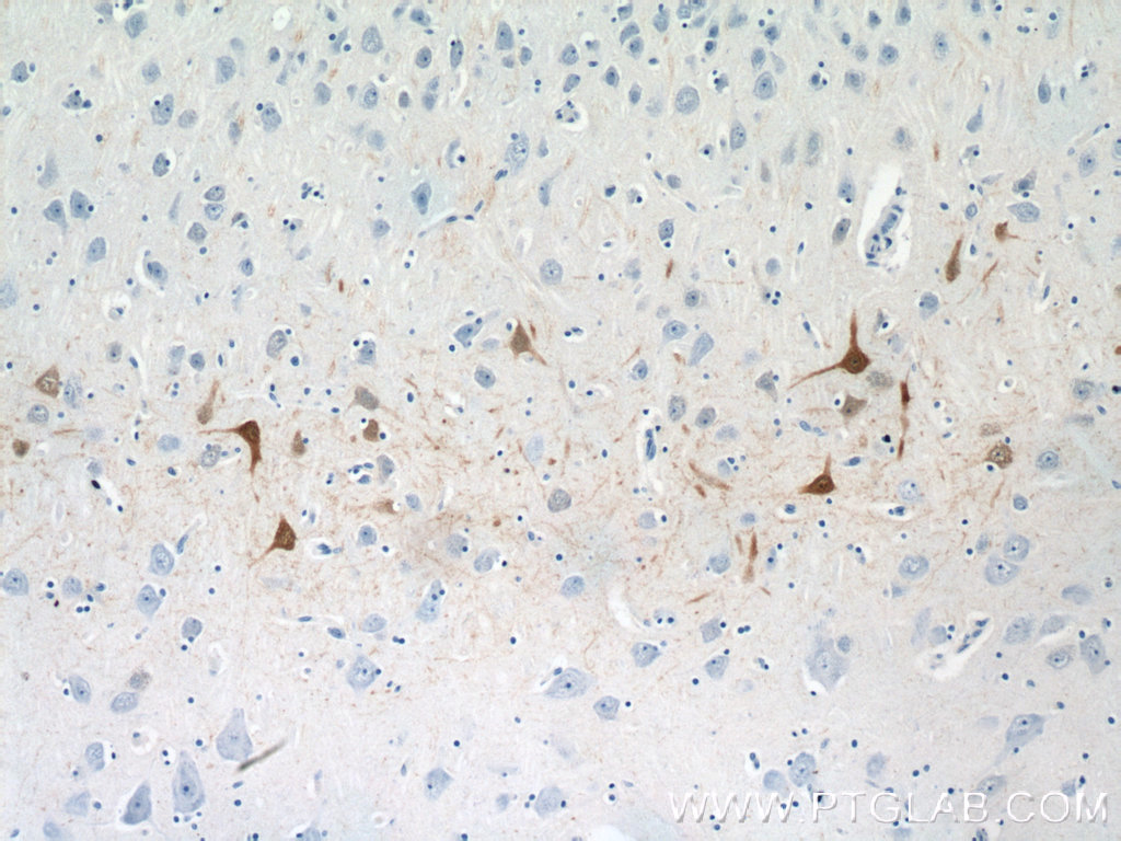
IHC staining of human brain using 14479-1-AP
Immunohistochemical analysis of paraffin-embedded human brain tissue slide using 14479-1-AP (Calbindin-D28k antibody at dilution of 1:200 (under 10x lens).
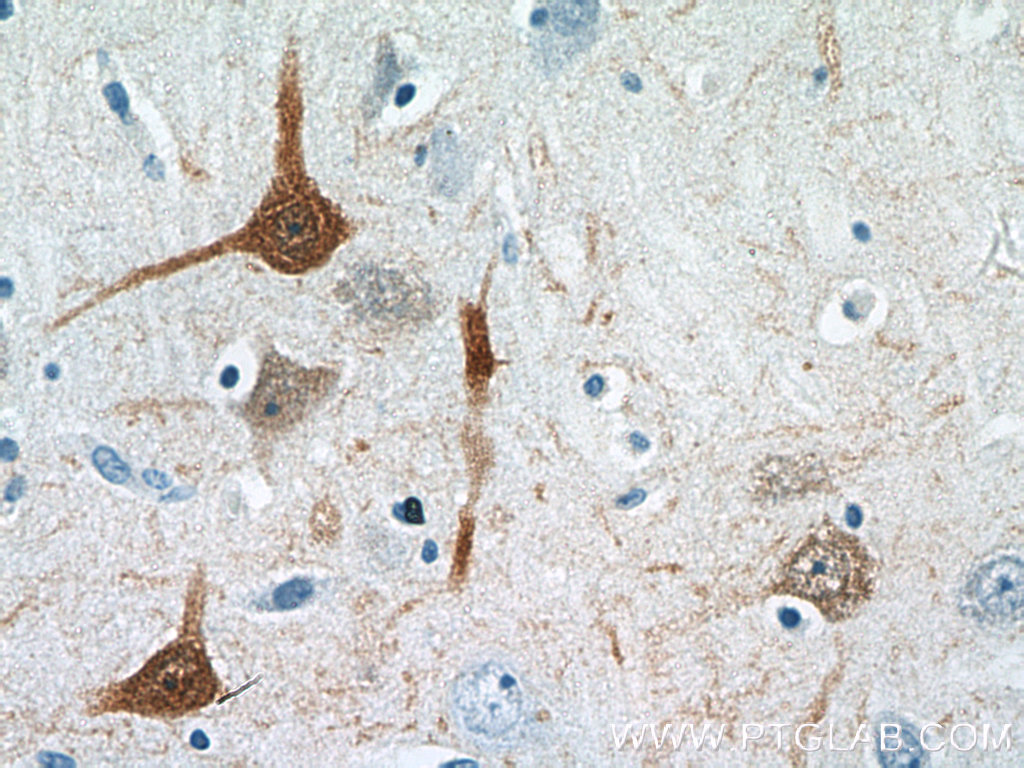
IHC staining of human brain using 14479-1-AP
Immunohistochemical analysis of paraffin-embedded human brain tissue slide using 14479-1-AP (Calbindin-D28k antibody at dilution of 1:200 (under 40x lens).
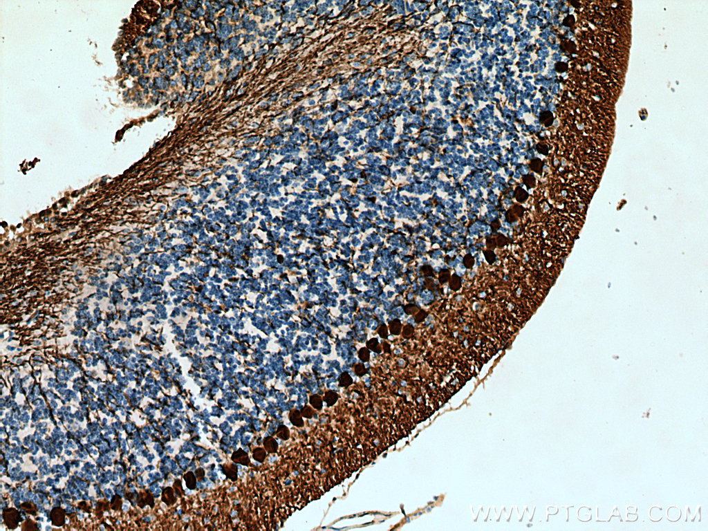
IHC staining of human cerebellum using 14479-1-AP
Immunohistochemical analysis of paraffin-embedded human cerebellum tissue slide using 14479-1-AP (Calbindin-D28k antibody) at dilution of 1:2000 (under 10x lens).
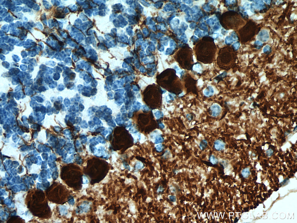
IHC staining of human cerebellum using 14479-1-AP
Immunohistochemical analysis of paraffin-embedded human cerebellum tissue slide using 14479-1-AP (Calbindin-D28k antibody) at dilution of 1:2000 (under 40x lens).
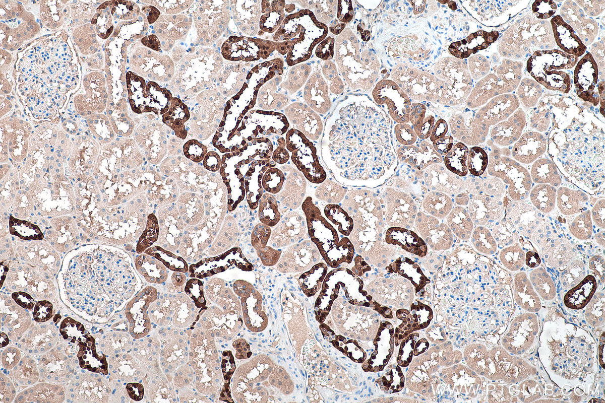
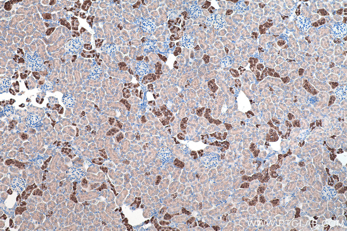
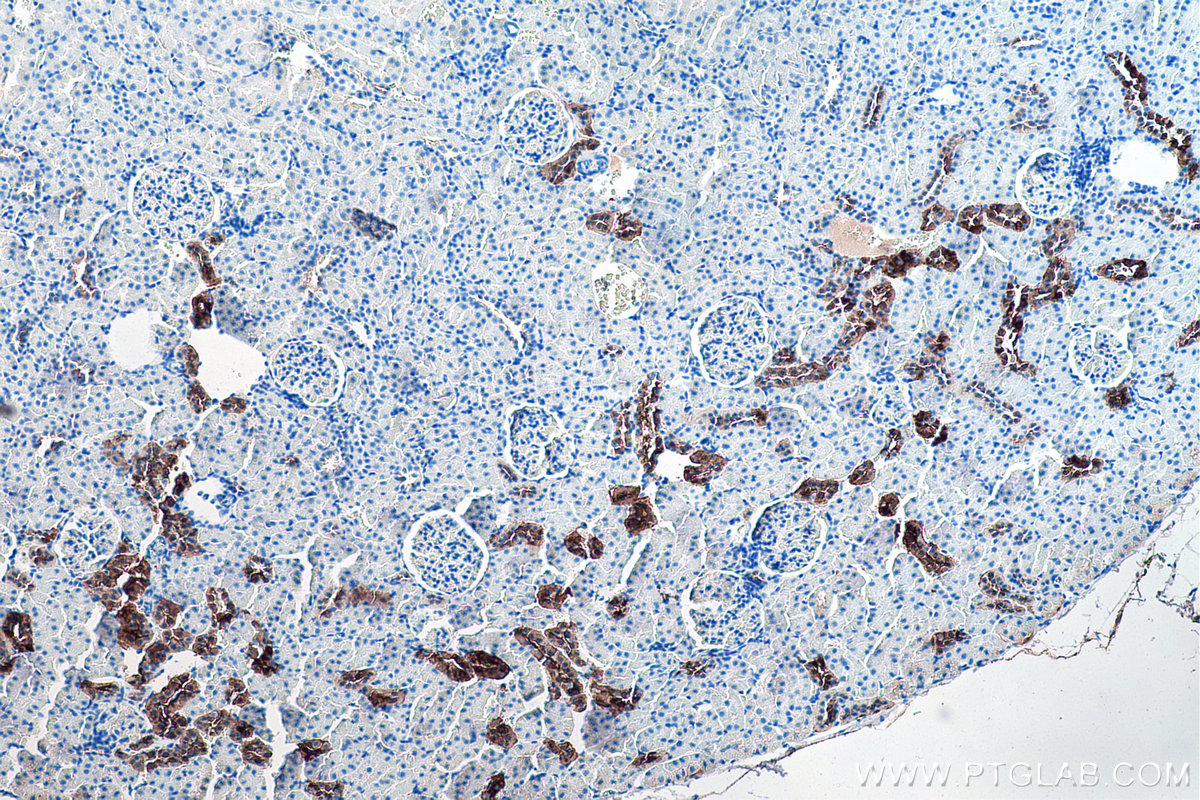
IHC staining of human kidney using 14479-1-AP
Immunohistochemical analysis of paraffin-embedded human kidney tissue slide using 14479-1-AP (Calbindin-D28k antibody) at dilution of 1:16000 (under 10x lens). Heat mediated antigen retrieval with Tris-EDTA buffer (pH 9.0).
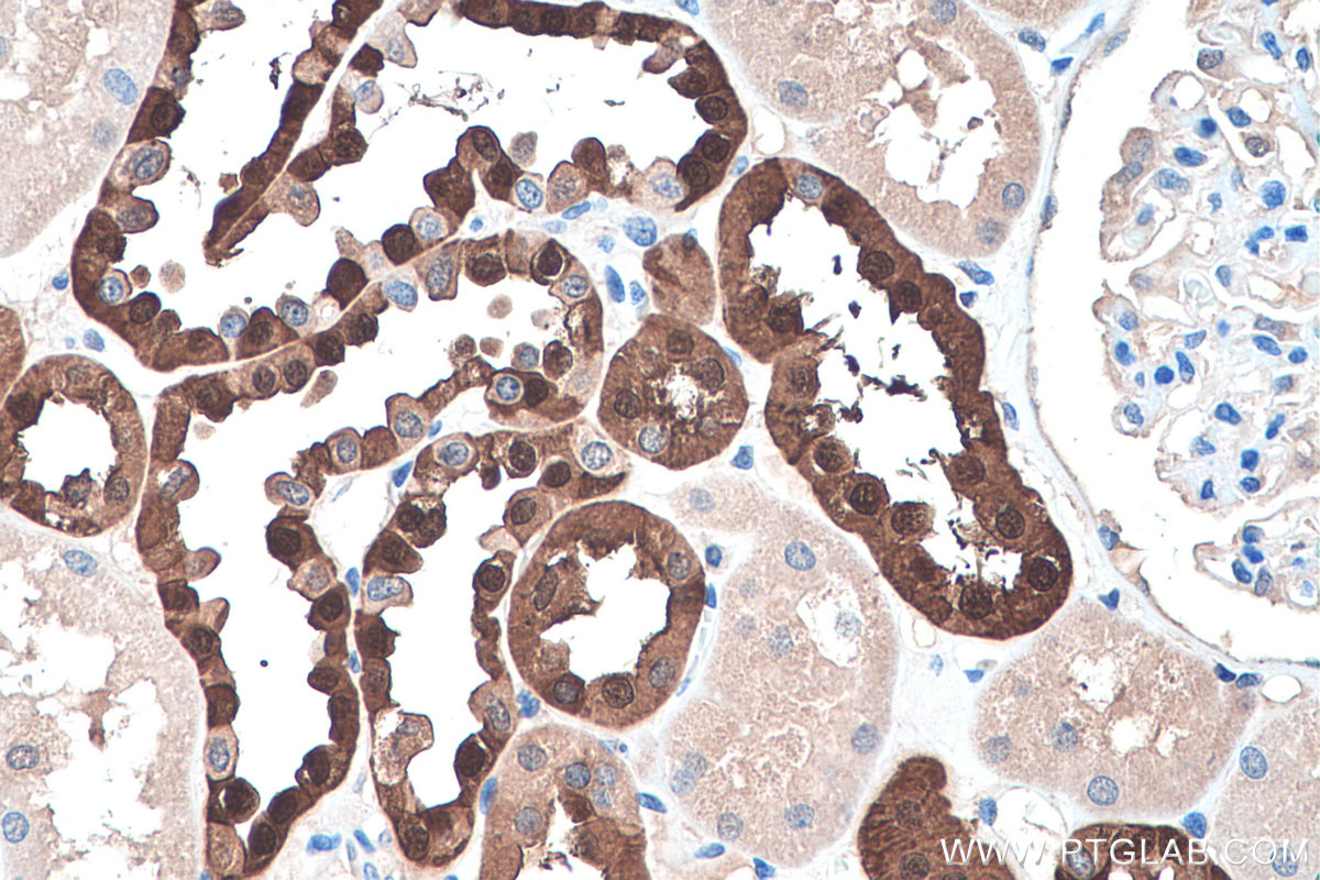
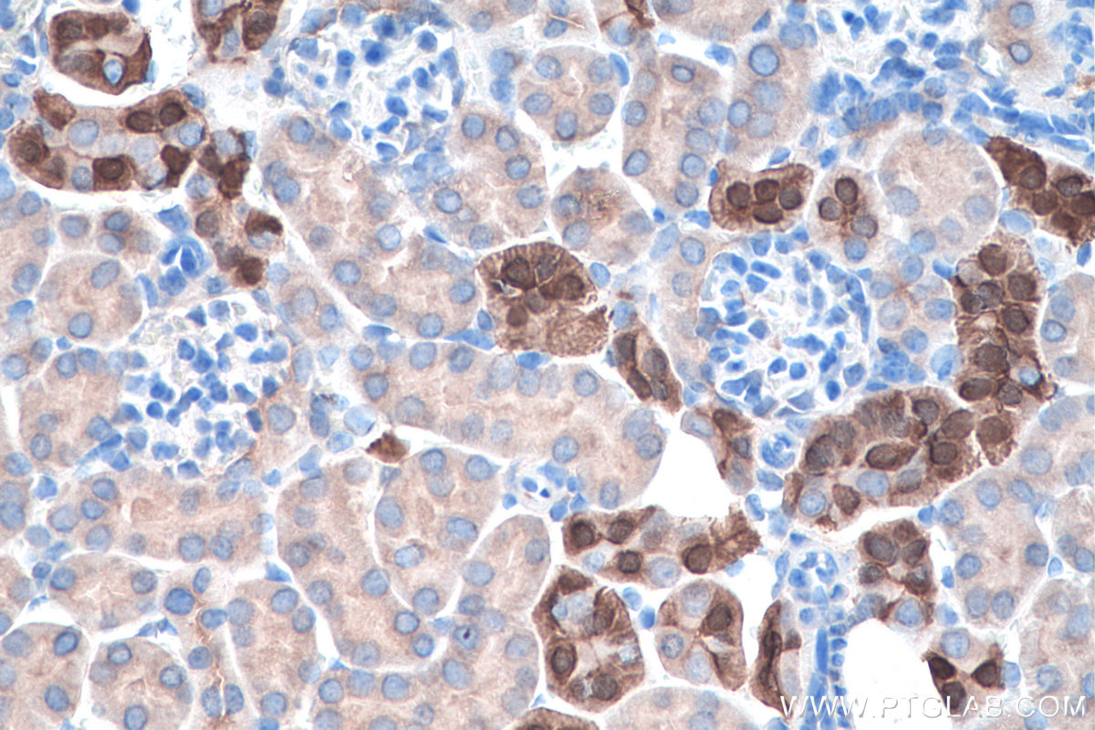
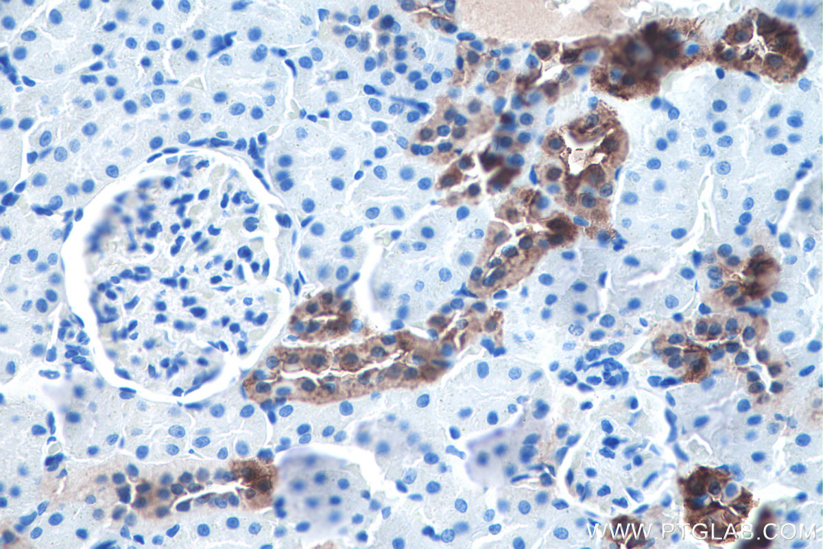
IHC staining of human kidney using 14479-1-AP
Immunohistochemical analysis of paraffin-embedded human kidney tissue slide using 14479-1-AP (Calbindin-D28k antibody) at dilution of 1:16000 (under 40x lens). Heat mediated antigen retrieval with Tris-EDTA buffer (pH 9.0).
IHC staining of mouse cerebellum using 14479-1-AP
Immunohistochemical analysis of paraffin-embedded mouse cerebellum tissue slide using 14479-1-AP (Calbindin-D28k antibody) at dilution of 1:2000 (under 10x lens). Heat mediated antigen retrieval with Tris-EDTA buffer (pH 9.0).
IHC staining of mouse cerebellum using 14479-1-AP
Immunohistochemical analysis of paraffin-embedded mouse cerebellum tissue slide using 14479-1-AP (Calbindin-D28k antibody) at dilution of 1:2000 (under 40x lens). Heat mediated antigen retrieval with Tris-EDTA buffer (pH 9.0).
IHC staining of mouse kidney using 14479-1-AP
Immunohistochemical analysis of paraffin-embedded mouse kidney tissue slide using 14479-1-AP (Calbindin-D28k antibody) at dilution of 1:16000 (under 10x lens). Heat mediated antigen retrieval with Tris-EDTA buffer (pH 9.0).
IHC staining of mouse kidney using 14479-1-AP
Immunohistochemical analysis of paraffin-embedded mouse kidney tissue slide using 14479-1-AP (Calbindin-D28k antibody) at dilution of 1:16000 (under 40x lens). Heat mediated antigen retrieval with Tris-EDTA buffer (pH 9.0).
IHC staining of mouse kidney using 14479-1-AP
Immunohistochemical analysis of paraffin-embedded mouse kidney tissue slide using 14479-1-AP (Calbindin-D28k antibody) at dilution of 1:2000 (under 10x lens). Heat mediated antigen retrieval with Tris-EDTA buffer (pH 9.0).
IHC staining of mouse kidney using 14479-1-AP
Immunohistochemical analysis of paraffin-embedded mouse kidney tissue slide using 14479-1-AP (Calbindin-D28k antibody) at dilution of 1:2000 (under 40x lens). Heat mediated antigen retrieval with Tris-EDTA buffer (pH 9.0).
IHC staining of rat cerebellum using 14479-1-AP
Immunohistochemical analysis of paraffin-embedded rat cerebellum tissue slide using 14479-1-AP (Calbindin-D28k antibody) at dilution of 1:16000 (under 10x lens). Heat mediated antigen retrieval with Tris-EDTA buffer (pH 9.0).
IHC staining of rat cerebellum using 14479-1-AP
Immunohistochemical analysis of paraffin-embedded rat cerebellum tissue slide using 14479-1-AP (Calbindin-D28k antibody) at dilution of 1:16000 (under 40x lens). Heat mediated antigen retrieval with Tris-EDTA buffer (pH 9.0).
IHC staining of rat kidney using 14479-1-AP
Immunohistochemical analysis of paraffin-embedded rat kidney tissue slide using 14479-1-AP (Calbindin-D28k antibody) at dilution of 1:16000 (under 10x lens). Heat mediated antigen retrieval with Tris-EDTA buffer (pH 9.0).
IHC staining of rat kidney using 14479-1-AP
Immunohistochemical analysis of paraffin-embedded rat kidney tissue slide using 14479-1-AP (Calbindin-D28k antibody) at dilution of 1:16000 (under 40x lens). Heat mediated antigen retrieval with Tris-EDTA buffer (pH 9.0).
IF-P Figures
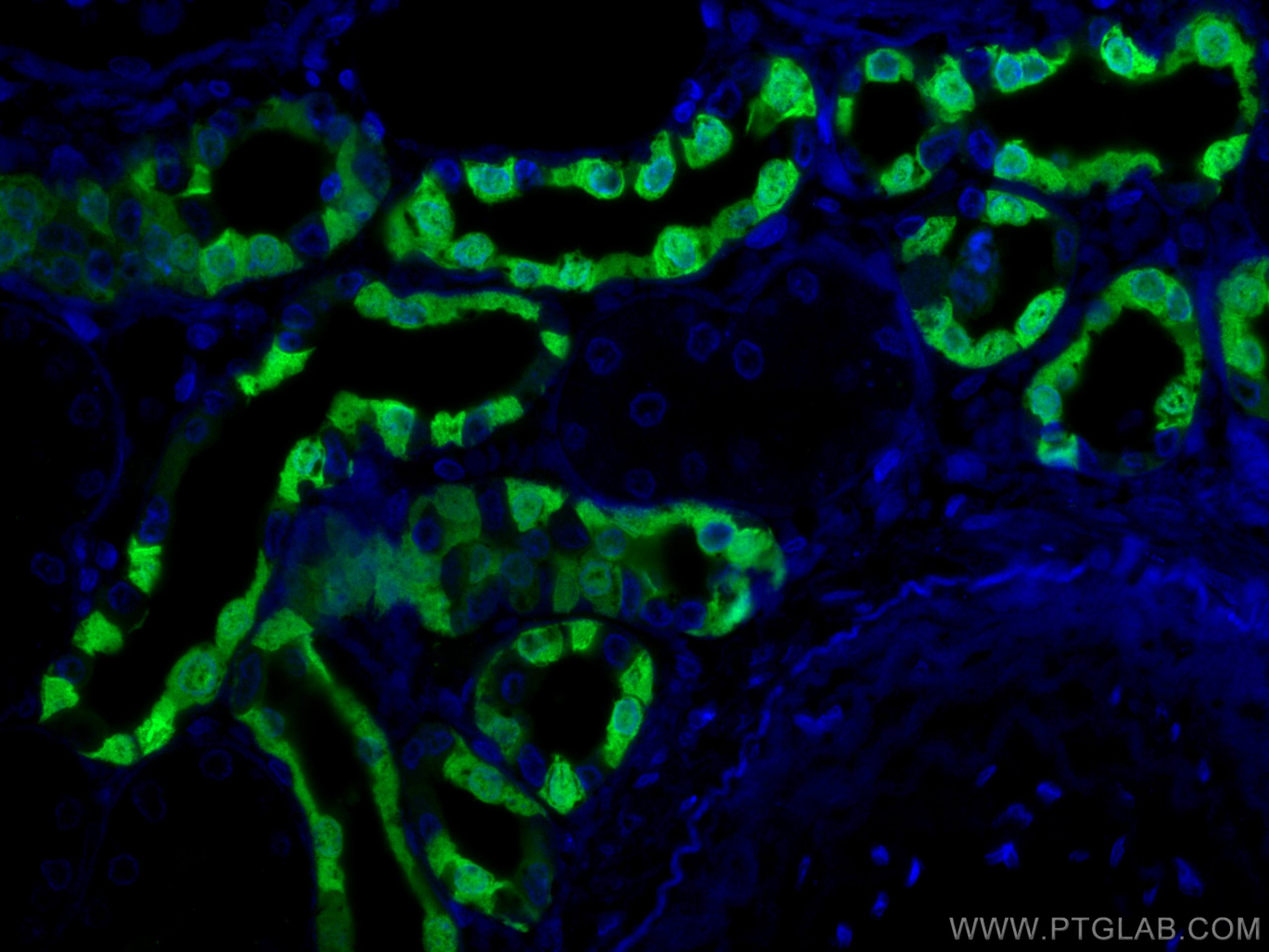
IF Staining of human kidney using 14479-1-AP
Immunofluorescent analysis of (4% PFA) fixed human kidney tissue using Calbindin-D28k antibody (14479-1-AP) at dilution of 1:200 and CoraLite®488-Conjugated AffiniPure Goat Anti-Rabbit IgG(H+L), PTPRO antibody (67000-1-Ig, Clone: 2F2B4, red).
IF Staining of human kidney using 14479-1-AP
Immunofluorescent analysis of (4% PFA) fixed human kidney tissue using Calbindin-D28k antibody (14479-1-AP) at dilution of 1:400 and CoraLite®488-Conjugated AffiniPure Goat Anti-Rabbit IgG(H+L).
IF Staining of human kidney using 14479-1-AP
Immunofluorescent analysis of (4% PFA) fixed human kidney tissue using Calbindin-D28k antibody (14479-1-AP) at dilution of 1:400 and CoraLite®488-Conjugated AffiniPure Goat Anti-Rabbit IgG(H+L).
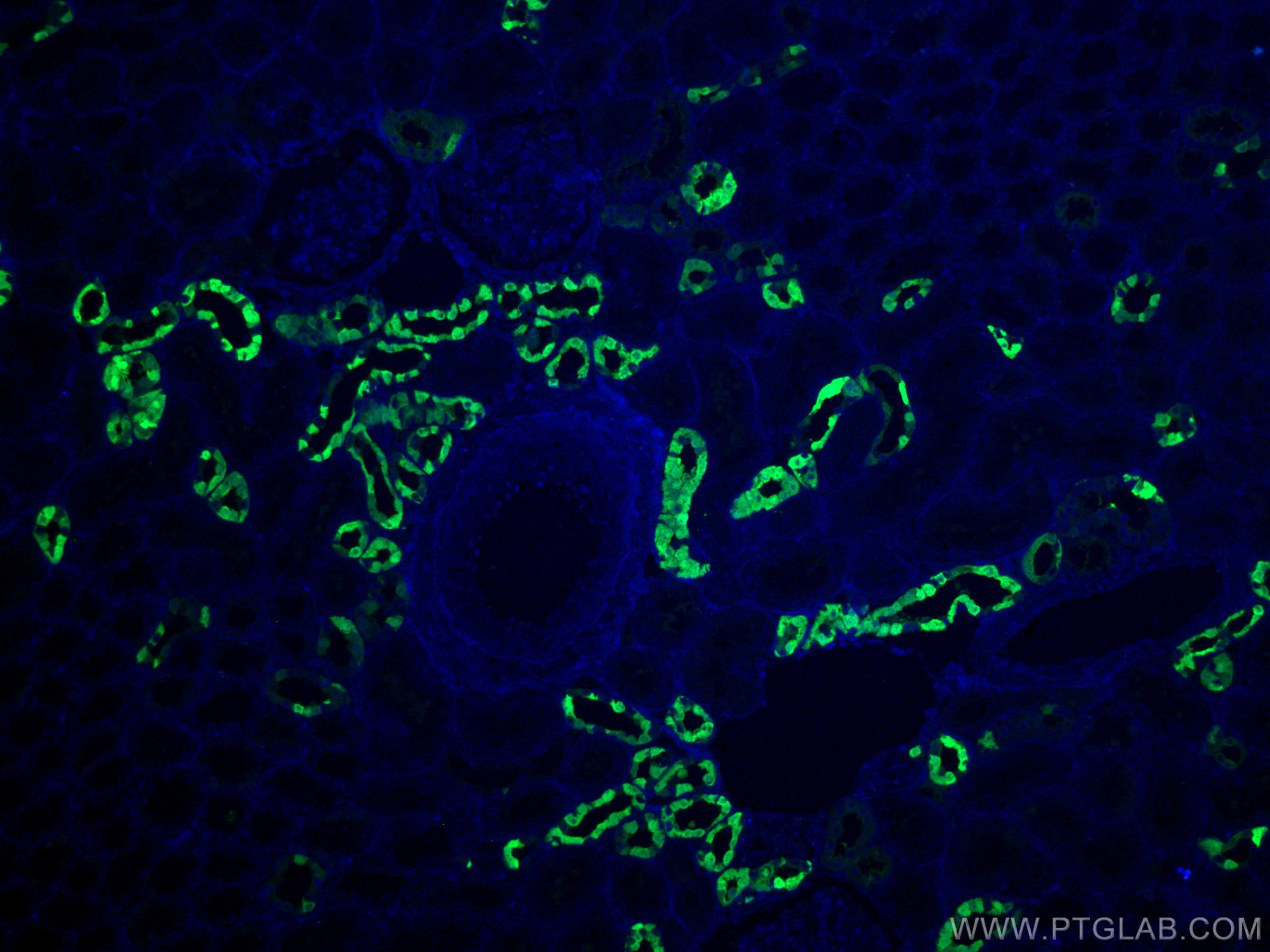
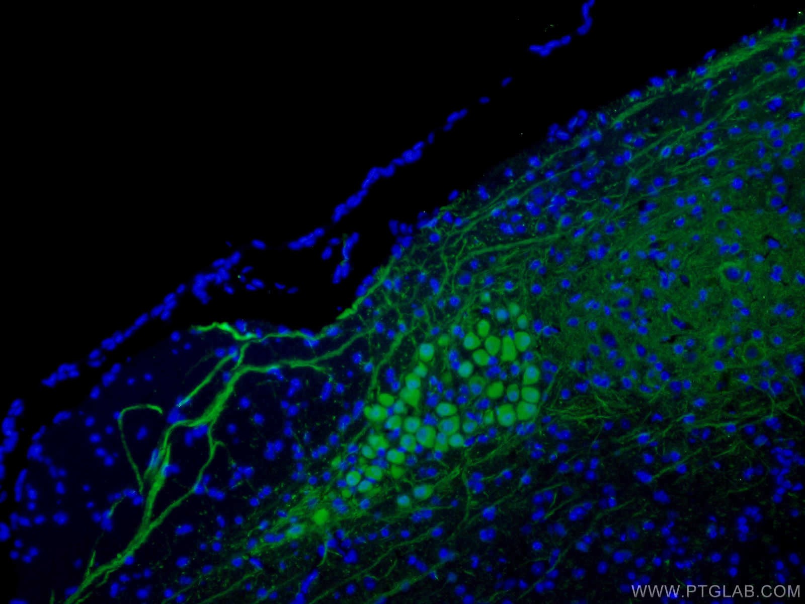
IF Staining of mouse cerebellum using 14479-1-AP
Immunofluorescent analysis of (4% PFA) fixed mouse cerebellum tissue using Calbindin-D28k antibody (14479-1-AP) at dilution of 1:200 and CoraLite®488-Conjugated AffiniPure Goat Anti-Rabbit IgG(H+L), PAX6 antibody (67529-1-Ig, Clone: 2C5A1, red).
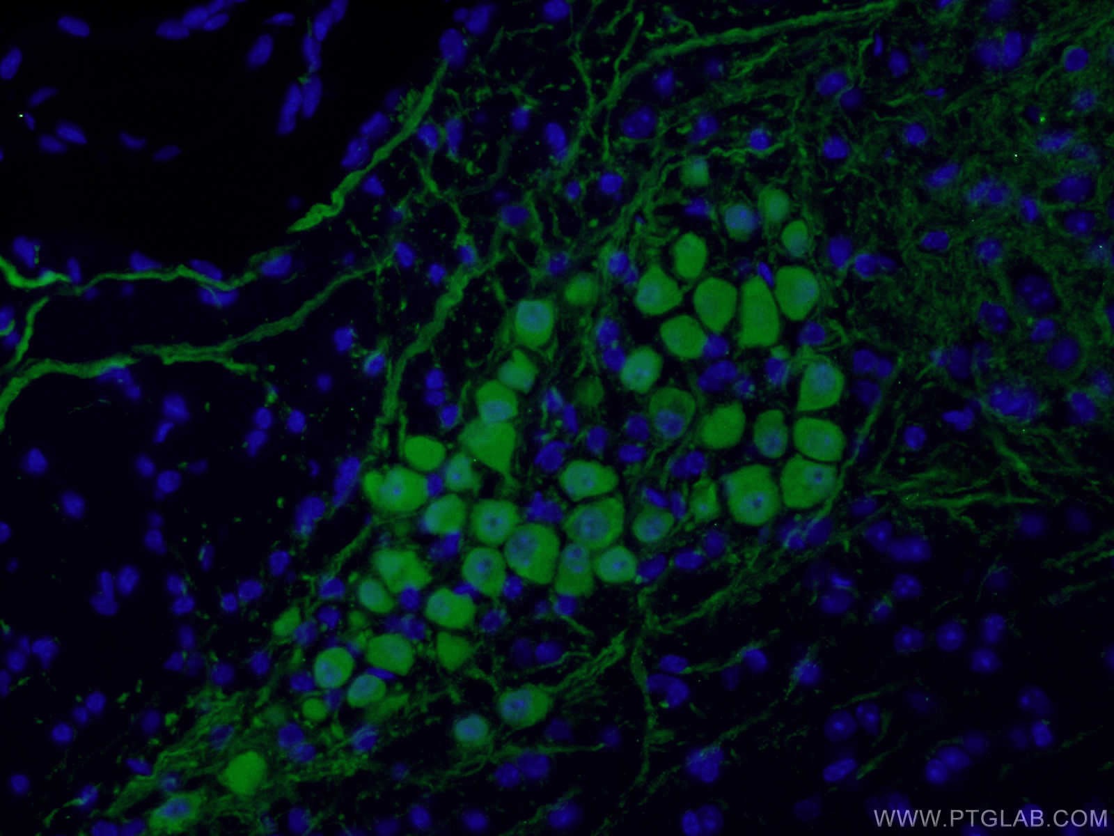
IF Staining of mouse cerebellum using 14479-1-AP
Immunofluorescent analysis of (4% PFA) fixed mouse cerebellum tissue using Calbindin-D28k antibody (14479-1-AP) at dilution of 1:200 and CoraLite®488-Conjugated AffiniPure Goat Anti-Rabbit IgG(H+L), PAX6 antibody (67529-1-Ig, Clone: 2C5A1, red).
IF Staining of mouse cerebellum using 14479-1-AP
Immunofluorescent analysis of (4% PFA) fixed paraffin-embedded mouse cerebellum tissue using Calbindin-D28k antibody (14479-1-AP) at dilution of 1:400 and CoraLite®488-Conjugated Goat Anti-Rabbit IgG(H+L) (SA00013-2). Heat mediated antigen retrieval with Tris-EDTA buffer (pH 9.0).
IF Staining of mouse cerebellum using 14479-1-AP
Immunofluorescent analysis of (4% PFA) fixed mouse cerebellum tissue using 14479-1-AP (Calbindin-D28k antibody) at dilution of 1:100 and Alexa Fluor 488-conjugated AffiniPure Goat Anti-Rabbit IgG(H+L).
IF Staining of mouse cerebellum using 14479-1-AP
Immunofluorescent analysis of (4% PFA) fixed mouse cerebellum tissue using 14479-1-AP (Calbindin-D28k antibody) at dilution of 1:100 and Alexa Fluor 488-conjugated AffiniPure Goat Anti-Rabbit IgG(H+L).
IF-FRO Figures
IF Staining of mouse cerebellum using 14479-1-AP
Immunofluorescent analysis of (4% PFA) fixed frozen OCT-embedded mouse cerebellum tissue using Calbindin-D28k antibody (14479-1-AP) at dilution of 1:200 and CoraLite®488-Conjugated Goat Anti-Rabbit IgG(H+L) (SA00013-2), FUS/TLS antibody (68262-1-Ig, Clone: 1B4F8, red).
IF Staining of mouse cerebellum tissue using 14479-1-AP
Immunofluorescent analysis of (4% PFA) fixed adult mouse cerebellum tissue using Calbindin-D28k antibody (14479-1-AP) at dilution of 1:1500 and Alexa Fluor 594 AffiniPure Donkey Anti-Rabbit IgG (H+L). (Image produced by Watt Lab at McGill University)
IF Staining of rat cerebellum using 14479-1-AP
Immunofluorescent analysis of (4% PFA) fixed frozen OCT-embedded rat cerebellum tissue using Calbindin-D28k antibody (14479-1-AP) at dilution of 1:200 and CoraLite®488-Conjugated Goat Anti-Rabbit IgG(H+L) (SA00013-2), FUS/TLS antibody (68262-1-Ig, Clone: 1B4F8, red).