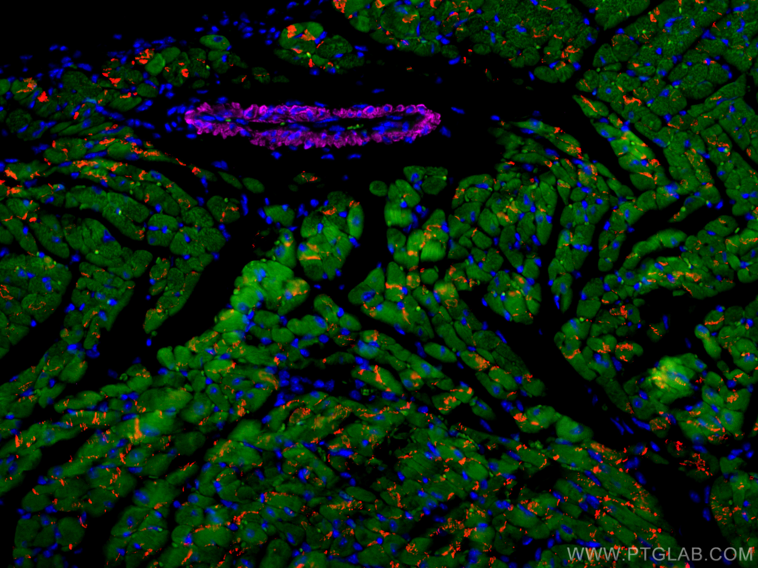
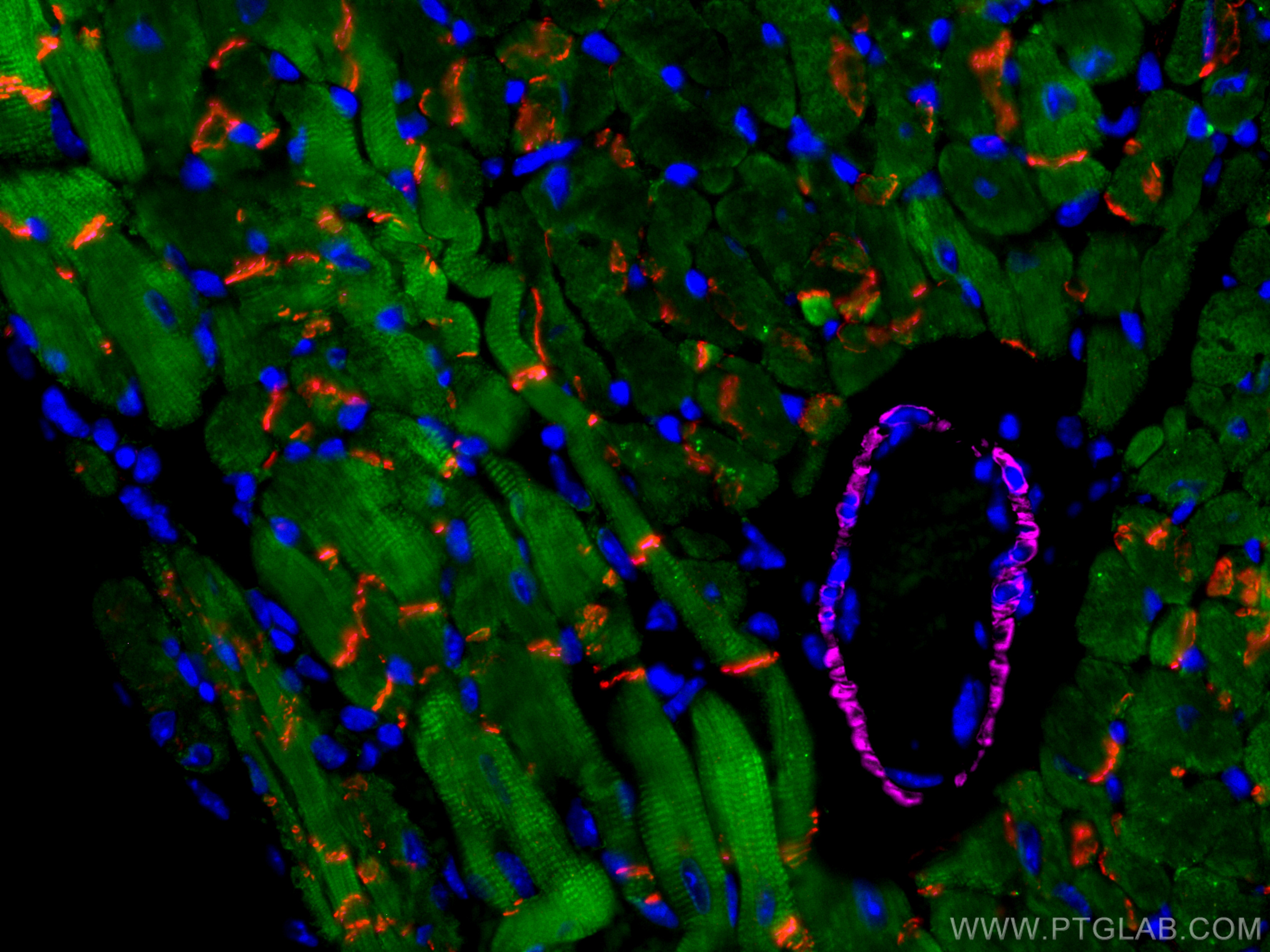
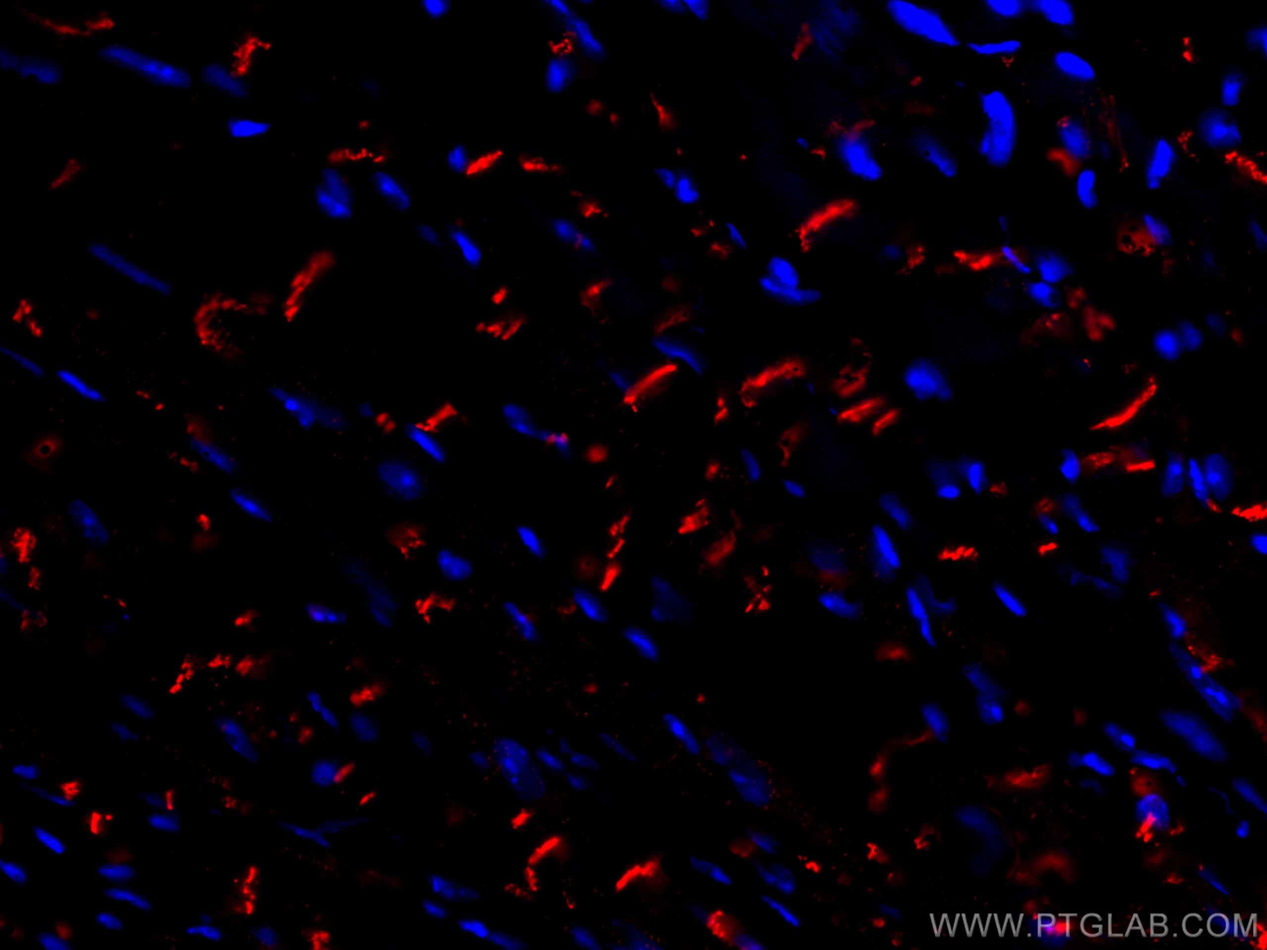
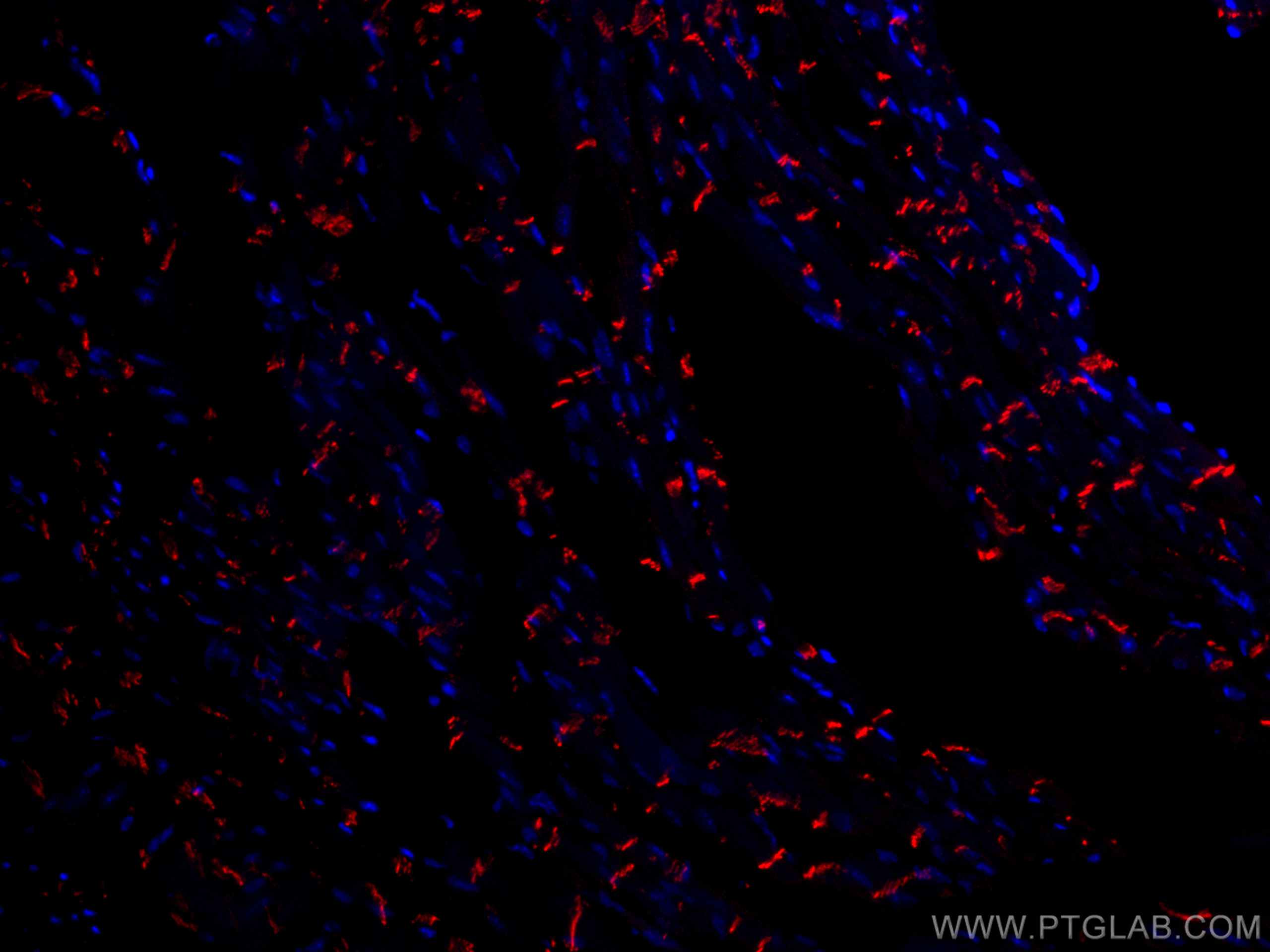
验证数据展示
经过测试的应用
| Positive IF-P detected in | mouse heart tissue |
推荐稀释比
| 应用 | 推荐稀释比 |
|---|---|
| Immunofluorescence (IF)-P | IF-P : 1:50-1:500 |
| It is recommended that this reagent should be titrated in each testing system to obtain optimal results. | |
| Sample-dependent, Check data in validation data gallery. | |
产品信息
CL594-22018 targets N-cadherin in IF-P applications and shows reactivity with human, mouse, rat samples.
| 经测试应用 | IF-P Application Description |
| 经测试反应性 | human, mouse, rat |
| 免疫原 |
CatNo: Ag16792 Product name: Recombinant human N-cadherin protein Source: e coli.-derived, PGEX-4T Tag: GST Domain: 421-535 aa of BC036470 Sequence: RISGGDPTGRFAIQTDQNSNDGLVTVVKPIDFEANRMFVLTVAAENQVPLAKGIQHPPQSTATMSVTVIDVNENPYFAPNPKIIRQEEGLHAGTMLTTFTAQDPDRYMQQNIRYT 种属同源性预测 |
| 宿主/亚型 | Rabbit / IgG |
| 抗体类别 | Polyclonal |
| 产品类型 | Antibody |
| 全称 | cadherin 2, type 1, N-cadherin (neuronal) |
| 别名 | Cadherin 2, CD325, CDH2, CDHN, CDw325, N cadherin, NCAD, N-cadherin, Neural cadherin |
| 计算分子量 | 906 aa, 100 kDa |
| 观测分子量 | 130 kDa |
| GenBank蛋白编号 | BC036470 |
| 基因名称 | N-cadherin |
| Gene ID (NCBI) | 1000 |
| RRID | AB_2919867 |
| 偶联类型 | CoraLite®594 Fluorescent Dye |
| 最大激发/发射波长 | 588 nm / 604 nm |
| 形式 | Liquid |
| 纯化方式 | Antigen affinity purification |
| UNIPROT ID | P19022 |
| 储存缓冲液 | PBS with 50% glycerol, 0.05% Proclin300, 0.5% BSA, pH 7.3. |
| 储存条件 | Store at -20°C. Avoid exposure to light. Stable for one year after shipment. Aliquoting is unnecessary for -20oC storage. |
背景介绍
Neuronal cadherin (N-cadherin), also known as cadherin-2 (CDH2), is a calcium-binding protein that mediates cell-cell adhesions of neuronal and some non-neuronal cell types.
What is the molecular weight of N-cadherin? Is N-cadherin post-translationally modified?
The molecular weight of mature N-cadherin is 127 kDa. N-cadherin is synthesized in a precursor form that undergoes proteolytic cleavage by furin at the Golgi apparatus. Additionally, it can be phosphorylated by casein kinase II and N-glycosylated, which affects its stability (PMID: 12604612 and 19846557).
What is the subcellular localization of N-cadherin? What is the tissue expression pattern of N-cadherin?
N-cadherin is an integral membrane protein present at the plasma membrane, forming adherens junctions. It is widely expressed in the nervous system, where it flanks the active zone of synapses and is important for synapse formation and remodeling. It is also present in the lens, skeletal, and cardiac muscles (PMID: 3857614). In the muscle, N-cadherin plays a role in myoblast differentiation, while in the heart it is required for the formation of intercalated discs. Additionally, N-cadherin is present in blood vessels, promoting angiogenesis by forming adhesive complexes between endothelial cells and pericytes (PMID: 24521477).
What is the role of N-cadherin during the epithelial-mesenchymal transition (EMT)?
EMT is a crucial process during gastrulation that leads to the formation of mesenchymal cells. It is marked by decreased expression of E-cadherin and upregulation of N-cadherin, which promotes cell migration (PMID: 23481201). Similarly, upregulation of N-cadherin is observed in many cancer cell types and is associated with increased invasiveness and metastasis.
实验方案
| Product Specific Protocols | |
|---|---|
| IF protocol for CL594 N-cadherin antibody CL594-22018 | Download protocol |
| Standard Protocols | |
|---|---|
| Click here to view our Standard Protocols |


