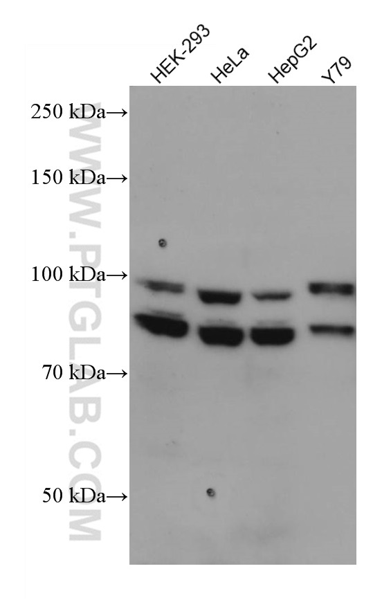
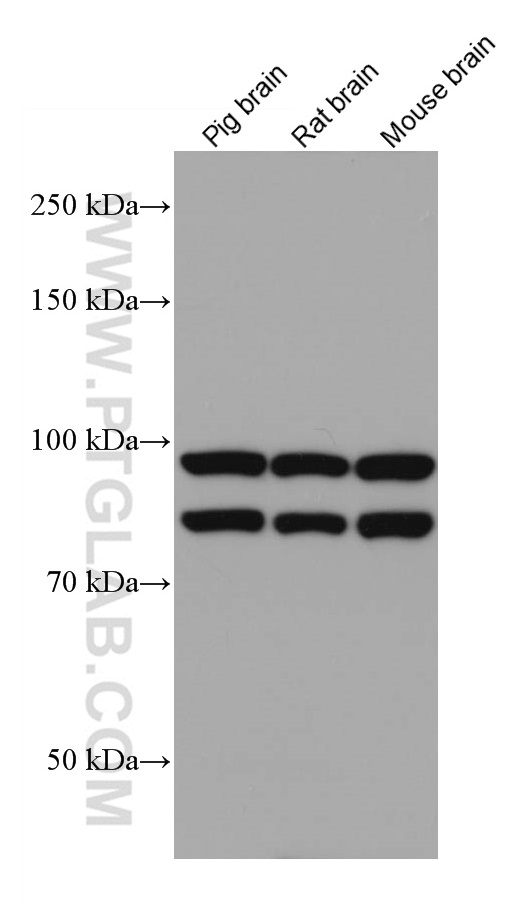
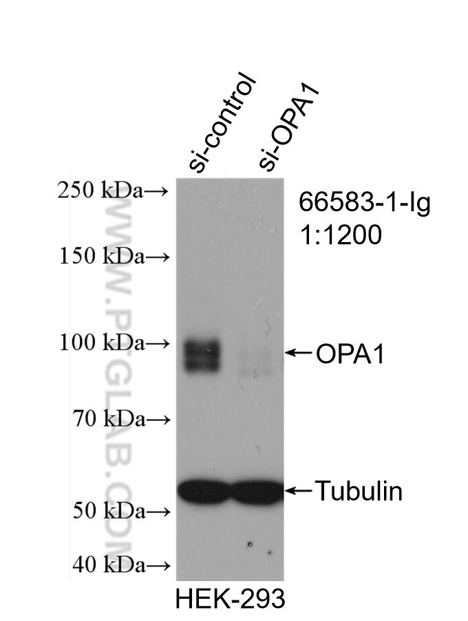
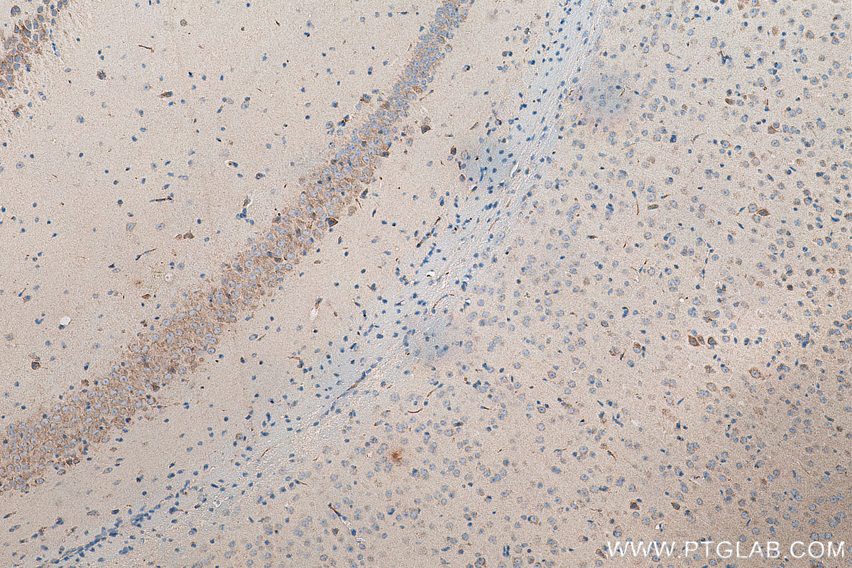
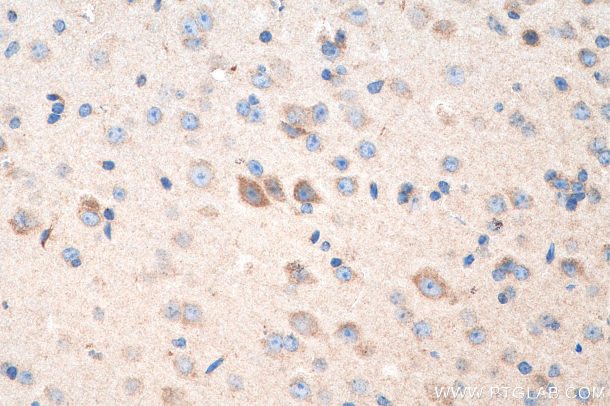
验证数据展示
产品信息
66583-1-PBS targets OPA1 in WB, IHC, ELISA applications and shows reactivity with Human, mouse, pig, rat samples.
| 经测试应用 | WB, IHC, ELISA Application Description |
| 经测试反应性 | Human, mouse, pig, rat |
| 免疫原 | OPA1 fusion protein Ag26868 种属同源性预测 |
| 宿主/亚型 | Mouse / IgG2b |
| 抗体类别 | Monoclonal |
| 产品类型 | Antibody |
| 全称 | optic atrophy 1 (autosomal dominant) |
| 别名 | FLJ12460, KIAA0567, largeG, MGM1, NTG, OPA1, Optic atrophy protein 1 |
| 计算分子量 | 960 aa, 112 kDa |
| 观测分子量 | 100 kDa and 80-90 kDa |
| GenBank蛋白编号 | BC075805 |
| 基因名称 | OPA1 |
| Gene ID (NCBI) | 4976 |
| RRID | AB_2881943 |
| 偶联类型 | Unconjugated |
| 形式 | Liquid |
| 纯化方式 | Protein A purification |
| UNIPROT ID | O60313 |
| 储存缓冲液 | PBS Only |
| 储存条件 | Store at -80°C. The product is shipped with ice packs. Upon receipt, store it immediately at -80°C |
背景介绍
OPA1 is a nuclear-encoded mitochondrial protein with similarity to dynamin-related GTPases. OPA1 localizes to the inner mitochondrial membrane and helps regulate mitochondrial stability and energy output. This protein also sequesters cytochrome c. OPA1 is associated with the inner membrane and protects cells from apoptosis by regulating inner membrane dynamics. Mutation of OPA1 causes the disease dominant optic atrophy, a degeneration of the retinal ganglion cells. OPA1 undergoes complex posttranscriptional regulation and posttranslational proteolysis. OPA1 is regulated by proteolytic cleavage, which degrades long OPA1 isoforms into short isoforms. The gene OPA1 can be cleaved into some chains with MW 100 kDa and 80-90 kDa.
