WB Figures
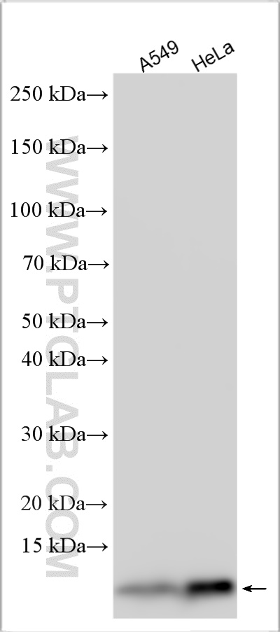
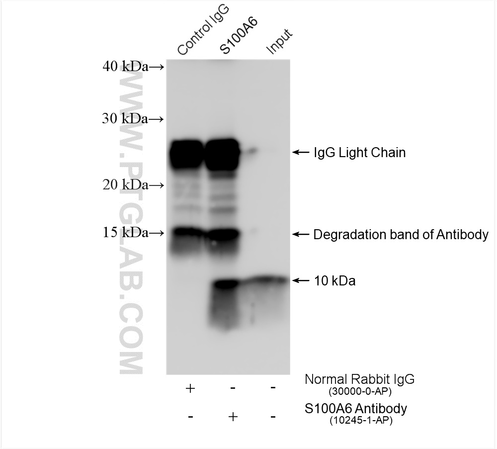
WB analysis of A549 using 10245-1-AP (same clone as 10245-1-PBS)
A549 cells were subjected to SDS PAGE followed by western blot with 10245-1-AP (S100A6 antibody) at dilution of 1:1000 incubated at room temperature for 1.5 hours. This data was developed using the same antibody clone with 10245-1-PBS in a different storage buffer formulation.
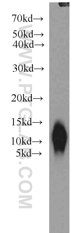
WB analysis using 10245-1-AP (same clone as 10245-1-PBS)
Various lysates were subjected to SDS PAGE followed by western blot with 10245-1-AP (S100A6 antibody) at dilution of 1:6000 incubated at room temperature for 1.5 hours. This data was developed using the same antibody clone with 10245-1-PBS in a different storage buffer formulation.
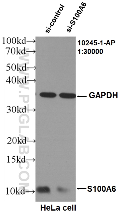
WB analysis of HeLa using 10245-1-AP (same clone as 10245-1-PBS)
WB result of S100A6 antibody (10245-1-AP, 1:30000) with si-Control and si-S100A6 transfected HeLa cells.. This data was developed using the same antibody clone with 10245-1-PBS in a different storage buffer formulation.
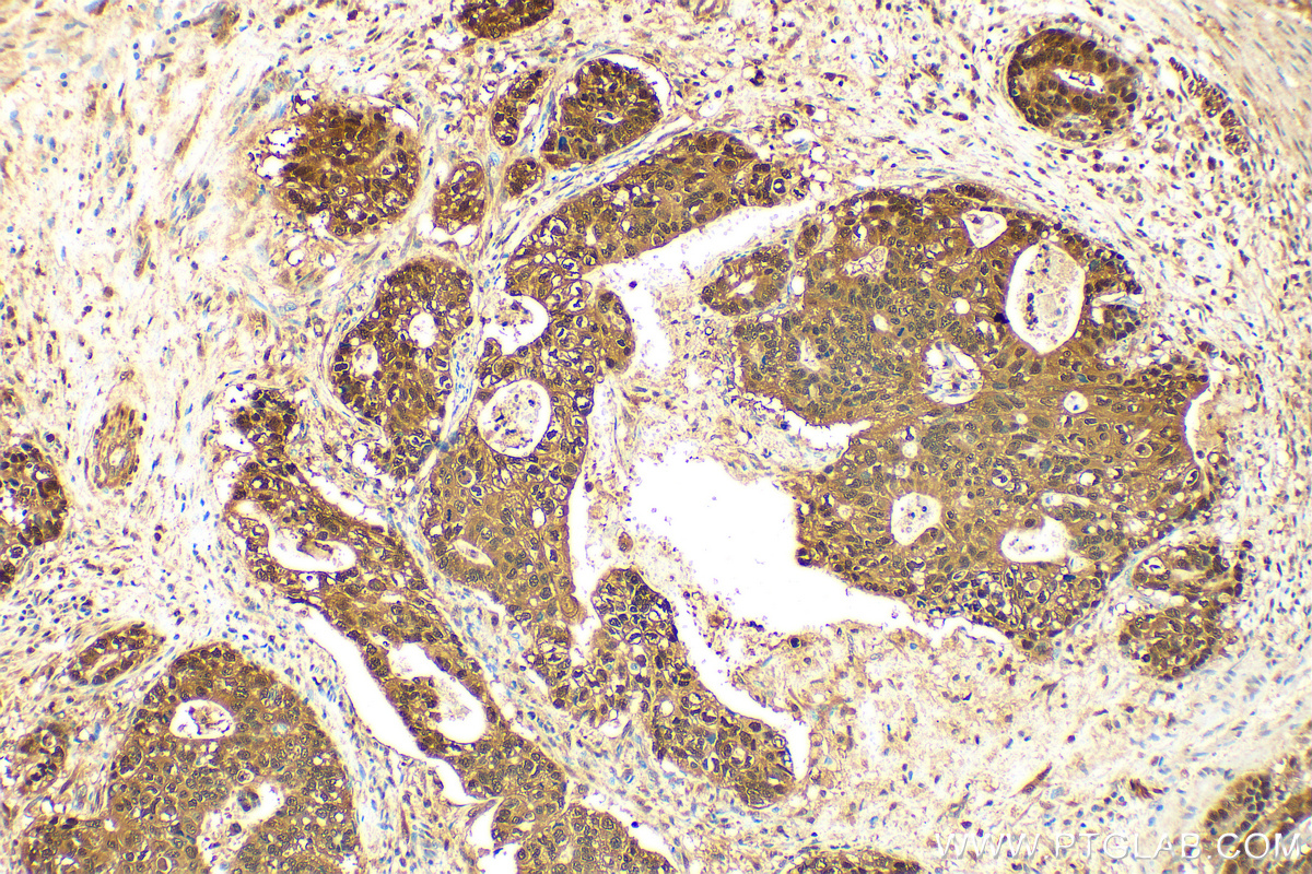
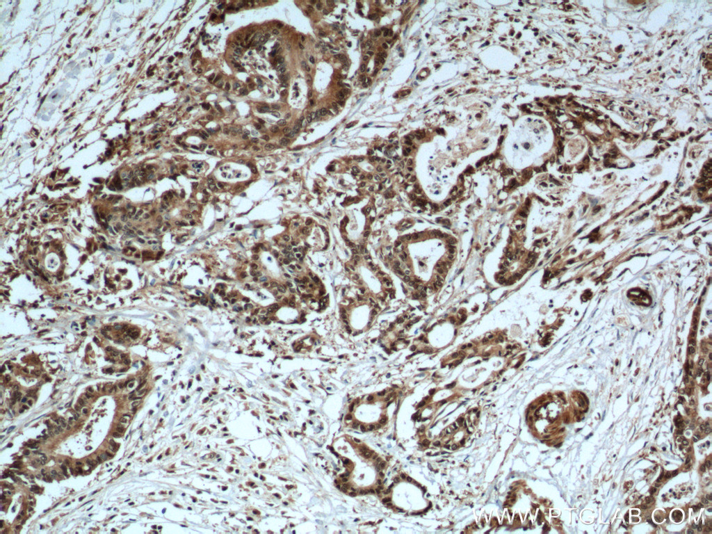
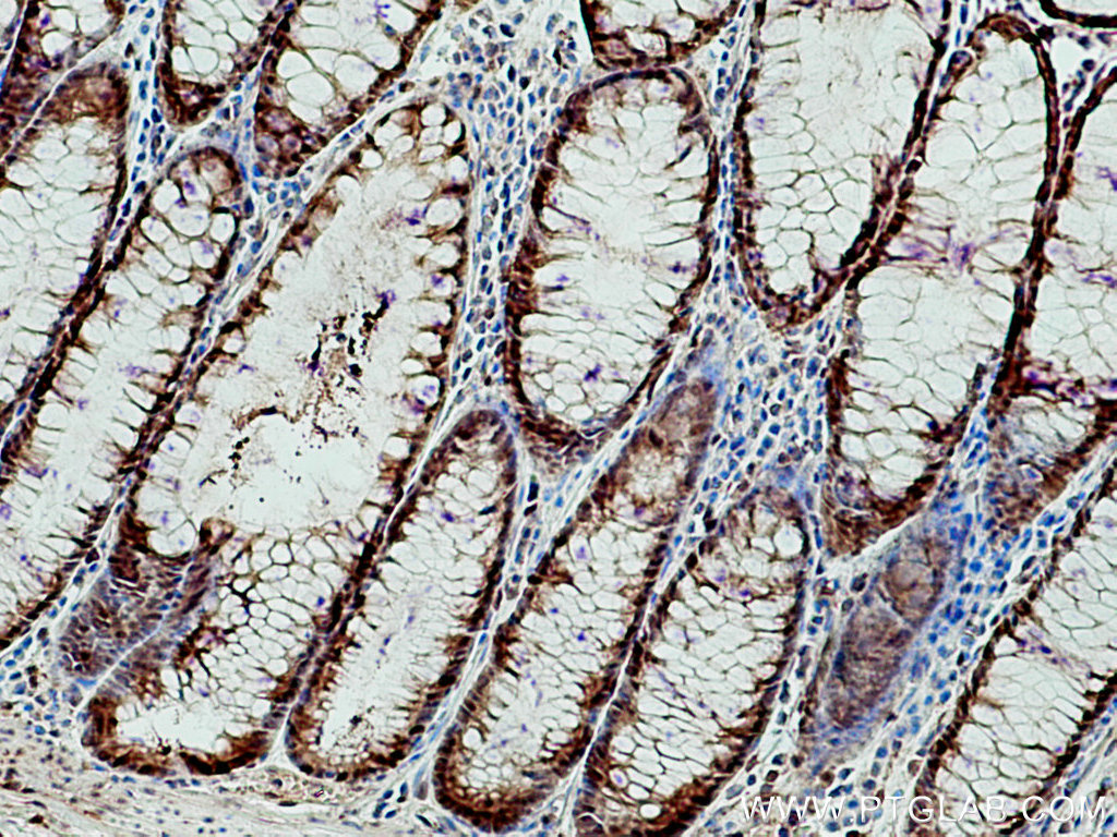
IHC staining of human colon cancer using 10245-1-AP (same clone as 10245-1-PBS)
Immunohistochemical analysis of paraffin-embedded human colon cancer tissue slide using 10245-1-AP (S100A6 antibody) at dilution of 1:800 (under 10x lens). Heat mediated antigen retrieval with Tris-EDTA buffer (pH 9.0). This data was developed using the same antibody clone with 10245-1-PBS in a different storage buffer formulation.
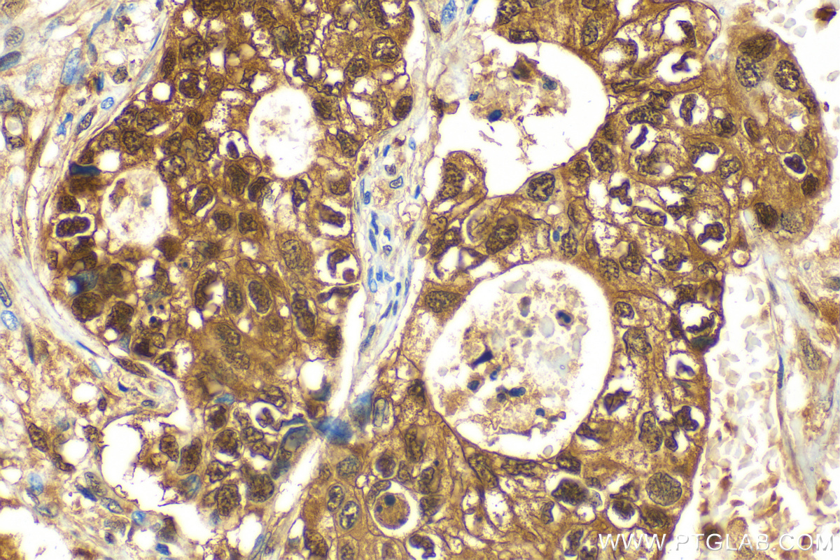
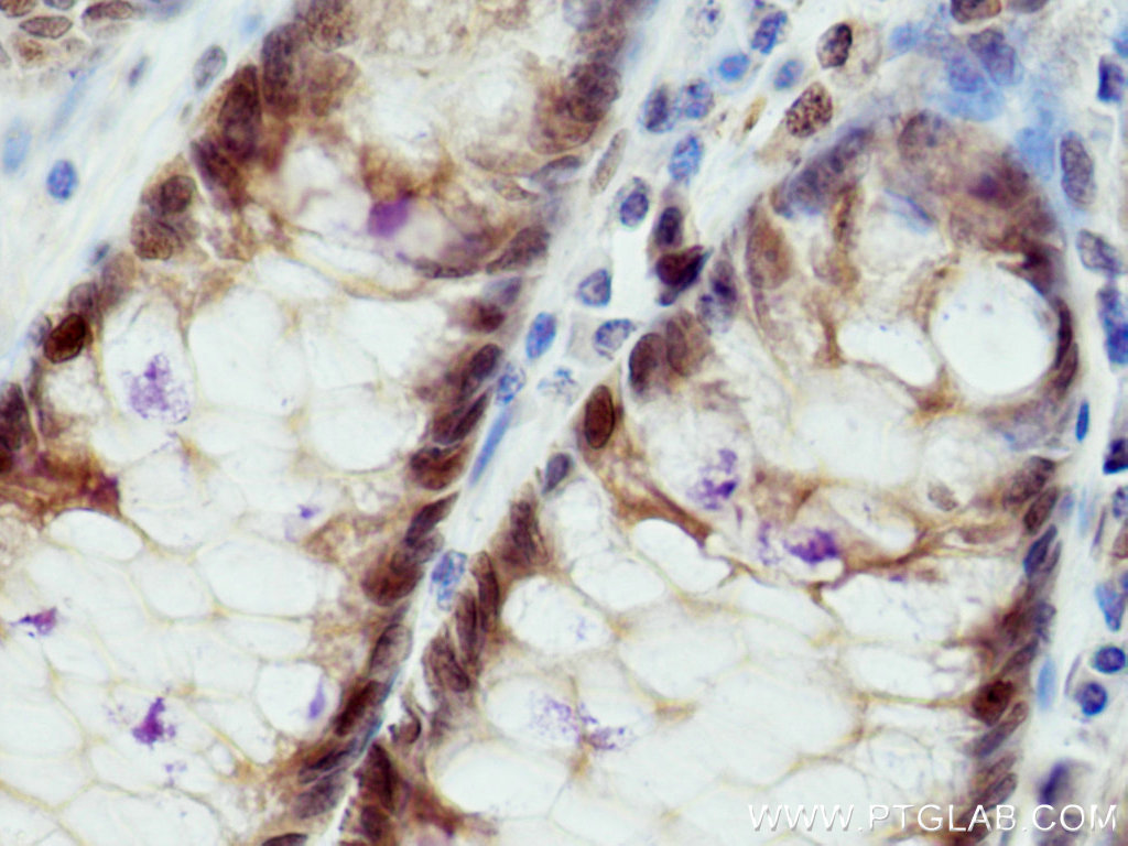
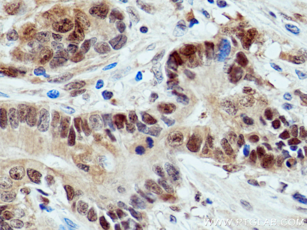
IHC staining of human colon cancer using 10245-1-AP (same clone as 10245-1-PBS)
Immunohistochemical analysis of paraffin-embedded human colon cancer tissue slide using 10245-1-AP (S100A6 antibody) at dilution of 1:800 (under 40x lens). Heat mediated antigen retrieval with Tris-EDTA buffer (pH 9.0). This data was developed using the same antibody clone with 10245-1-PBS in a different storage buffer formulation.
IHC staining of human liver cancer using 10245-1-AP (same clone as 10245-1-PBS)
Immunohistochemical analysis of paraffin-embedded human liver cancer tissue slide using 10245-1-AP (S100A6 antibody) at dilution of 1:800 (under 10x lens). Heat mediated antigen retrieval with Tris-EDTA buffer (pH 9.0). This data was developed using the same antibody clone with 10245-1-PBS in a different storage buffer formulation.
IHC staining of human liver cancer using 10245-1-AP (same clone as 10245-1-PBS)
Immunohistochemical analysis of paraffin-embedded human liver cancer tissue slide using 10245-1-AP (S100A6 antibody) at dilution of 1:800 (under 40x lens). Heat mediated antigen retrieval with Tris-EDTA buffer (pH 9.0). This data was developed using the same antibody clone with 10245-1-PBS in a different storage buffer formulation.
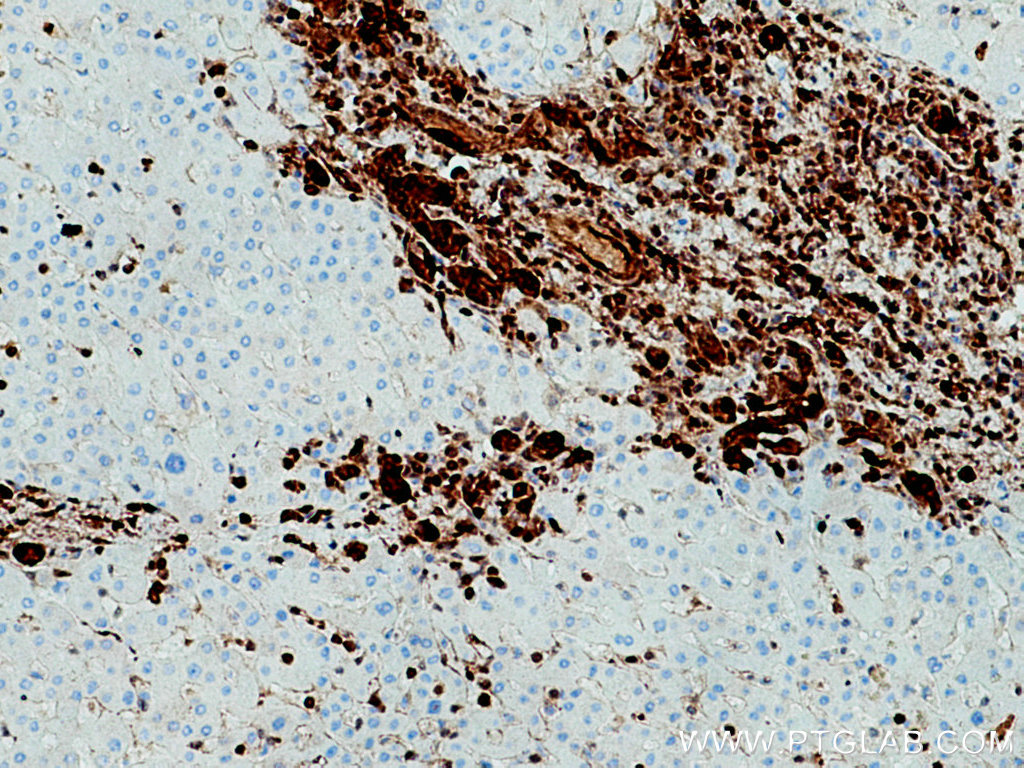
IHC staining of human liver cancer using 10245-1-AP (same clone as 10245-1-PBS)
Immunohistochemical analysis of paraffin-embedded human liver cancer tissue slide using 10245-1-AP (S100A6 Antibody) at dilution of 1:200 (under 10x lens). This data was developed using the same antibody clone with 10245-1-PBS in a different storage buffer formulation.
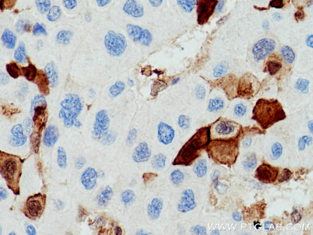
IHC staining of human liver cancer using 10245-1-AP (same clone as 10245-1-PBS)
Immunohistochemical analysis of paraffin-embedded human liver cancer tissue slide using 10245-1-AP (S100A6 Antibody) at dilution of 1:200 (under 40x lens). This data was developed using the same antibody clone with 10245-1-PBS in a different storage buffer formulation.
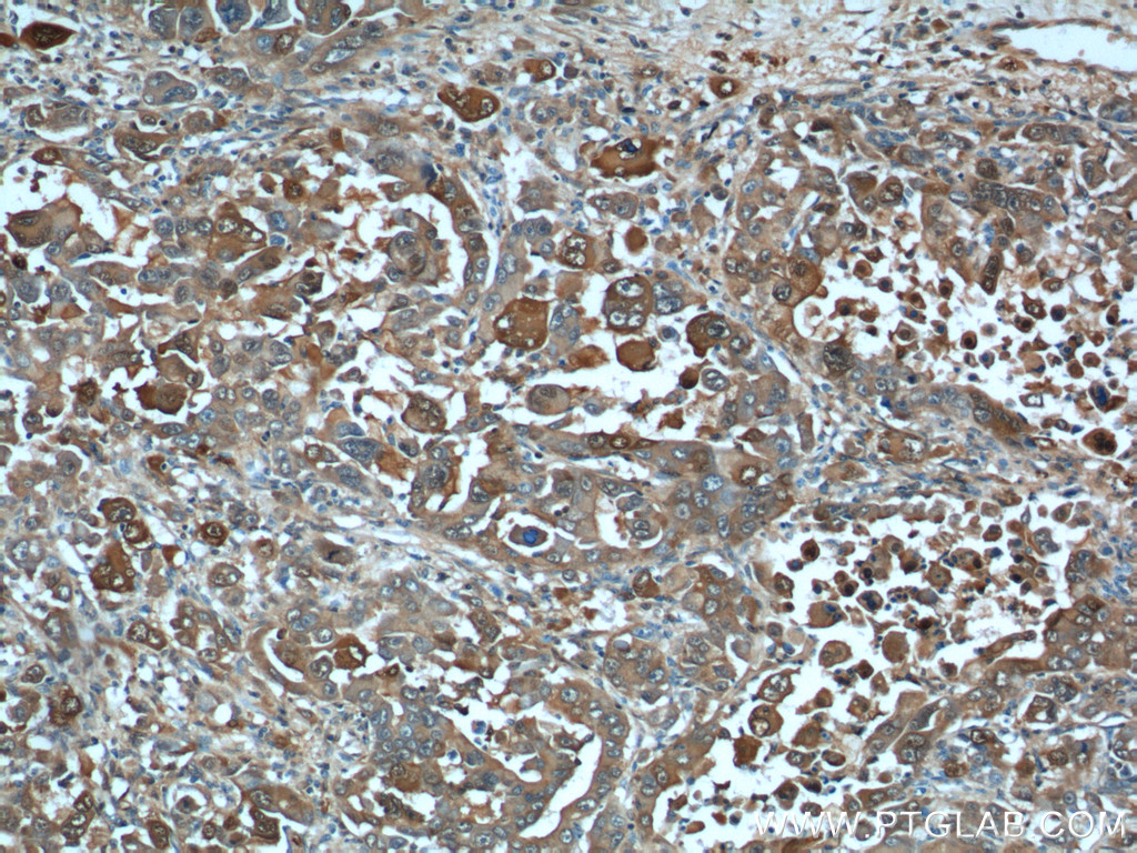
IHC staining of human pancreas cancer using 10245-1-AP (same clone as 10245-1-PBS)
Immunohistochemical analysis of paraffin-embedded human pancreas cancer tissue slide using 10245-1-AP (S100A6 Antibody) at dilution of 1:200 (under 10x lens). Heat mediated antigen retrieval with Tris-EDTA buffer (pH 9.0). This data was developed using the same antibody clone with 10245-1-PBS in a different storage buffer formulation.
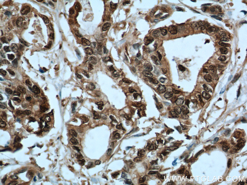
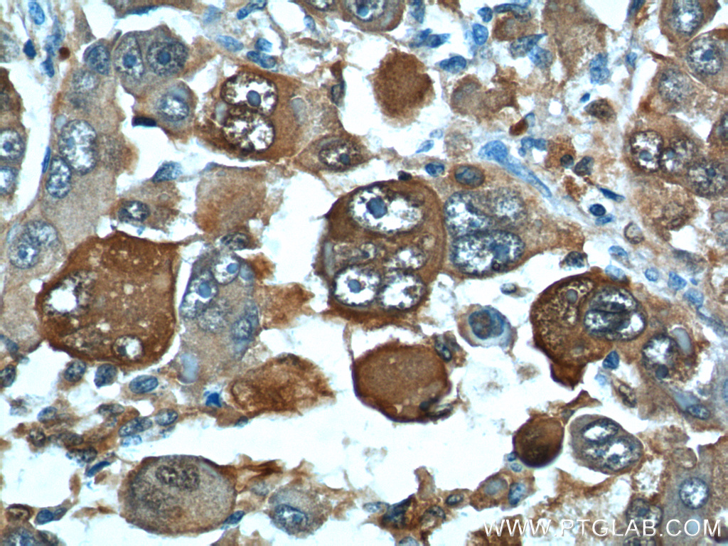
IHC staining of human pancreas cancer using 10245-1-AP (same clone as 10245-1-PBS)
Immunohistochemical analysis of paraffin-embedded human pancreas cancer tissue slide using 10245-1-AP (S100A6 Antibody) at dilution of 1:200 (under 40x lens). Heat mediated antigen retrieval with Tris-EDTA buffer (pH 9.0). This data was developed using the same antibody clone with 10245-1-PBS in a different storage buffer formulation.
IHC staining of human stomach cancer using 10245-1-AP (same clone as 10245-1-PBS)
Immunohistochemical analysis of paraffin-embedded human stomach cancer tissue slide using 10245-1-AP (S100A6 antibody) at dilution of 1:800 (under 10x lens). Heat mediated antigen retrieval with Tris-EDTA buffer (pH 9.0). This data was developed using the same antibody clone with 10245-1-PBS in a different storage buffer formulation.
IHC staining of human stomach cancer using 10245-1-AP (same clone as 10245-1-PBS)
Immunohistochemical analysis of paraffin-embedded human stomach cancer tissue slide using 10245-1-AP (S100A6 antibody) at dilution of 1:800 (under 40x lens). Heat mediated antigen retrieval with Tris-EDTA buffer (pH 9.0). This data was developed using the same antibody clone with 10245-1-PBS in a different storage buffer formulation.
IHC staining of human stomach cancer using 10245-1-AP (same clone as 10245-1-PBS)
Immunohistochemical analysis of paraffin-embedded human stomach cancer tissue slide using 10245-1-AP (S100A6 antibody) at dilution of 1:800 (under 10x lens). Heat mediated antigen retrieval with Tris-EDTA buffer (pH 9.0). This data was developed using the same antibody clone with 10245-1-PBS in a different storage buffer formulation.
IF/ICC Figures
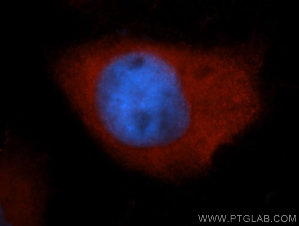
IF Staining of MCF-7 using 10245-1-AP (same clone as 10245-1-PBS)
Immunofluorescent analysis of MCF-7 cells, using S100A6 antibody 10245-1-AP at 1:50 dilution and Rhodamine-labeled goat anti-rabbit IgG (red). Blue pseudocolor = DAPI (fluorescent DNA dye). This data was developed using the same antibody clone with 10245-1-PBS in a different storage buffer formulation.
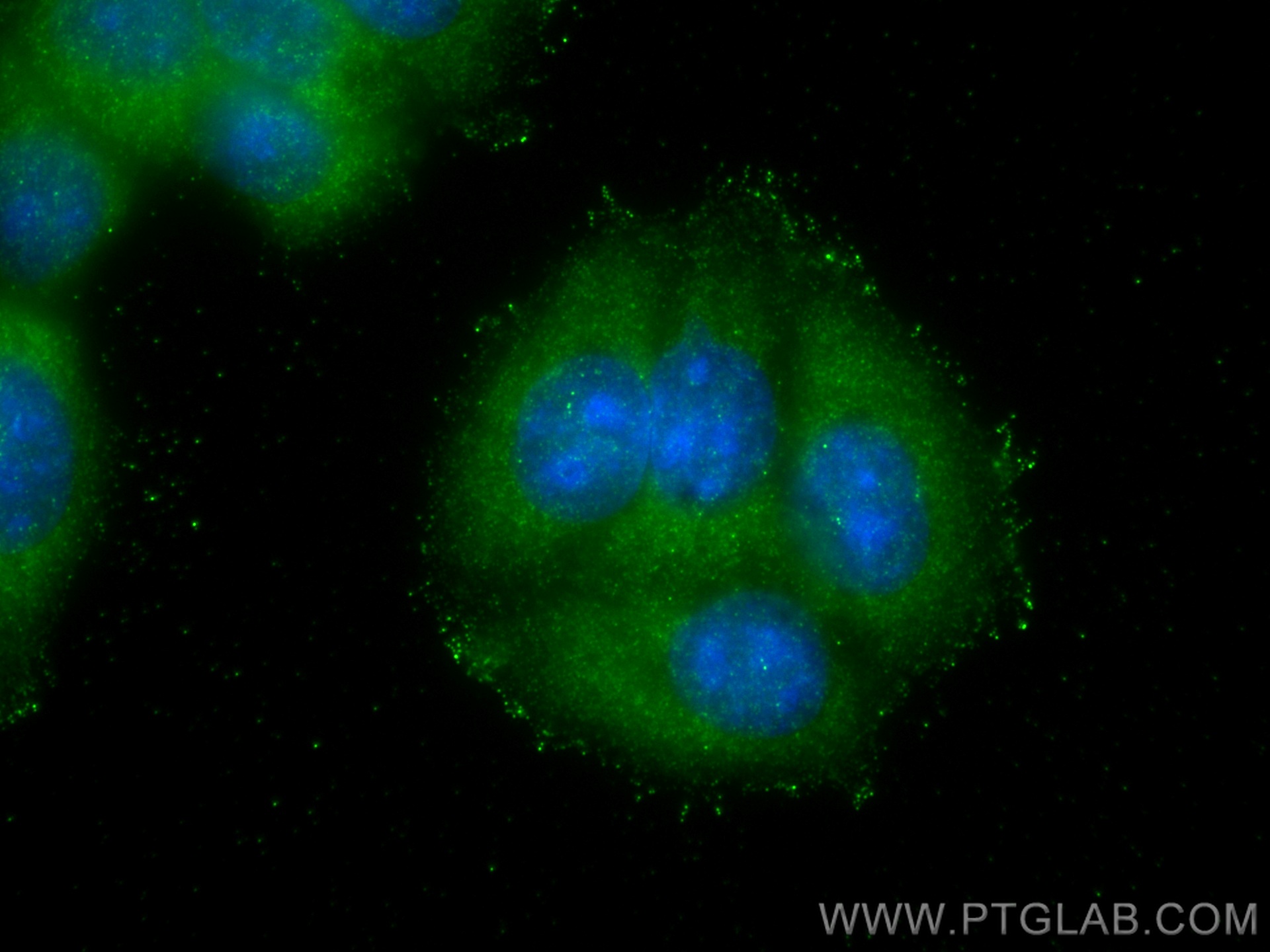
IF Staining of MCF-7 using 10245-1-AP (same clone as 10245-1-PBS)
Immunofluorescent analysis of (-20°C Methanol) fixed MCF-7 cells using S100A6 antibody (10245-1-AP) at dilution of 1:400 and CoraLite®488-Conjugated AffiniPure Goat Anti-Rabbit IgG(H+L). This data was developed using the same antibody clone with 10245-1-PBS in a different storage buffer formulation.
