验证数据展示
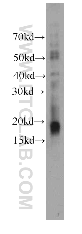
 with si-Control and si-Stathmin transfected Jurkat cells.")
 at dilution of 1:1000 incubated at room temperature for 1.5 hours.")

. Basal cell staining and dendritic processes as expected. By Dr. Brian Lin (Schwob Lab).")
经过测试的应用
| Positive WB detected in | Jurkat cells, K-562 cells, human brain tissue |
| Positive IHC detected in | human testis tissue Note: suggested antigen retrieval with TE buffer pH 9.0; (*) Alternatively, antigen retrieval may be performed with citrate buffer pH 6.0 |
推荐稀释比
| 应用 | 推荐稀释比 |
|---|---|
| Western Blot (WB) | WB : 1:500-1:2000 |
| Immunohistochemistry (IHC) | IHC : 1:20-1:200 |
| It is recommended that this reagent should be titrated in each testing system to obtain optimal results. | |
| Sample-dependent, Check data in validation data gallery. | |
产品信息
66090-1-Ig targets Stathmin 1 in WB, IF, IHC, ELISA applications and shows reactivity with human, mouse samples.
| 经测试应用 | WB, IF, IHC, ELISA Application Description |
| 经测试反应性 | human, mouse |
| 免疫原 | Stathmin 1 fusion protein Ag19102 种属同源性预测 |
| 宿主/亚型 | Mouse / IgG1 |
| 抗体类别 | Monoclonal |
| 产品类型 | Antibody |
| 全称 | stathmin 1/oncoprotein 18 |
| 别名 | |
| 计算分子量 | 18 kDa |
| 观测分子量 | 18 kDa |
| GenBank蛋白编号 | BC014353 |
| 基因名称 | Stathmin 1 |
| Gene ID (NCBI) | 3925 |
| 偶联类型 | Unconjugated |
| 形式 | Liquid |
| 纯化方式 | Protein G purification |
| UNIPROT ID | P16949 |
| 储存缓冲液 | PBS with 0.02% sodium azide and 50% glycerol , pH 7.3 |
| 储存条件 | Store at -20°C. Stable for one year after shipment. Aliquoting is unnecessary for -20oC storage. |
背景介绍
Stathmin 1 (STMN1) normally regulates microtubule dynamics either by sequestering free tubulin heterodimers or by promoting microtubule catastrophe. STMN1 is highly expressed in fetal and adult brain, spinal cord, and cerebellum. Many different phosphorylated forms are observed depending on specific combinations among the sites which can be phosphorylated. Phosphorylation of stathmin is involved in response to NGF, neuron polarization and microtubule polymerization inhibition activity. Increased expression of STMN1 has been observed in a variety of human malignancies, such as colorectal primary tumors and metastatic tissues, but its association with melanoma is so far not well known.