Popular antibodies for flow cytometry
Jump to:
- Introduction to flow cytometry
- Flow Cytometry Applications
- Flow Cytometry Fluorophores and Dyes
- Flow Cytometry Multicolor Panel Design
- Flow Cytometry Sample Preparation
- Protocol for studying extracellular and intracellular proteins
- Experimental protocol to study cell viability and apoptosis
- Popular antibodies for flow cytometry
Popular antibodies for flow cytometry
FITC-Anti-Human CD3
| Type | Mouse Monoclonal |
| Catalog number | FITC-65151 |
| Clone | UCHT1 |
CD3 is a complex of proteins (gamma, delta, and two epsilon chains) that directly associates with the T-cell receptor (TCR) to regulate T-cell activation. The TCR-CD3 complex is responsible for recognizing antigens bound to major histocompatibility complex (MHC) molecules. Once TCR-mediated peptide-MHC binding occurs, the signal is transmitted to the CD3 complex, leading to intracellular signal transduction. CD3 is a pan-T cell marker for the detection of normal and neoplastic T cells.

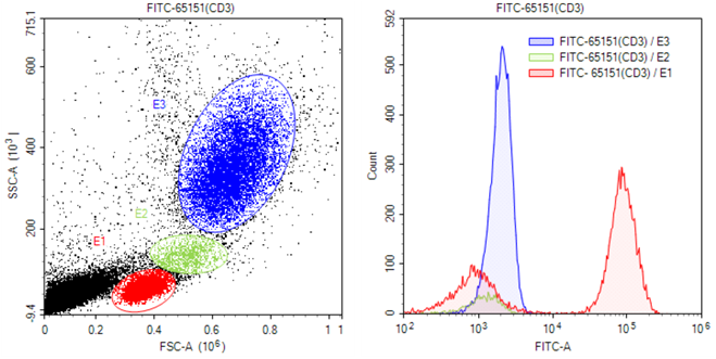
Figure 7. FITC-Anti-Human CD3 (FITC-65151, clone UCHT1) in flow cytometry.
0.1 mL human whole blood cells were surface-stained with FITC-Anti-Human CD3 (FITC-65151, clone UCHT1). Samples were then treated with red blood cell lysis buffer and were gated for analysis of CD3 staining. Cells in the lymphocyte (red histogram), monocyte (green histogram), or granulocyte (blue histogram) gates (E1, E2, E3) were used to compare CD3 staining.
APC Anti-Human CD19
| Type | Mouse Monoclonal |
| Catalog number | APC-65145 |
| Clone | SJ25C1 |
CD19 is a B cell-specific molecule that controls B cell activation by complexing with the B cell receptor (BCR). CD19 is a member of the Ig superfamily and is the dominant component for the signaling complex on B cells that includes CD21, CD81, and CD22. CD19 deficiency leads to reduced proliferative responses to BCR stimulation in vitro and reduced antibody production following vaccination.

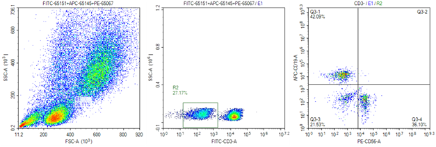
Figure 8. APC-anti-human CD19 (APC-65145, clone SJ25C1) in flow cytometry
0.1 mL human whole blood cells were surface-stained with 0.20 ug FITC-Anti-Human CD3 (FITC-65151, clone UCHT1), 0.20 ug APC-anti-human CD19 (APC-65145, clone SJ25C1) and PE-Anti-Human CD56 (PE-65067, clone MEM 188). Samples were then treated with red blood cell lysis buffer and were gated for CD3 negative lymphocytes for analysis of CD19 and CD56 staining.
APC-Anti-Human CD4
| Type | Mouse Monoclonal |
| Catalog number | APC-65134 |
| Clone | OKT4 |
CD4 is a 55-kDa transmembrane glycoprotein expressed on T helper cells, the majority of thymocytes, monocytes, macrophages, and dendritic cells. CD4 is an accessory protein for major histocompatibility complex (MHC) class-II antigen/T-cell receptor interaction. It plays an important role in T helper cell development and activation. CD4 serves as a receptor for human immunodeficiency virus (HIV).

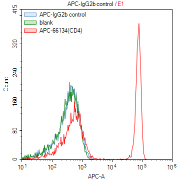
Figure 9. APC-Anti-Human CD4 (APC-65134, clone OKT4) in flow cytometry.
1X10^6 human peripheral blood lymphocytes were surface-stained with 0.20 ug APC-Anti-Human CD4 (APC-65134, clone OKT4) (red) or 0.20 ug APC-Mouse IgG2b isotype control (blue) and blank control (green). Samples were not fixed.


