Loading Control Antibodies for Western Blotting
Technical tips and related products
By Deborah Grainger
The proteins and peptides that regularly serve as endogenous or internal controls may not always take center stage in your research, but they are indispensable to your conducting meaningful experiments and are essential for publication.
Western blotting requires such controls: it is widely used for the semi-quantification of protein levels under of a set of different experimental parameters. Protein standards are required to make sense of Western blotting results, and check that any increases and decreases in target proteins are actually due to experimental manipulations and not, for example, because the sample went wandering during gel loading. Internal control proteins — i.e. those with constant, unchanging levels — are usually detected in a second round of blotting, following primary detection of your protein of interest. This step is used to standardize results and normalize for any errors that creep into a Western blot experiment, such as sample loss through loading at SDS-PAGE or Western blot transfer.
The loading control candidates for Western blotting are usually proteins with high and constitutive expression. As mentioned above, the most basic criterion for a loading control is that its level remains unchanged throughout an experiment, regardless of tissues or cell types used and how they are handled. This means control candidates require careful selection; even bastions of the loading control repertoire — such as β-actin and α-tubulin — can be affected by the conditions of your experiment (so be sure to double check your chosen manipulations do not impact them.)
Here we’ve provided background information on each of the internal control proteins targeted by control antibodies we offer, helping you choose the best control for your personal needs…
View our summary table below to help select a loading control for your sample type with a different molecular weight to your protein of interest.
| Whole cell/cytoplasmic | Nuclear | Mitochondrial | Serum | ||||
| Loading control | MW (kDa) | Loading control | MW (kDa) | Loading control | MW (kDa) | Loading control | MW (kDa) |
| Vinculin | 117 | ||||||
| Transferrin | 77 | ||||||
| Lamin B1 | 66 | ||||||
| HDAC1 | 65 | ||||||
| α-tubulin | 52 | ||||||
| β-tubulin | 50 | ||||||
| β-actin | 42 | ||||||
| GAPDH | 36 | TBP (rodent) | 33-36 | ||||
| PCNA | 36 | VDAC1/Porin | 36 | ||||
| TBP (human) | 37-43 | ||||||
| COXIV | 19 | ||||||
| Histone H3 | 16 | ||||||
Actin
Type: Whole cell/cytoplasm
Molecular weight: ~42kDa
The six isoforms of actin constitute a family of highly conserved globular proteins comprised of three main isoform groups, alpha, beta, and gamma. The alpha actins — alpha C1 and alpha 1 and 2 — are a major constituent of the contractile apparatus in muscle tissues. The beta (β) and gamma 1 and 2 (γ1 and γ2) actins co-exist in most cell types and are an integral part of the cytoskeleton; they are mediators of cell trafficking, structural integrity and cell motility. Together, actins are the most abundant proteins in the typical eukaryotic cell, accounting for about 15 percent of total protein in some cell types. As such, actin is widely used as an internal control in Western blotting experiments.
 |
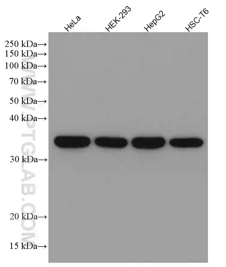
| Western blot analysis of ACTB in multiple cell line and tissue lysates using anti-beta actin mouse monoclonal (60008-1-Ig) at a dilution of 1:5000. |
 |
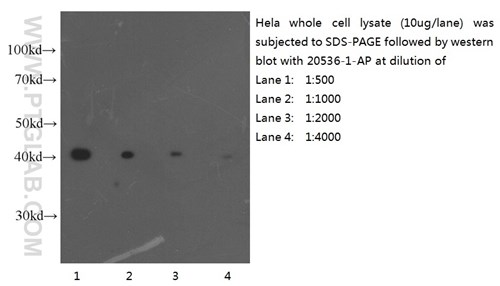
| HeLa cell lysate (10 ug/lane) was separated by SDS-PAGE and actin was detected by anti-ACTB antibody 20536-1-AP at varying dilutions. (L-R) 1:500, 1:1,000, 1:2,000 and 1:4,000. |
Proteintech’s polyclonal ACTB antibody (20536-1-AP) was generated using a β-actin protein antigen (amino acids 14–167) and recognizes all forms of actin, making it a pan-actin antibody. Many studies use β-actin antibodies recognizing all actin isoforms to probe for this loading control collective. However, if your studies involve work with skeletal muscle samples, or you are working with conditions that see changes in cell growth or altered interactions with the extracellular matrix, another loading control may be better suited to your needs.
| Related antibodies | Catalog number |
| Rabbit polyclonal ACTB antibody | 20536-1-AP |
| Mouse monoclonal ACTB antibody | 60008-1-Ig |
| HRP-conjugated ACTB antibody | HRP-60008 |
| Rabbit polyclonal ACTA1 antibody | 17521-1-AP |
| Rabbit polyclonal ACTA2 antibody | 55135-1-AP |
COX-4
Type: Mitochondrial
Molecular weight: 17kDa
COX-4, or COXIV (cytochrome c oxidase subunit IV), is a nuclear-encoded subunit of the human mitochondrial respiratory chain enzyme cytochrome c oxidase (COX). The COX-4 subunit can be expressed as either of two isoforms, isoform 1 and 2 named COX4I1 and COX4I2 respectively. COX4I1 expression is ubiquitous throughout all tissues, whilst COX4I2 is lung-specific. Because of its dependably high level, COX4I1 is commonly detected as an effective loading control for mitochondrial proteins. However, some caution is advised when selecting this protein for Western blot detection as many other proteins run at its 17kDa size during SDS-PAGE (make sure your band of interest won’t be obscured). It is also advisory to double check that any experimental manipulations do not affect its levels. For an alternative mitochondrial protein loading control see our entry on VDAC1 below.
Proteintech’s COX4I1 antibody (11242-1-AP) was generated against a COX4I1 whole-protein antigen (amino acids 1–169) and also recognizes COX4I2.
| Related antibodies | Catalog number |
| Rabbit polyclonal COX4I1 antibody | 11242-1-AP |
| Rabbit polyclonal COX4I2 antibody | 11463-1-AP |
GAPDH
Type: Nuclear
Molecular weight: 36kDa
Proliferating Cell Nuclear Antigen (PCNA) is a processivity factor for DNA polymerase δ; it helps control eukaryotic DNA replication by increasing polymerase nucleotide processing ability during elongation of the leading strand. PCNA protein has been highly conserved throughout evolution — the amino acid sequences of rats and humans differ by only 4 of 261 amino acids — meaning whole-protein raised antibodies targeting PCNA should work across multiple species.
Levels of PCNA do not vary with cell cycle status in mammalian cells, but, as it is more abundant in proliferating cells, PCNA is mostly used as loading control in cell populations undergoing proliferation. PCNA is best avoided if your experiments induce DNA damage as this protein is quickly degraded when DNA damage pathways are activated.
Proteintech’s PCNA antibody is a rabbit polyclonal antibody raised against an internal region of human PCNA, encompassing amino acids 8-256.
 |
| Various lysates were subjected to SDS PAGE followed by western blot with 60004-1-Ig (GAPDH antibody) at dilution of 1:50000 incubated at room temperature for 1.5 hours. |
 |
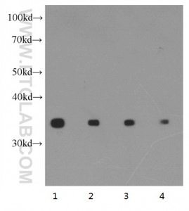
| Western blot with Hela cell lysate using anti-GAPDH (10494-1-AP) at various dilutions (L-R: 1:2000, 1:4000, 1:8000 and 1:16000). |
| GAPDH antibodies | Catalog number |
| rabbit polyclonal GAPDH antibody | 10494-1-AP |
| Mouse monoclonal GAPDH antibody | 60004-1-Ig |
| HRP-conjugated GAPDH antibody | HRP-60004 |
Lamin B1
Type: Nuclear
Molecular weight: 66kDa
Lamins are integral components of the nuclear lamina, a dense, fibrous layer underlying the nuclear envelope on its nucleoplasmic side. Lamins play an important role in the structural integrity of the nucleus and its traffic control, as well as interacting with chromatin and gene expression. Vertebrate lamins consist of two types, A and B. The LMNB1 gene encodes one of the two B type proteins, lamin B1 and can be used as a loading control for those working with nuclear fractions; however, this protein is not suitable for samples where the nuclear envelope has been removed. It is also worthwhile to note that lamins become phosphorylated during mitosis when the lamina matrix is reversibly disassembled. Proteintech’s LMNB1 antibody (12987-1-AP) has been raised against a protein antigen (amino acids 236 to 586 at the C-terminus) and is validated for use in Western blot, IHC, ELISA and immunofluorescence.
| Lamin antibodies | Catalog number |
| Rabbit polyclonal LMNB1 antibody | 12987-2-AP |
| Rabbit polyclonal LMNA/C antibody | 10298-1-AP |
PCNA
Type: Nuclear
Molecular weight: 36kDa
Proliferating Cell Nuclear Antigen (PCNA) is a processivity factor for DNA polymerase δ; it helps control eukaryotic DNA replication by increasing polymerase nucleotide processing ability during elongation of the leading strand. PCNA protein has been highly conserved throughout evolution — the amino acid sequences of rats and humans differ by only 4 of 261 amino acids — meaning whole-protein raised antibodies targeting PCNA should work across multiple species.
Levels of PCNA do not vary with cell cycle status in mammalian cells, but, as it is more abundant in proliferating cells, PCNA is mostly used as loading control in cell populations undergoing proliferation. PCNA is best avoided if your experiments induce DNA damage as this protein is quickly degraded when DNA damage pathways are activated.
Proteintech’s PCNA antibody is a rabbit polyclonal antibody raised against an internal region of human PCNA, encompassing amino acids 8-256.
 |
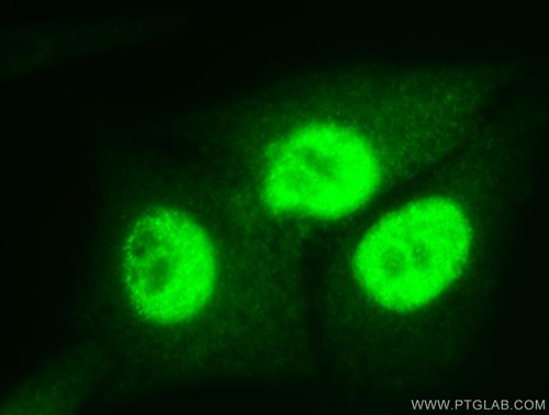
| Immunofluorescence analysis of PCNA in HepG2 cells, using PCNA antibody 10205-2-AP at 1:50 dilution and FITC-labeled donkey anti-rabbit IgG (green). |
| PCNA antibodies | Catalog number |
| Rabbit polyclonal PCNA antibody | 10205-2-AP |
| Mouse monoclonal PCNA antibody | 60097-1-Ig |
Tubulin
Type: Whole cell/cytoplasmic
Molecular weight: 50-55kDa
Tubulins are the major components of microtubules, the major transport network in cells. The microtubules are involved in a wide variety of cellular activities ranging from mitosis and transport events to cell movement and the maintenance of cell shape. Highly and stably expressed and conserved across the species, tubulins make excellent whole cell or cytoplasmic fraction loading controls in Western blotting. However, tubulin expression may vary according to resistance to antimicrobial and antimitotic drugs.
Proteintech has polyclonal antibodies against several tubulin subunits including an α-tubulin antibody (11224-1-AP) and two β-tubulin antibodies 10068-1-AP and 10094-1-Ap.
| Tubulin antibodies | Catalog number |
| Rabbit polyclonal alpha tubulin antibody | 11224-1-AP |
| Rabbit polyclonal beta tubulin antibody (antigen: amino acids 43-258) | 10068-1-AP |
| Rabbit polyclonal beta tubulin antibody (antigen: amino acids 57-294) | 10094-1-AP |
| HRP-conjugated TUB1A antibody | HRP-66031 |


