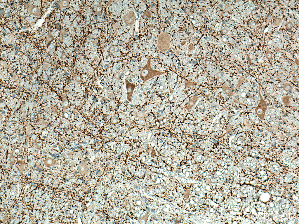
IHC staining of human brain using 67015-1-Ig (same clone as 67015-1-PBS)
Immunohistochemical analysis of paraffin-embedded human brain tissue slide using 67015-1-Ig (MAP2 antibody) at dilution of 1:2000 (under 10x lens. Heat mediated antigen retrieval with Tris-EDTA buffer (pH 9.0). This data was developed using the same antibody clone with 67015-1-PBS in a different storage buffer formulation.
IHC staining of human brain using 67015-1-Ig (same clone as 67015-1-PBS)
Immunohistochemical analysis of paraffin-embedded human brain tissue slide using 67015-1-Ig (MAP2 antibody) at dilution of 1:2000 (under 40x lens. Heat mediated antigen retrieval with Tris-EDTA buffer (pH 9.0). This data was developed using the same antibody clone with 67015-1-PBS in a different storage buffer formulation.
IHC staining of mouse brain using 67015-1-Ig (same clone as 67015-1-PBS)
Immunohistochemical analysis of paraffin-embedded mouse brain tissue slide using 67015-1-Ig (MAP2 antibody) at dilution of 1:2000 (under 40x lens. Heat mediated antigen retrieval with Tris-EDTA buffer (pH 9.0). This data was developed using the same antibody clone with 67015-1-PBS in a different storage buffer formulation.
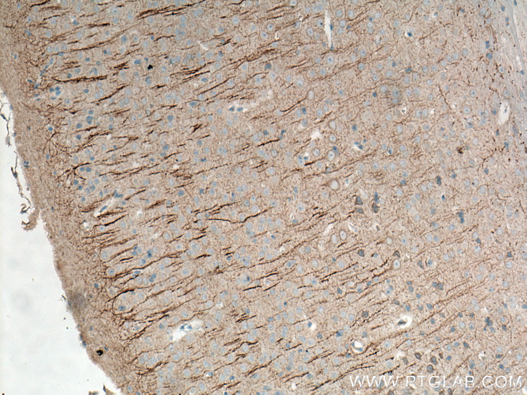
IHC staining of mouse brain using 67015-1-Ig (same clone as 67015-1-PBS)
Immunohistochemical analysis of paraffin-embedded mouse brain tissue slide using 67015-1-Ig (MAP2 antibody) at dilution of 1:2000 (under 10x lens. Heat mediated antigen retrieval with Tris-EDTA buffer (pH 9.0). This data was developed using the same antibody clone with 67015-1-PBS in a different storage buffer formulation.
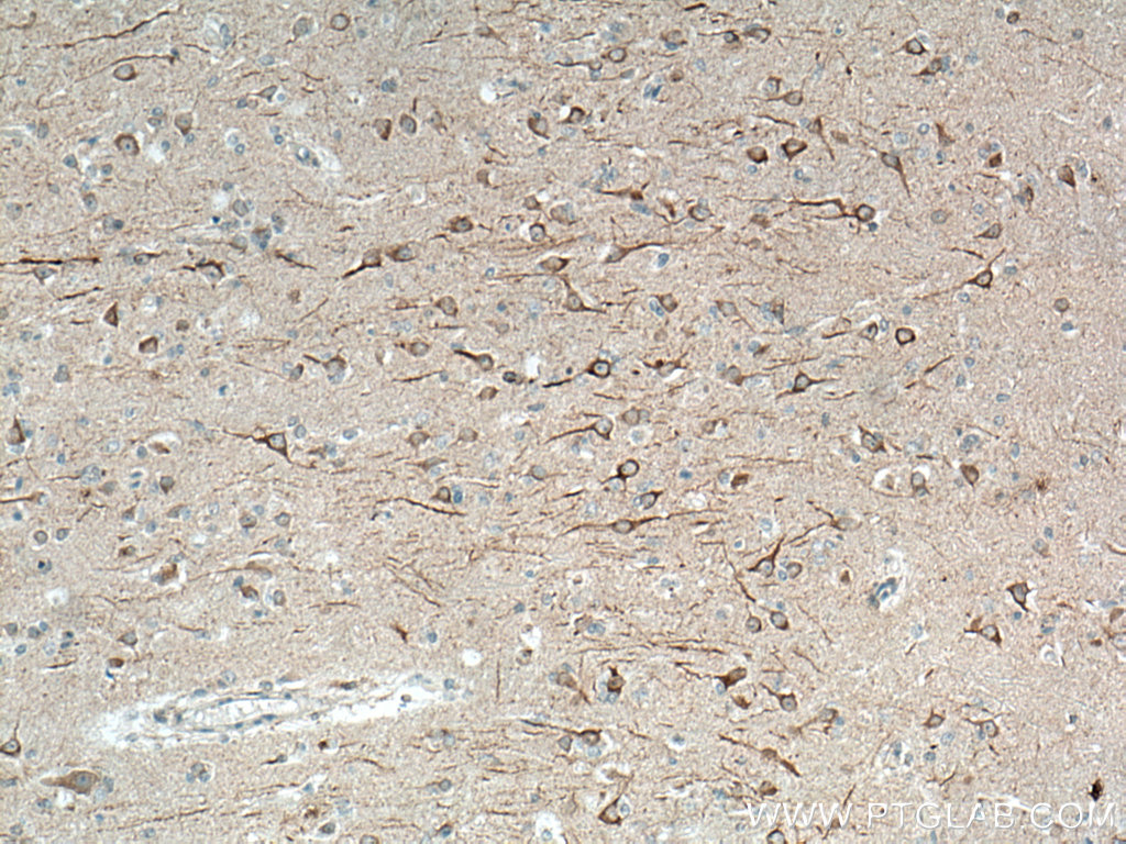
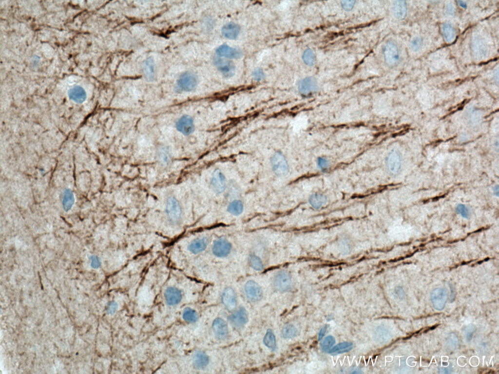
IHC staining of mouse brain using 67015-1-Ig (same clone as 67015-1-PBS)
Immunohistochemical analysis of paraffin-embedded mouse brain tissue slide using 67015-1-Ig (MAP2 antibody) at dilution of 1:2000 (under 10x lens). Heat mediated antigen retrieval with Tris-EDTA buffer (pH 9.0). This data was developed using the same antibody clone with 67015-1-PBS in a different storage buffer formulation.
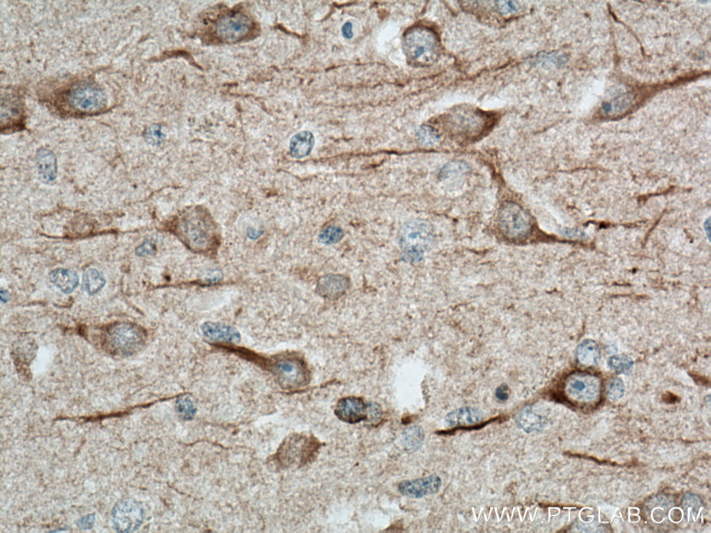
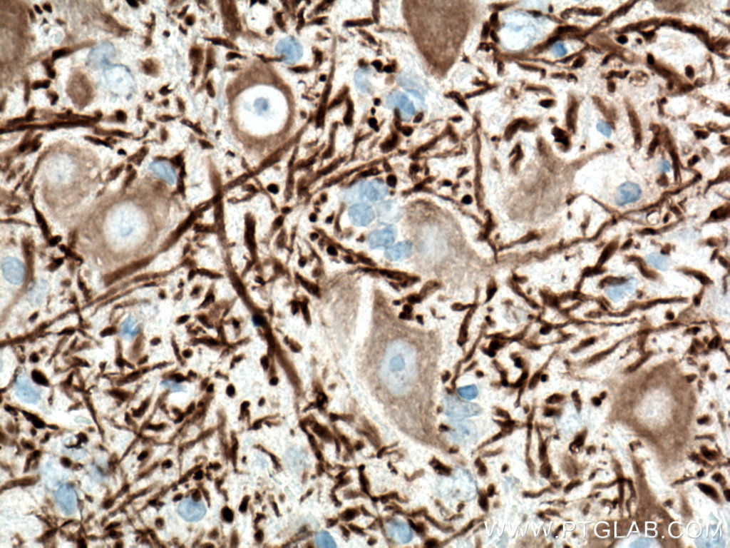
IHC staining of mouse brain using 67015-1-Ig (same clone as 67015-1-PBS)
Immunohistochemical analysis of paraffin-embedded mouse brain tissue slide using 67015-1-Ig (MAP2 antibody) at dilution of 1:2000 (under 40x lens). Heat mediated antigen retrieval with Tris-EDTA buffer (pH 9.0). This data was developed using the same antibody clone with 67015-1-PBS in a different storage buffer formulation.
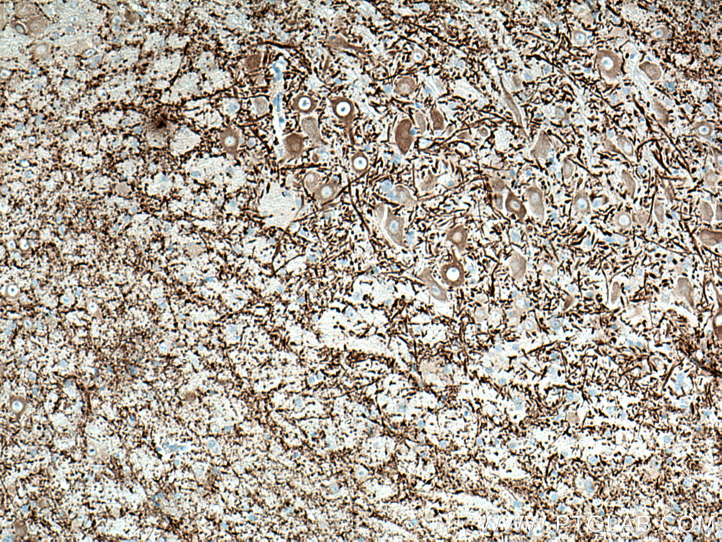
IHC staining of mouse brain using 67015-1-Ig (same clone as 67015-1-PBS)
Immunohistochemical analysis of paraffin-embedded mouse brain tissue slide using 67015-1-Ig (MAP2 antibody) at dilution of 1:2000 (under 10x lens. Heat mediated antigen retrieval with Tris-EDTA buffer (pH 9.0). This data was developed using the same antibody clone with 67015-1-PBS in a different storage buffer formulation.
IHC staining of mouse brain using 67015-1-Ig (same clone as 67015-1-PBS)
Immunohistochemical analysis of paraffin-embedded mouse brain tissue slide using 67015-1-Ig (MAP2 antibody) at dilution of 1:2000 (under 40x lens. Heat mediated antigen retrieval with Tris-EDTA buffer (pH 9.0). This data was developed using the same antibody clone with 67015-1-PBS in a different storage buffer formulation.
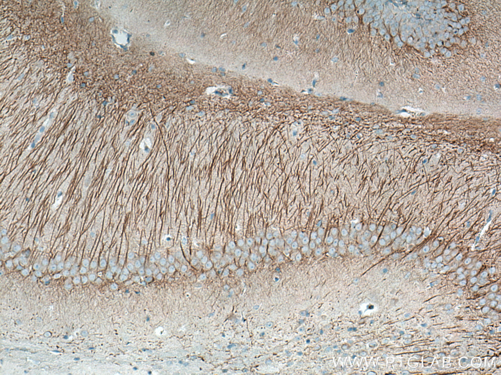
IHC staining of mouse cerebellum using 67015-1-Ig (same clone as 67015-1-PBS)
Immunohistochemical analysis of paraffin-embedded mouse cerebellum tissue slide using 67015-1-Ig (MAP2 antibody) at dilution of 1:2000 (under 40x lens. Heat mediated antigen retrieval with Tris-EDTA buffer (pH 9.0). This data was developed using the same antibody clone with 67015-1-PBS in a different storage buffer formulation.
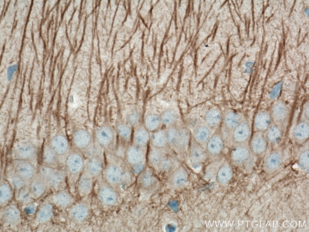
IHC staining of mouse cerebellum using 67015-1-Ig (same clone as 67015-1-PBS)
Immunohistochemical analysis of paraffin-embedded mouse cerebellum tissue slide using 67015-1-Ig (MAP2 antibody) at dilution of 1:2000 (under 10x lens. Heat mediated antigen retrieval with Tris-EDTA buffer (pH 9.0). This data was developed using the same antibody clone with 67015-1-PBS in a different storage buffer formulation.
IHC staining of rat brain using 67015-1-Ig (same clone as 67015-1-PBS)
Immunohistochemical analysis of paraffin-embedded rat brain tissue slide using 67015-1-Ig (MAP2 antibody) at dilution of 1:2000 (under 10x lens). Heat mediated antigen retrieval with Tris-EDTA buffer (pH 9.0). This data was developed using the same antibody clone with 67015-1-PBS in a different storage buffer formulation.
IHC staining of rat brain using 67015-1-Ig (same clone as 67015-1-PBS)
Immunohistochemical analysis of paraffin-embedded rat brain tissue slide using 67015-1-Ig (MAP2 antibody) at dilution of 1:2000 (under 40x lens). Heat mediated antigen retrieval with Tris-EDTA buffer (pH 9.0). This data was developed using the same antibody clone with 67015-1-PBS in a different storage buffer formulation.
IHC staining of rat cerebellum using 67015-1-Ig (same clone as 67015-1-PBS)
Immunohistochemical analysis of paraffin-embedded rat cerebellum tissue slide using 67015-1-Ig (MAP2 antibody) at dilution of 1:2000 (under 10x lens). Heat mediated antigen retrieval with Tris-EDTA buffer (pH 9.0). This data was developed using the same antibody clone with 67015-1-PBS in a different storage buffer formulation.
IF-P Figures
IF Staining of mouse brain using 67015-1-Ig (same clone as 67015-1-PBS)
Immunofluorescent analysis of (4% PFA) fixed mouse brain tissue using MAP2 antibody (67015-1-Ig, Clone: 1C3E6 ) at dilution of 1:400 and CoraLite®488-Conjugated AffiniPure Goat Anti-Mouse IgG(H+L). This data was developed using the same antibody clone with 67015-1-PBS in a different storage buffer formulation.
IF Staining of rat brain using 67015-1-Ig (same clone as 67015-1-PBS)
Immunofluorescent analysis of (4% PFA) fixed rat brain tissue using MAP2 antibody (67015-1-Ig, Clone: 1C3E6 ) at dilution of 1:400 and CoraLite®488-Conjugated AffiniPure Goat Anti-Mouse IgG(H+L). This data was developed using the same antibody clone with 67015-1-PBS in a different storage buffer formulation.
IF-FRO Figures
IF Staining of mouse brain using 67015-1-Ig (same clone as 67015-1-PBS)
Immunofluorescent analysis of (4% PFA) fixed frozen OCT-embedded mouse brain tissue using MAP2 antibody (67015-1-Ig, Clone: 1C3E6 ) at dilution of 1:2000 and CoraLite®488-Conjugated Goat Anti-Mouse IgG(H+L) (SA00013-1), CoraLite®594 GFAP antibody (CL594-16825, red), TDP-43 antibody (10782-2-AP, Magenta). This data was developed using the same antibody clone with 67015-1-PBS in a different storage buffer formulation.
IF Staining of rat brain using 67015-1-Ig (same clone as 67015-1-PBS)
Immunofluorescent analysis of (4% PFA) fixed frozen OCT-embedded rat brain tissue using MAP2 antibody (67015-1-Ig, Clone: 1C3E6 ) at dilution of 1:2000 and CoraLite®488-Conjugated Goat Anti-Mouse IgG(H+L) (SA00013-1), GFAP antibody (81063-1-RR, Clone: 4C6, red). This data was developed using the same antibody clone with 67015-1-PBS in a different storage buffer formulation.

")
")
")
")
")
")
")
")
")
")