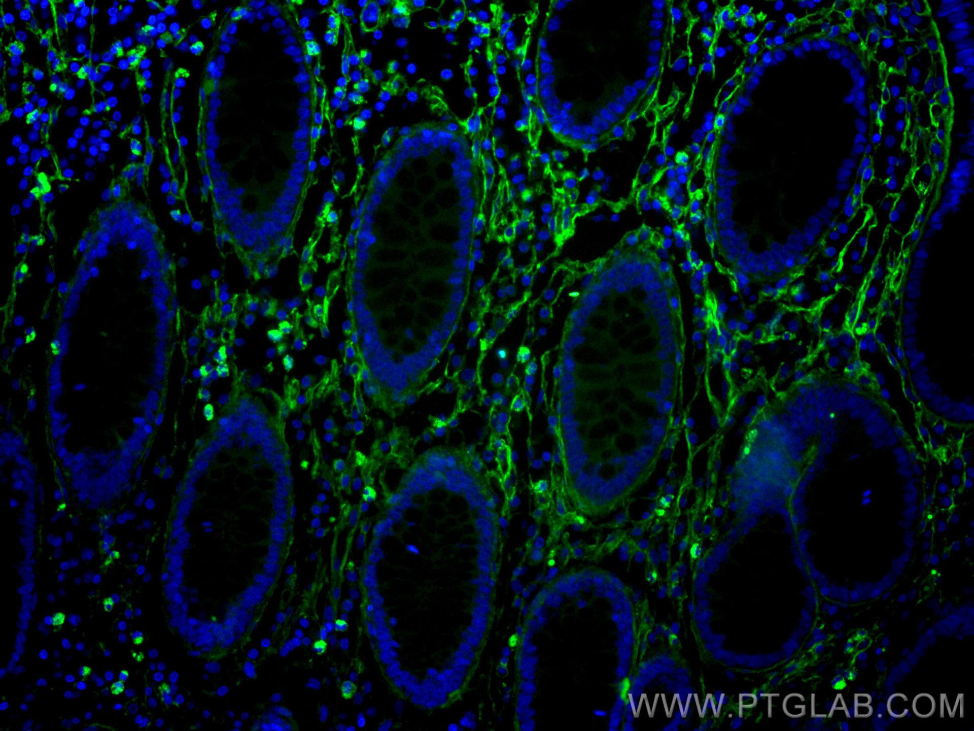
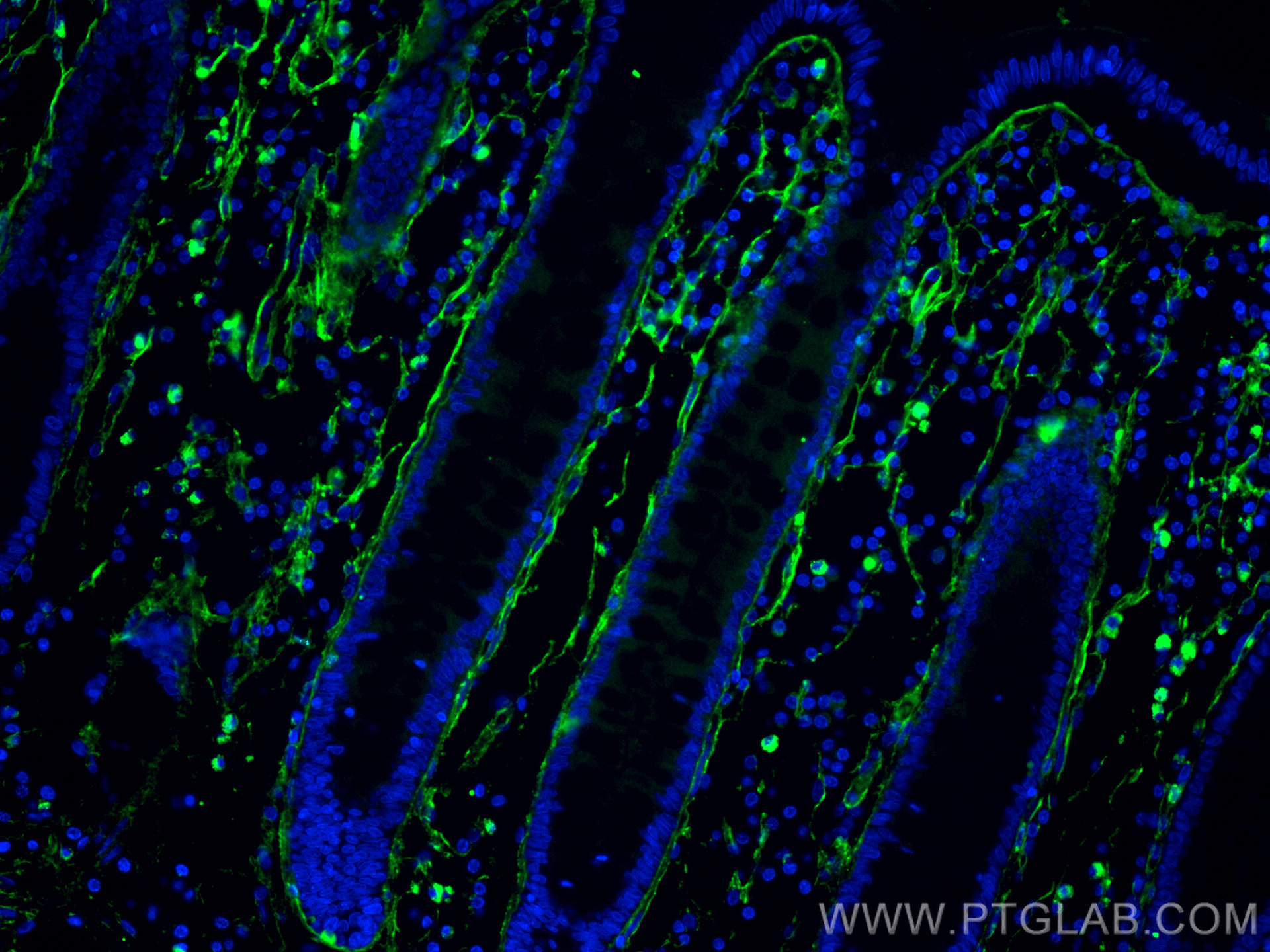
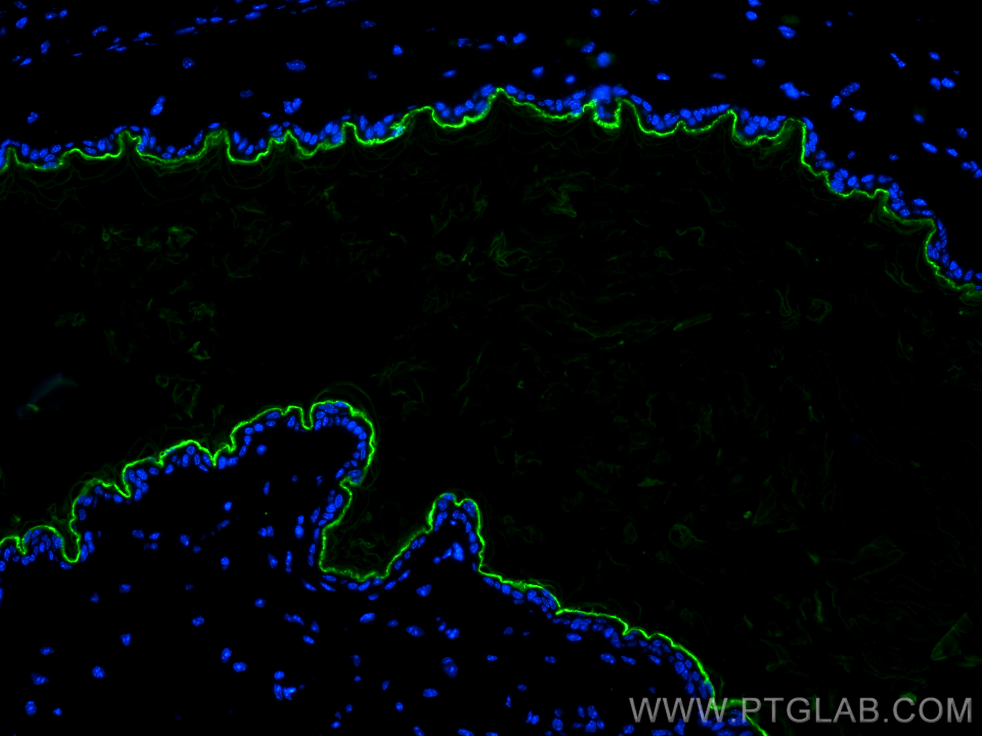
验证数据展示
经过测试的应用
| Positive IF-P detected in | human colon cancer tissue, mouse skin tissue |
推荐稀释比
| 应用 | 推荐稀释比 |
|---|---|
| Immunofluorescence (IF)-P | IF-P : 1:50-1:500 |
| It is recommended that this reagent should be titrated in each testing system to obtain optimal results. | |
| Sample-dependent, Check data in validation data gallery. | |
发表文章中的应用
| IF | See 1 publications below |
产品信息
CL488-67288 targets Collagen Type I in IF-P applications and shows reactivity with human, mouse, pig samples.
| 经测试应用 | IF-P Application Description |
| 文献引用应用 | IF |
| 经测试反应性 | human, mouse, pig |
| 文献引用反应性 | human |
| 免疫原 | Peptide 种属同源性预测 |
| 宿主/亚型 | Mouse / IgG1 |
| 抗体类别 | Monoclonal |
| 产品类型 | Antibody |
| 全称 | collagen, type I, alpha 1 |
| 别名 | COL1A1, col1, COL I, Alpha-1 type I collagen, Alpha 1 type I collagen |
| 计算分子量 | 139 kDa |
| 观测分子量 | 120-130 kDa |
| GenBank蛋白编号 | NM_000088 |
| 基因名称 | COL1A1 |
| Gene ID (NCBI) | 1277 |
| RRID | AB_2919445 |
| 偶联类型 | CoraLite® Plus 488 Fluorescent Dye |
| 最大激发/发射波长 | 493 nm / 522 nm |
| 形式 | Liquid |
| 纯化方式 | Protein G purification |
| UNIPROT ID | P02452 |
| 储存缓冲液 | PBS with 50% glycerol, 0.05% Proclin300, 0.5% BSA , pH 7.3 |
| 储存条件 | Store at -20°C. Avoid exposure to light. Stable for one year after shipment. Aliquoting is unnecessary for -20oC storage. |
背景介绍
Type I collagen, the major structural component of connective tissues such as skin, tendon and bone, is the most abundant and widely expressed collagen in humans (PMID: 7620364; 8645190; 9016532). Type I collagen is a heterotrimer comprising one alpha 2(I) and two alpha 1(I) chains which are encoded by the unlinked loci COL1A2 and COL1A1 respectively. Mutations in COL1A1 are associated with osteogenesis imperfecta types I-IV, Ehlers-Danlos syndrome type VIIA, Ehlers-Danlos syndrome Classical type, Caffey Disease and idiopathic osteoporosis. This antibody raised against a synthesized peptide corresponding to 1206-1218 aa of human pro-alpha 1 chain of type I collagen recognize collagen alpha-1(I) chain.
实验方案
| Product Specific Protocols | |
|---|---|
| IF protocol for CL Plus 488 Collagen Type I antibody CL488-67288 | Download protocol |
| Standard Protocols | |
|---|---|
| Click here to view our Standard Protocols |


