
Leukemia markers
Leukemia is a cancer that affects white blood cells (WBC).
Leukemia typically starts in the bone marrow. Cells undergo neoplastic transformation where their proliferation is abnormal and their expression of markers is aberrant. There are many types of leukemia, but the disease is broadly grouped into myeloid or lymphoid, depending on the WBC type that is affected, with the main subtypes being chronic and acute.
Myeloid
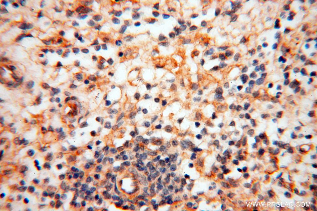
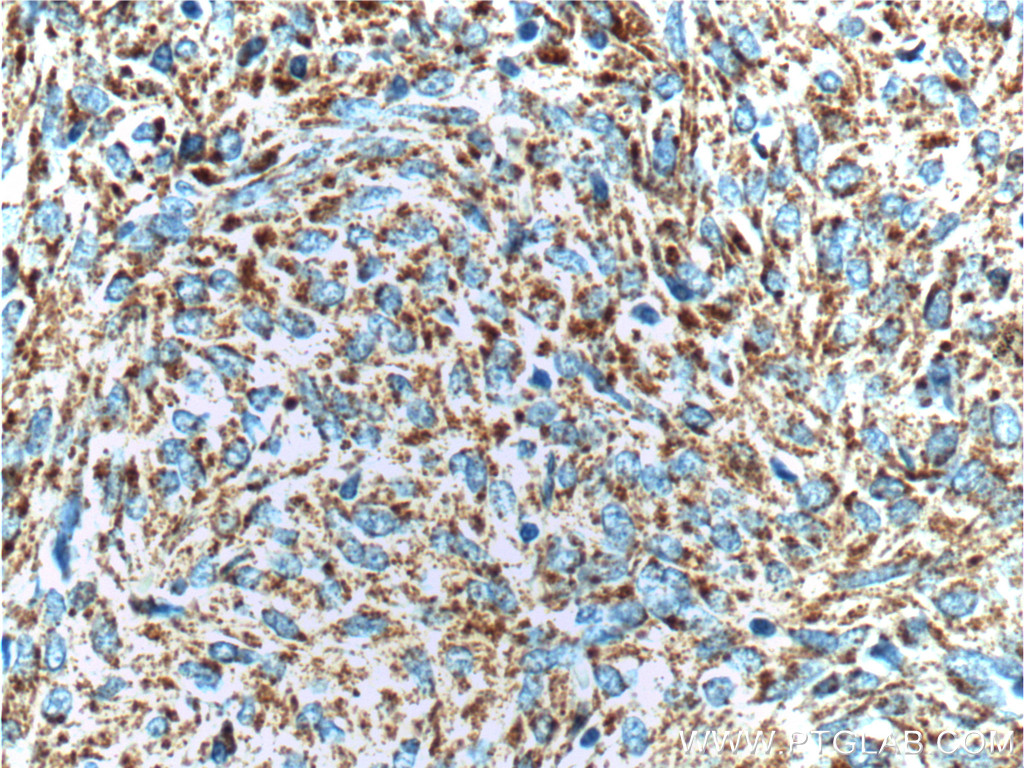
To study leukemia affecting cells of the innate immune system, a number of markers that label myeloid lineage cells can be used. One example is CD11b, which is expressed at low levels by natural killer cells and at high levels by monocytes, neutrophils, and eosinophils.CD13 (Figure 1) is a commonly used marker that is persistently and specifically expressed by myeloid precursor cells, monocytes, and granulocytes. Particularly in acute myeloid leukemia, CD33 (Figure 2) is broadly expressed on affected myeloid cells and is a common target for therapies1.
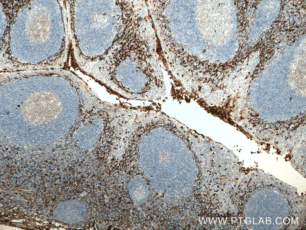 |
| Figure 1. Immunohistochemistry of paraffin-embedded human tonsillitis tissue slide using 66211-1-Ig (CD13 antibody) at a dilution of 1:5000 (under 4x lens). Heat mediated antigen retrieved with Tris-EDTA buffer(pH9). |
 |
|
Figure 2 Immunohistochemistry of paraffin-embedded human spleen using 17425-1-AP (CD33 antibody) at a dilution of 1:100 (under 40x lens) |

Lymphoid
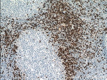
To study leukemias that affect cells of the adaptive immune system, cells of the lymphoid lineage such as B cells and T cells are labeled. The expression of CD19 (Figure 3) is specific to the B cell lineage, and is maintained throughout differentiation and even neoplastic transformation, making it useful in the diagnosis of leukaemia2. The expression of CD4 and CD8 (Figure 4) marks the two populations of T cells specifically, and expression continues in leukemic cells. The relative numbers of these cell populations can help in the diagnosis of acute and chronic lymphoid leukemia and can have implications for prognosis of the disease3.
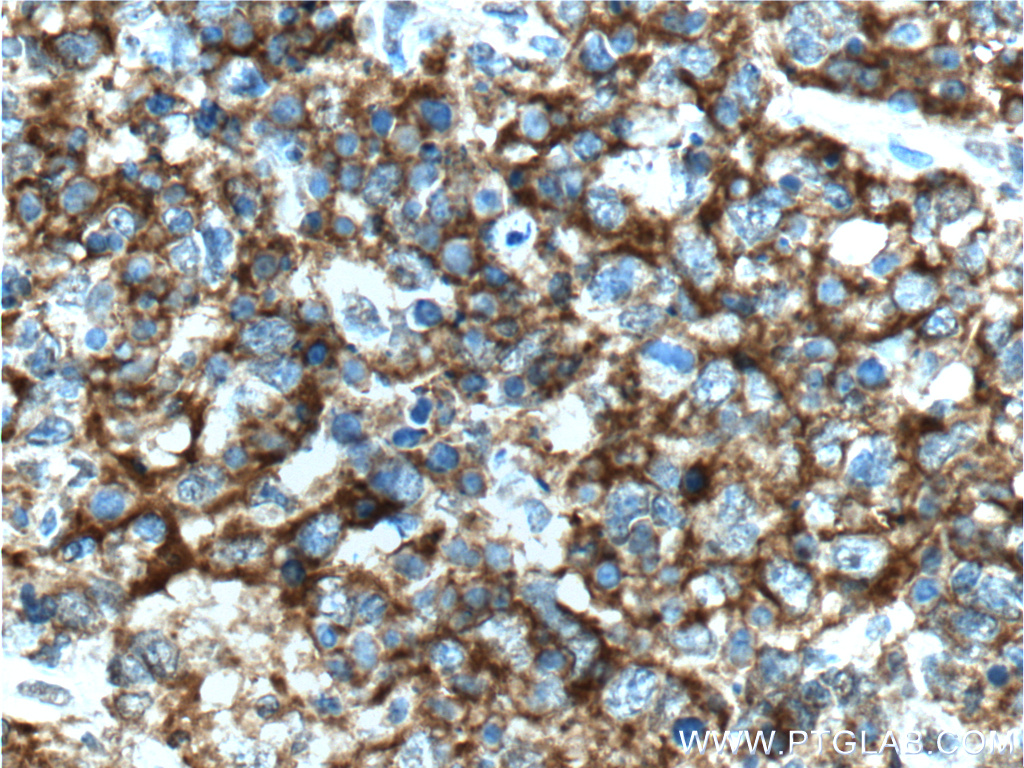 |
| Figure 3. Immunohistochemistry of paraffin-embedded human follicular lymphoma tissue slide using 66298-1-Ig(CD19 Antibody) at a dilution of 1:400 (under 40x lens). Heat mediated antigen retrieved with Tris-EDTA buffer(pH9). |
 |
|
Figure 4. Immunohistochemistry of paraffin-embedded human tonsillitis tissue slide using 66868-1-Ig (CD8a antibody) at a dilution of 1:16000 (under 10x lens) heat mediated antigen retrieved with Tris-EDTA buffer(pH9). |
Leukemic Stem Cells
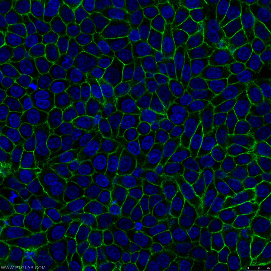
An important and rare subpopulation of leukemia cells have the stem cell properties of quiescence, self-renewal, and chemoresistance. They are particularly associated with relapse and treatment resistance, making them an important target in research. Hematopoietic stem cells (HSCs) are often identified with CD34, although this marker is also found on other progenitor cell types such as multipotent mesenchymal stromal cells (MSCs). A common way to stain for these leukemic stem cells is the aberrant expression of CD117 (Figure 5), also called proto-oncogene c-Kit or stem cell growth factor receptor. CD117 marks HSCs as well as progenitors from the myeloid and lymphoid lineages, but it is lost in the differentiation process4. Markers like CD44 (Figure 6) are maintained throughout development and mark both stem cells and differentiated cells. This cell surface molecule is key in processes such as migration, proliferation, and differentiation, as well as in the presentation of different factors to receptors.
 |
|
Figure 5. Immunohistochemistry of paraffin-embedded stromal tumor tissue slide using 18696-1-AP(c-Kit/CD117 antibody) at a dilution of 1:200 (under 10x lens). |
 |
|
Figure 6. Immunofluorescent analysis of (-20°C Ethanol) fixed HeLa cells using 15675-1-AP (CD44 antibody) at a dilution of 1:200 and Alexa Fluor 488-conjugated AffiniPure Goat Anti-Rabbit IgG(H+L). |
A combinatorial approach is advised when determining which markers to select for leukemia, as well as careful consideration of the specific disease sub-type.
Relevant leukemia markers
|
Marker |
Description |
Antibody |
|
Myeloid |
||
|
CD11b |
integrin family member |
|
|
CD33 |
Transmembrane receptor, cell adhesion and endocytic properties |
|
|
CD13 |
Extracellular peptidase |
|
|
Lymphoid |
||
|
CD4 |
Co-receptor for TCR, glycoprotein interacting with MHC Class II |
|
|
CD8 |
Co-receptor for TCR, glycoprotein interacting with MHC Class I |
|
|
CD19 |
Co-receptor for BCR, controls B cell activation |
|
|
Leukemic stem cells |
||
|
CD117 |
Receptor tyrosine kinase important for mast-cell survival, proliferation, activation, and chemotaxis. |
|
|
CD34 |
Phosphoglycoprotein involved in cell adhesion |
|
|
CD44 |
Cell surface glycoprotein involved in migration and adhesion |
|
References
- Laszlo, G. S., Estey, E. H. & Walter, R. B. The past and future of CD33 as a therapeutic target in acute myeloid leukemia. Blood Rev. 28, 143–153 (2014).
- Scheuermann, R. H. & Racila, E. CD19 Antigen in Leukemia and Lymphoma Diagnosis and Immunotherapy. Leuk. Lymphoma 18, 385–397 (1995).
- Gonzalez-Rodriguez, A. P. et al. Prognostic significance of CD8 and CD4 T cells in chronic lymphocytic leukemia. Leuk. Lymphoma 51, 1829–1836 (2010).
- Hanekamp, D., Cloos, J. & Schuurhuis, G. J. Leukemic stem cells: identification and clinical application. Int. J. Hematol. 105, 549–557 (2017).


