Multi-rAb 重组二抗
下一代二抗领潮未来
Multi-rAb重组二抗是精确设计的重组单克隆抗体的混合物,可识别同一IgG上的多个互补表位。这些多克隆二抗混合物中的每个重组单克隆都经过严格的鉴定和筛选,以确保最佳的性能。
Multi-rAb重组二抗是精确设计的重组单克隆抗体的混合物,可识别同一IgG上的多个互补表位。这些多克隆二抗混合物中的每个重组单克隆都经过严格的鉴定和筛选,以确保最佳的性能。
-
超强信号、超低背景
-
批间高度稳定
-
超高性价比
-
经过内源性免疫学实验验证
-
极小的物种交叉反应性
-
可大量稳定供应
Multi-rAb重组二抗产品列表
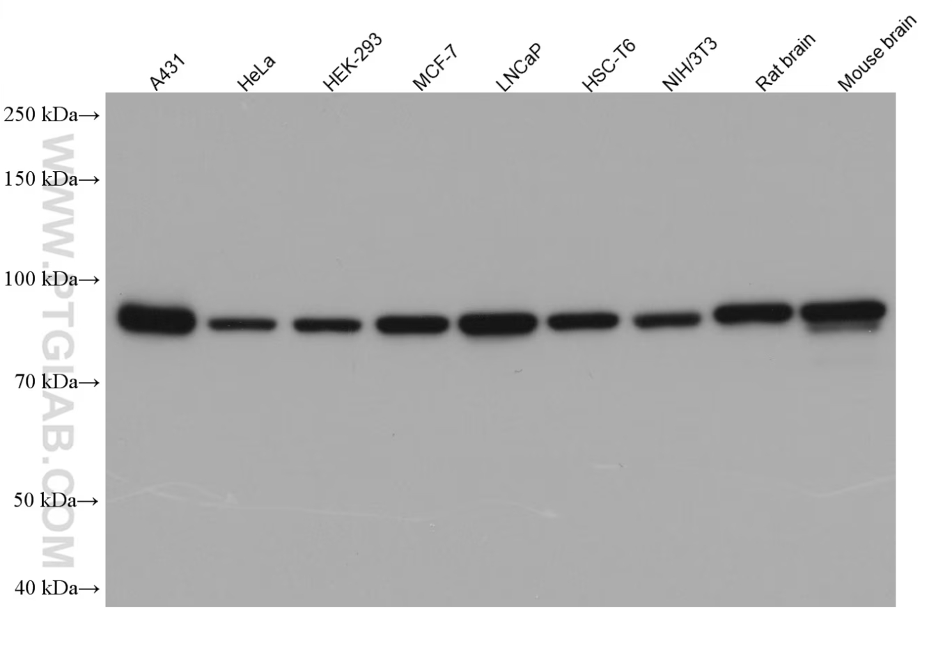
Various lysates were subjected to SDS-PAGE followed by western blot with anti-beta catenin rabbit polyclonal antibody (51067-2-AP) at a dilution of 1:50000. Multi-rAb HRP-Goat Anti-Rabbit Recombinant Secondary Antibody (H+L) (RGAR001) was used at a dilution of 1:20000 for detection.
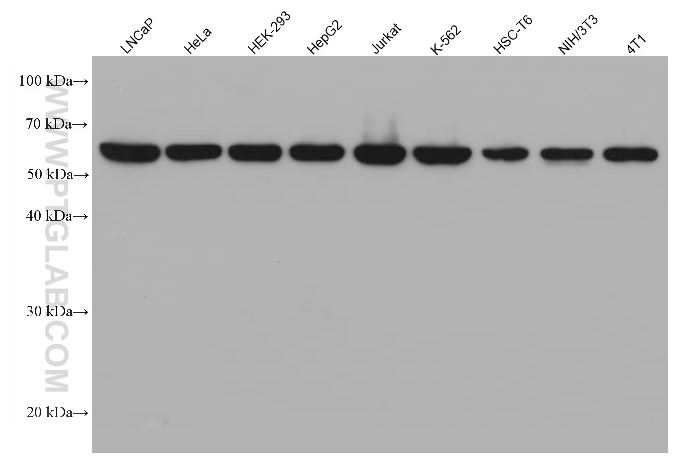
Various lysates were subjected to SDS-PAGE followed by western blot with U2AF2 mouse monoclonal antibody (68166-1-Ig) at a dilution of 1:20000. Multi-rAb HRP-Goat Anti-Mouse Recombinant Secondary Antibody (H+L) (RGAM001) was used at a dilution of 1:20000 for detection.
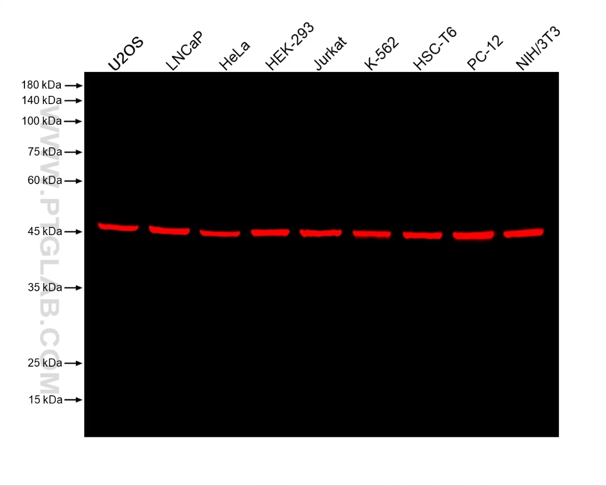
Various lysates were subjected to SDS-PAGE followed by western blot with anti-beta tubulin rabbit recombinant antibody (80713-1-RR) at a dilution of 1:20000. Multi-rAb CoraLite® Plus 750-Goat Anti-Rabbit Recombinant Secondary Antibody (H+L) (RGAR006) was used at a dilution of 1:10000 for detection.
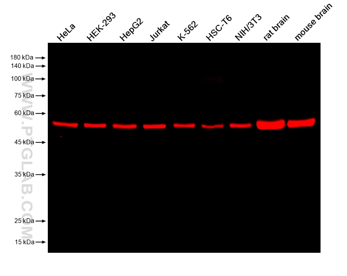
Various lysates were subjected to SDS-PAGE followed by western blot with anti-EIF3E mouse monoclonal antibody (67095-1-Ig, isotype IgG1) at a dilution of 1:50000. Multi-rAb CoraLite® Plus 750-Goat Anti-Mouse Recombinant Secondary Antibody (H+L) (RGAM006) was used at a dilution of 1:10000 for detection.
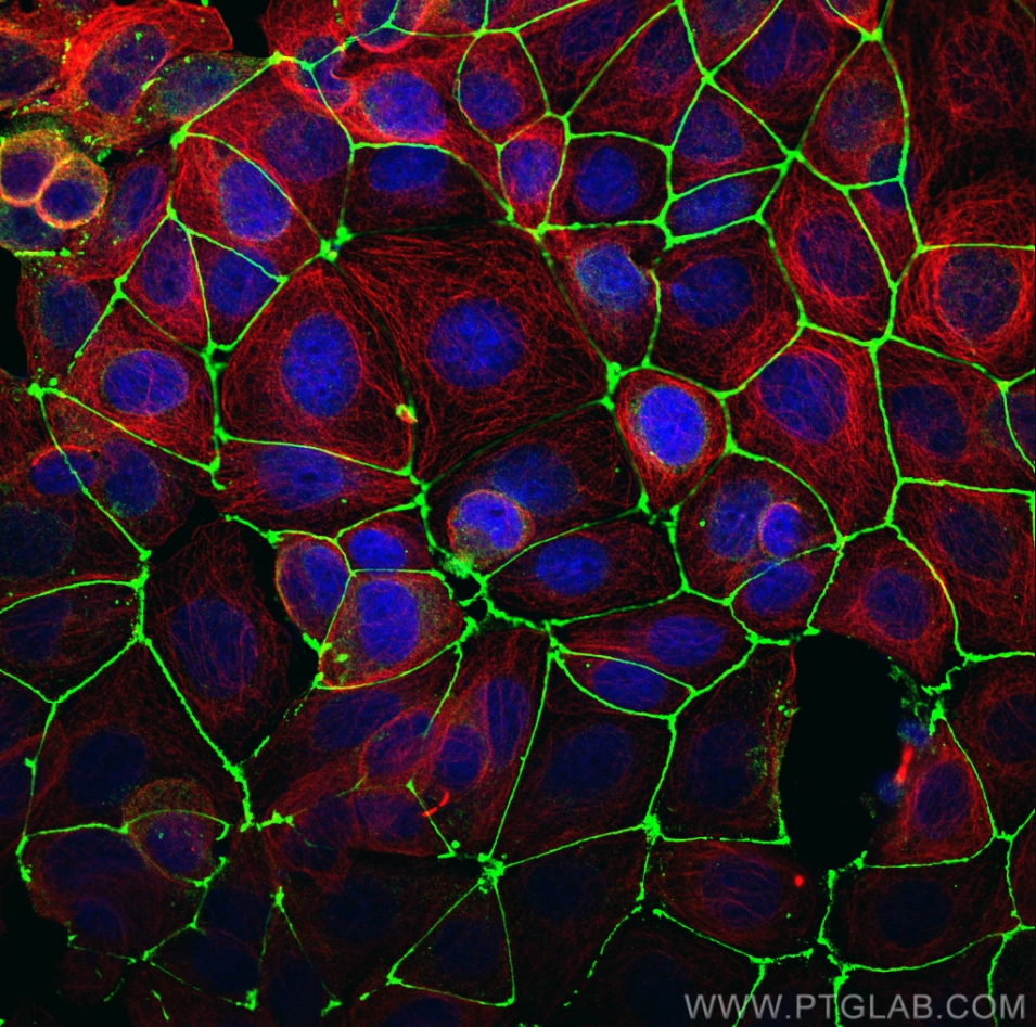
Immunofluorescence analysis of MCF-7 cells stained with rabbit anti-ZO1 polyclonal antibody (21773-1-AP, green) and mouse anti-Alpha Tubulin monoclonal antibody (66031-1-Ig, red). Multi-rAb CoraLite® Plus 488-Goat Anti-Rabbit Recombinant Secondary Antibody (H+L) (RGAR002, 1:500) and Multi-rAb CoraLite® Plus 594-Goat Anti-Mouse Recombinant Secondary Antibody (H+L) (RGAM004, 1:500) were used for detection.
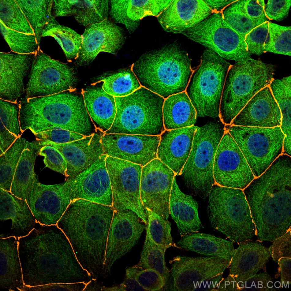
Immunofluorescence analysis of MCF-7 cells stained with rabbit anti-ZO1 polyclonal antibody (21773-1-AP, orange) and mouse anti-Alpha Tubulin monoclonal antibody (66031-1-Ig, green). Multi-rAb CoraLite® Plus 555-Goat Anti-Rabbit Recombinant Secondary Antibody (H+L) (RGAR003, 1:500) and Multi-rAb CoraLite® Plus 488-Goat Anti-Mouse Recombinant Secondary Antibody (H+L) (RGAM002, 1:500) were used for detection.
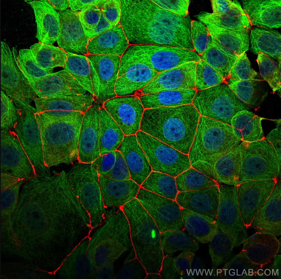
Immunofluorescence analysis of MCF-7 cells stained with rabbit anti-ZO1 polyclonal antibody (21773-1-AP, red) and mouse anti-Alpha Tubulin monoclonal antibody (66031-1-Ig, green). Multi-rAb CoraLite® Plus 594-Goat Anti-Rabbit Recombinant Secondary Antibody (H+L) (RGAR004, 1:500) and Multi-rAb CoraLite® Plus 488-Goat Anti-Mouse Recombinant Secondary Antibody (H+L) (RGAM002, 1:500) were used for detection.
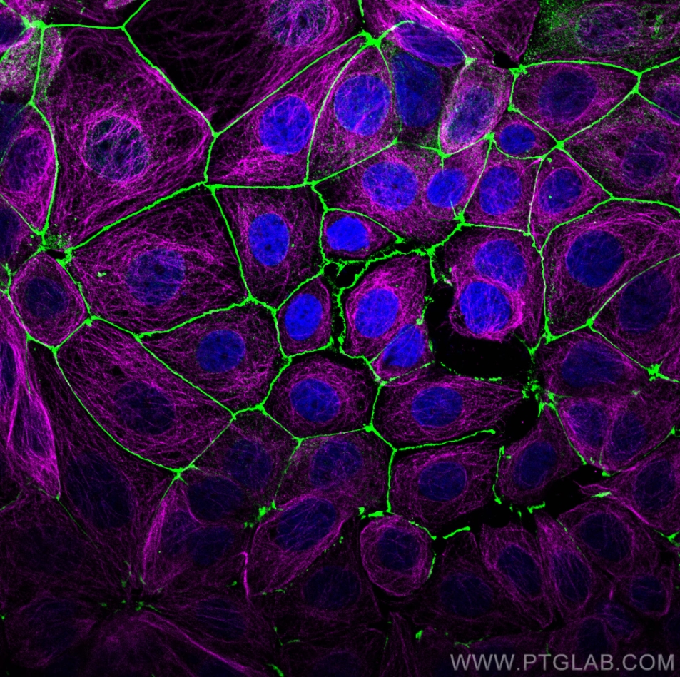
Immunofluorescence analysis of MCF-7 cells stained with rabbit anti-ZO1 polyclonal antibody (21773-1-AP, green) and mouse anti-Alpha Tubulin monoclonal antibody (66031-1-Ig, magenta). Multi-rAb CoraLite® Plus 488-Goat Anti-Rabbit Recombinant Secondary Antibody (H+L) (RGAR002, 1:500) and Multi-rAb CoraLite® Plus 647-Goat Anti-Mouse Recombinant Secondary Antibody (H+L) (RGAM005, 1:500) were used for detection.
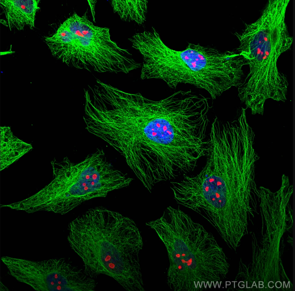
Immunofluorescence analysis of Hela cells stained with rabbit anti-Alpha Tubulin polyclonal antibody (11224-1-AP, green) and mouse anti-NPM1 monoclonal antibody (60096-1-Ig, red). Multi-rAb CoraLite® Plus 488-Goat Anti-Rabbit Recombinant Secondary Antibody (H+L) (RGAR002, 1:500) and Multi-rAb CoraLite® Plus 594-Goat Anti-Mouse Recombinant Secondary Antibody (H+L) (RGAM004, 1:500) were used for detection.
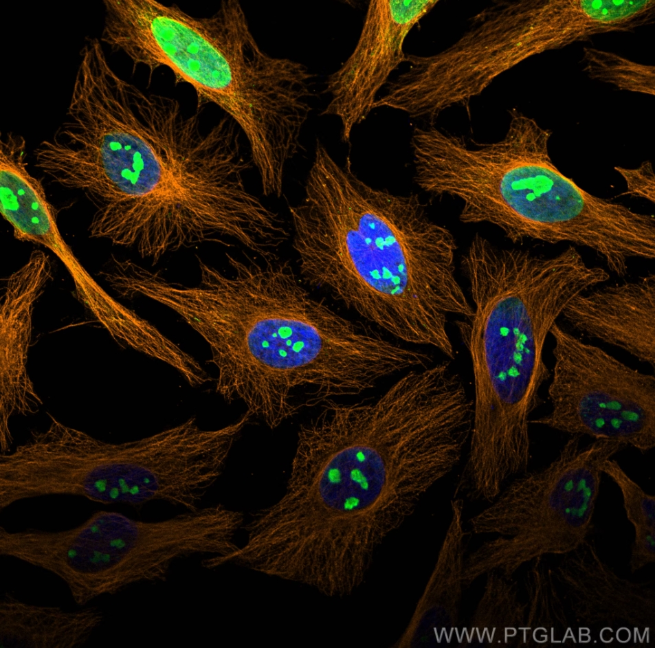
Immunofluorescence analysis of Hela cells stained with rabbit anti-Alpha Tubulin polyclonal antibody (11224-1-AP, orange) and mouse anti-NPM1 monoclonal antibody (60096-1-Ig, green). Multi-rAb CoraLite® Plus 555-Goat Anti-Rabbit Recombinant Secondary Antibody (H+L) (RGAR003, 1:500) and Multi-rAb CoraLite® Plus 488-Goat Anti-Mouse Recombinant Secondary Antibody (H+L) (RGAM002, 1:500) were used for detection.
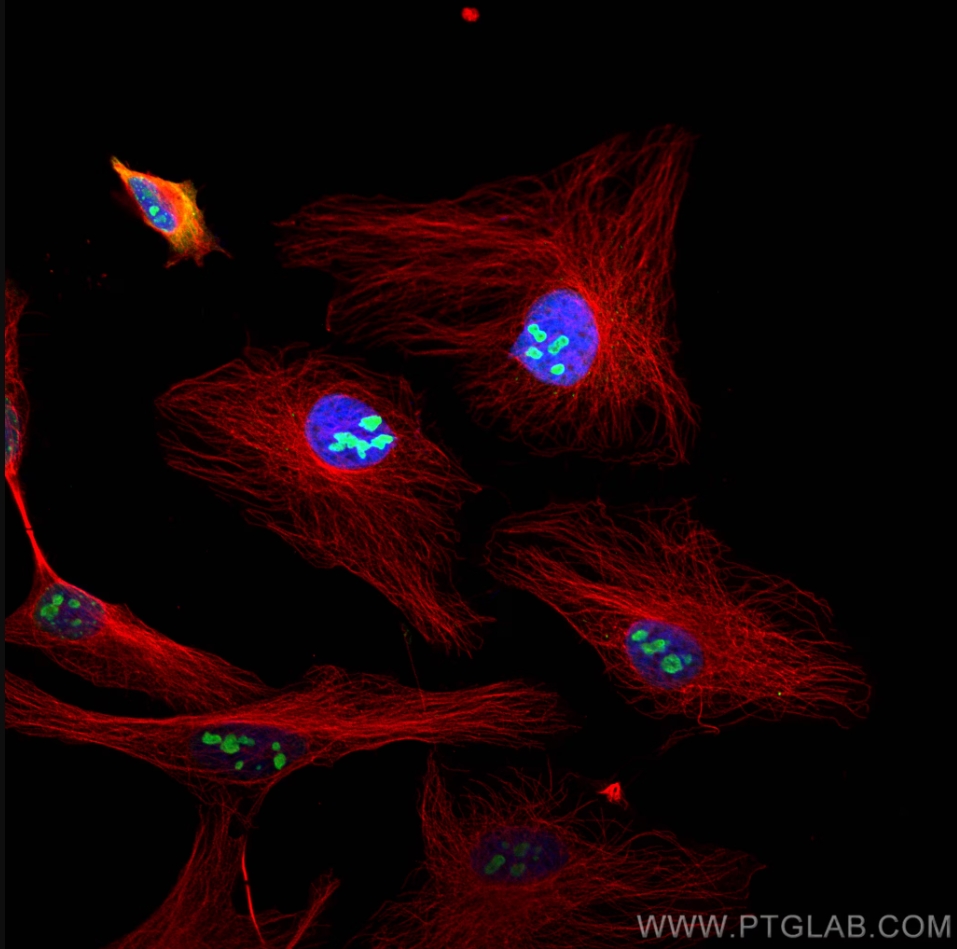
Immunofluorescence analysis of Hela cells stained with rabbit anti-Alpha Tubulin polyclonal antibody (11224-1-AP, red) and mouse anti-NPM1 monoclonal antibody (60096-1-Ig, green). Multi-rAb CoraLite® Plus 594-Goat Anti-Rabbit Recombinant Secondary Antibody (H+L) (RGAR004, 1:500) and Multi-rAb CoraLite® Plus 488-Goat Anti-Mouse Recombinant Secondary Antibody (H+L) (RGAM002, 1:500) were used for detection.
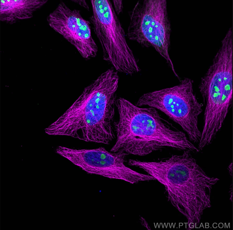
Immunofluorescence analysis of Hela cells stained with rabbit anti-Alpha Tubulin polyclonal antibody (11224-1-AP, magenta) and mouse anti-NPM1 monoclonal antibody (60096-1-Ig, green). Multi-rAb CoraLite® Plus 647-Goat Anti-Rabbit Recombinant Secondary Antibody (H+L) (RGAR005, 1:500) and Multi-rAb CoraLite® Plus 488-Goat Anti-Mouse Recombinant Secondary Antibody (H+L) (RGAM002, 1:500) were used for detection.
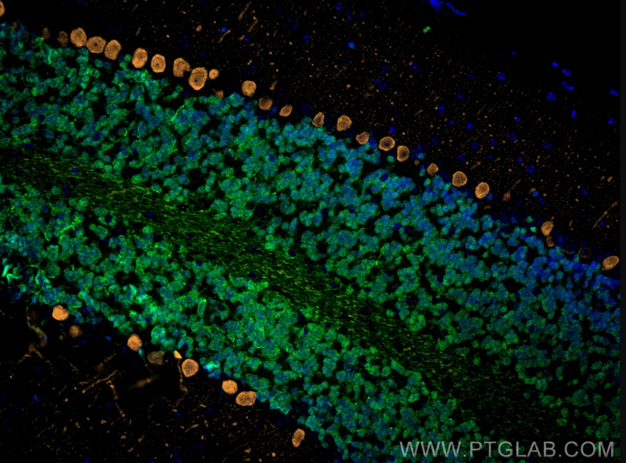
Immunofluorescence analysis of mouse cerebellum FFPE tissue stained with rabbit anti-NeuN polyclonal antibody (26975-1-AP, green) and mouse anti-Calbindin-D28k monoclonal antibody (66394-1-Ig, orange). Multi-rAb CoraLite® Plus 488-Goat Anti-Rabbit Recombinant Secondary Antibody (H+L) (RGAR002, 1:500) and Multi-rAb CoraLite® Plus 555-Goat Anti-Mouse Recombinant Secondary Antibody (H+L) (RGAM003, 1:500) were used for detection.
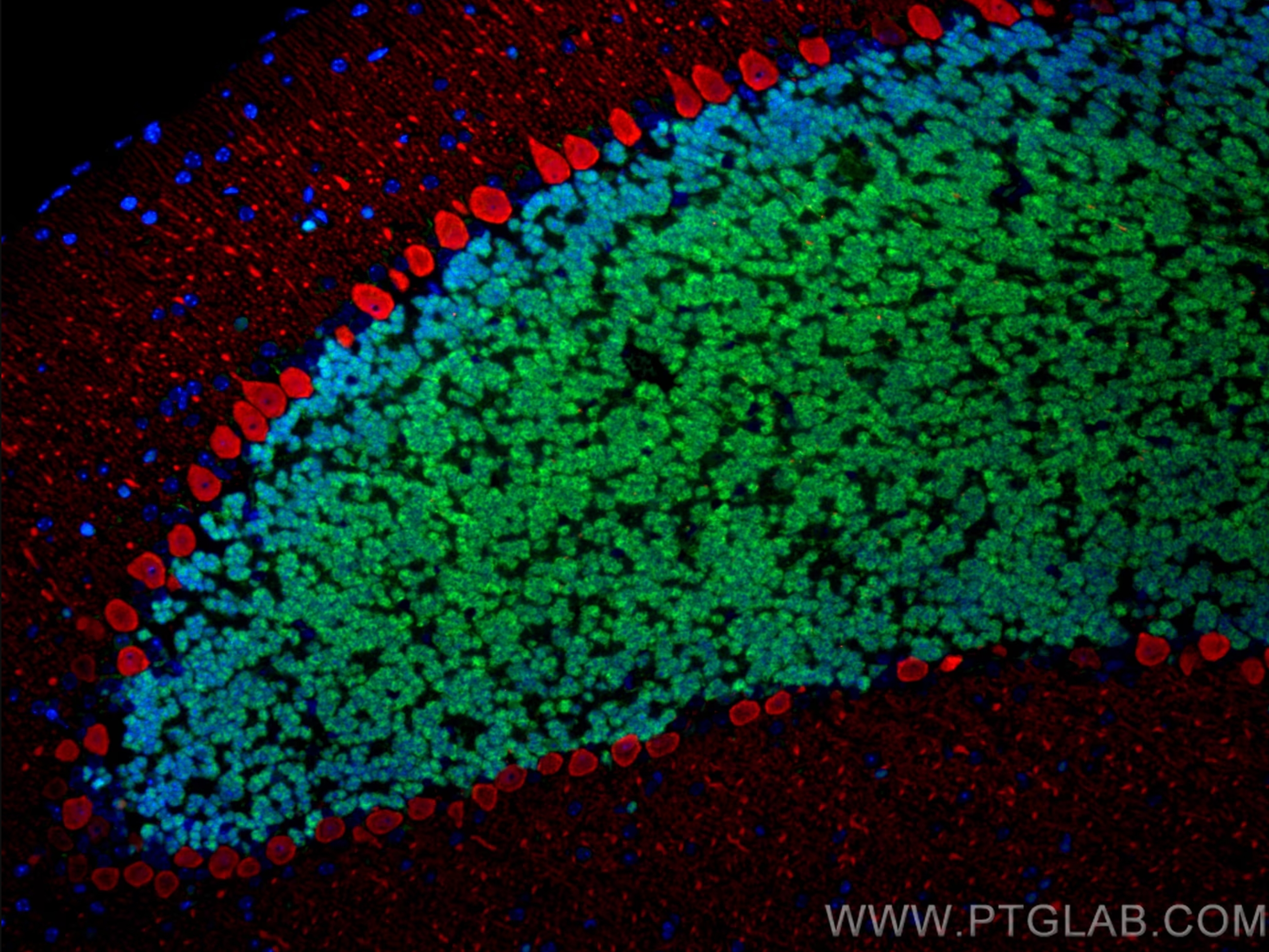
Immunofluorescence analysis of mouse cerebellum FFPE tissue stained with rabbit anti-NeuN polyclonal antibody (26975-1-AP, green) and mouse anti-Calbindin-D28k monoclonal antibody (66394-1-Ig, red). Multi-rAb CoraLite® Plus 488-Goat Anti-Rabbit Recombinant Secondary Antibody (H+L) (RGAR002, 1:500) and Multi-rAb CoraLite® Plus 594-Goat Anti-Mouse Recombinant Secondary Antibody (H+L) (RGAM004, 1:500) were used for detection.
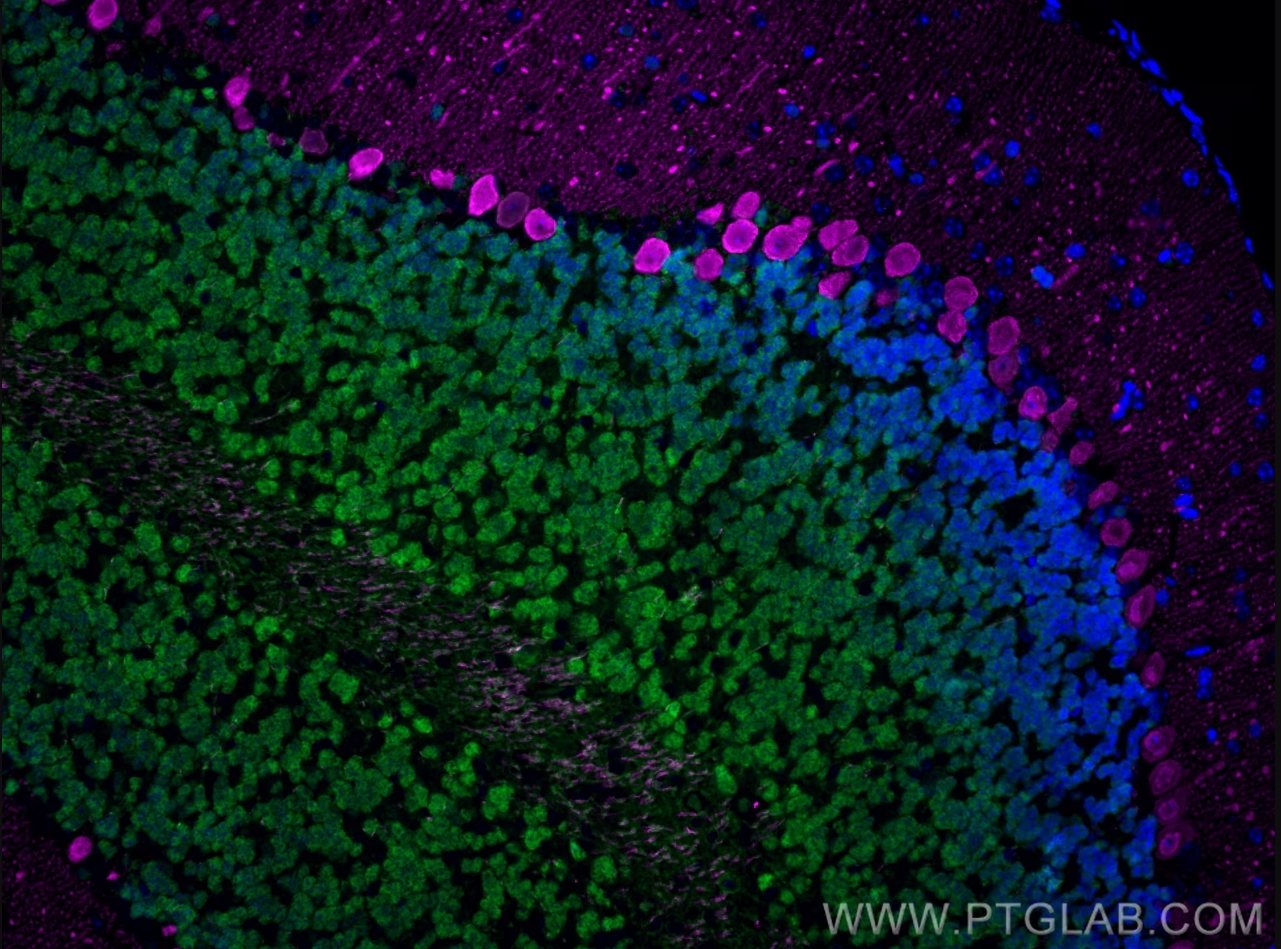
Immunofluorescence analysis of mouse cerebellum FFPE tissue stained with rabbit anti-NeuN polyclonal antibody (26975-1-AP, green) and mouse anti-Calbindin-D28k monoclonal antibody (66394-1-Ig, magenta). Multi-rAb CoraLite® Plus 488-Goat Anti-Rabbit Recombinant Secondary Antibody (H+L) (RGAR002, 1:500) and Multi-rAb CoraLite® Plus 647-Goat Anti-Mouse Recombinant Secondary Antibody (H+L) (RGAM005, 1:500) were used for detection.
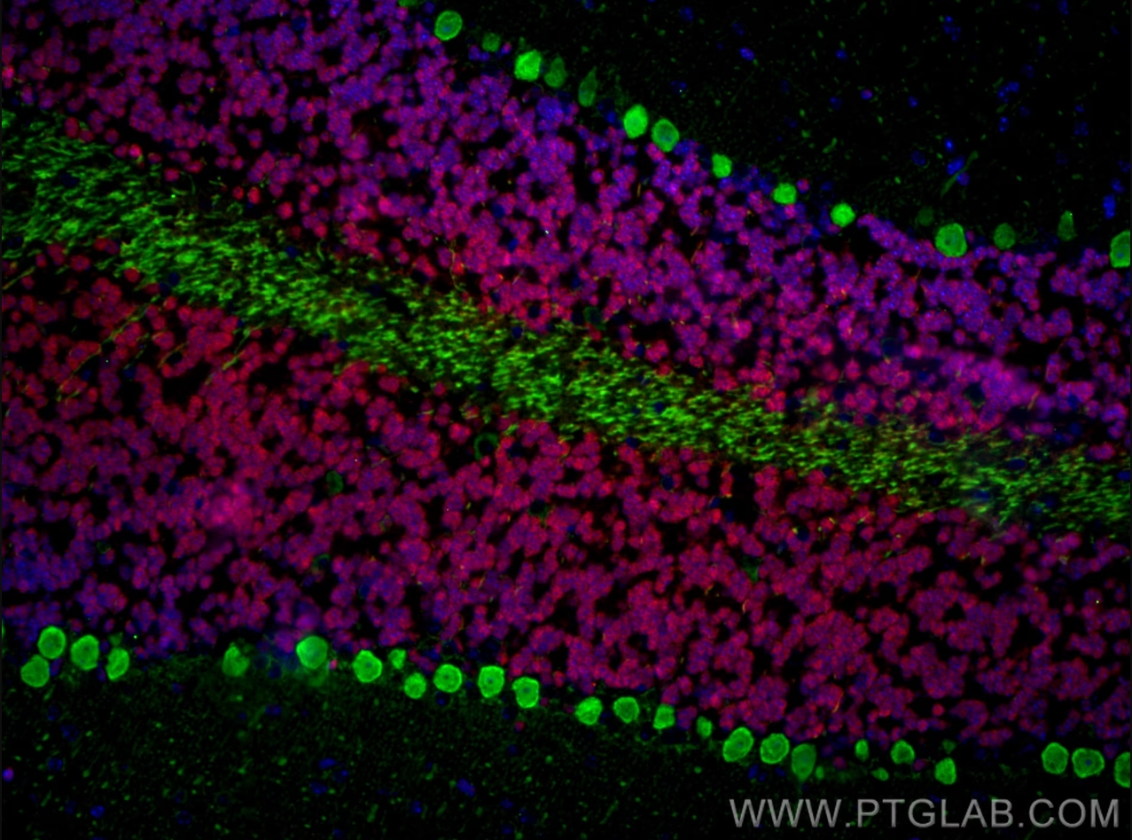
Immunofluorescence analysis of mouse cerebellum FFPE tissue stained with rabbit anti-NeuN polyclonal antibody (26975-1-AP, red) and mouse anti-Calbindin-D28k monoclonal antibody (66394-1-Ig, green). Multi-rAb CoraLite® Plus 594-Goat Anti-Rabbit Secondary Antibody (H+L) (RGAR004, 1:500) and Multi-rAb CoraLite® Plus 488-Goat Anti-Mouse Recombinant Secondary Antibody (H+L) (RGAM002, 1:500) were used for detection.
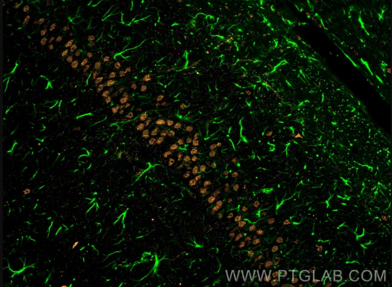
Immunofluorescence analysis of rat brain FFPE tissue stained with rabbit anti-GFAP polyclonal antibody (16825-1-AP, green) and mouse anti-NeuN monoclonal antibody (66836-1-Ig, orange). Multi-rAb CoraLite® Plus 488-Goat Anti-Rabbit Recombinant Secondary Antibody (H+L) (RGAR002, 1:500) and Multi-rAb CoraLite® Plus 555-Goat Anti-Mouse Recombinant Secondary Antibody (H+L) (RGAM003, 1:500) were used for detection.
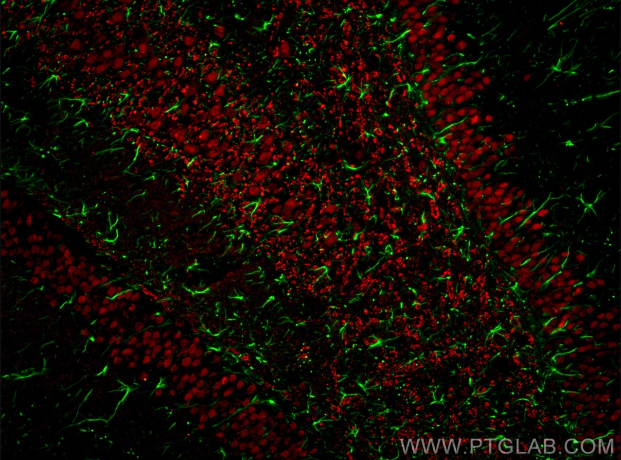
Immunofluorescence analysis of rat brain FFPE tissue stained with rabbit anti-GFAP polyclonal antibody (16825-1-AP, green) and mouse anti-NeuN monoclonal antibody (66836-1-Ig, red). Multi-rAb CoraLite® Plus 488-Goat Anti-Rabbit Recombinant Secondary Antibody (H+L) (RGAR002, 1:500) and Multi-rAb CoraLite® Plus 594-Goat Anti-Mouse Recombinant Secondary Antibody (H+L) were (RGAM004, 1:500) used for detection.
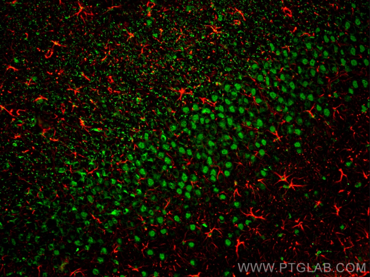
Immunofluorescence analysis of rat brain FFPE section stained with rabbit anti-GFAP polyclonal antibody (16825-1-AP, red) and mouse anti-NeuN monoclonal antibody (66836-1-Ig, green). Multi-rAb CoraLite® Plus 594-Goat Anti-Rabbit Recombinant Secondary Antibody (H+L) (RGAR004, 1:500) and Multi-rAb CoraLite® Plus 488-Goat Anti-Mouse Recombinant Secondary Antibody (H+L) (RGAM002, 1:500) were used for detection.
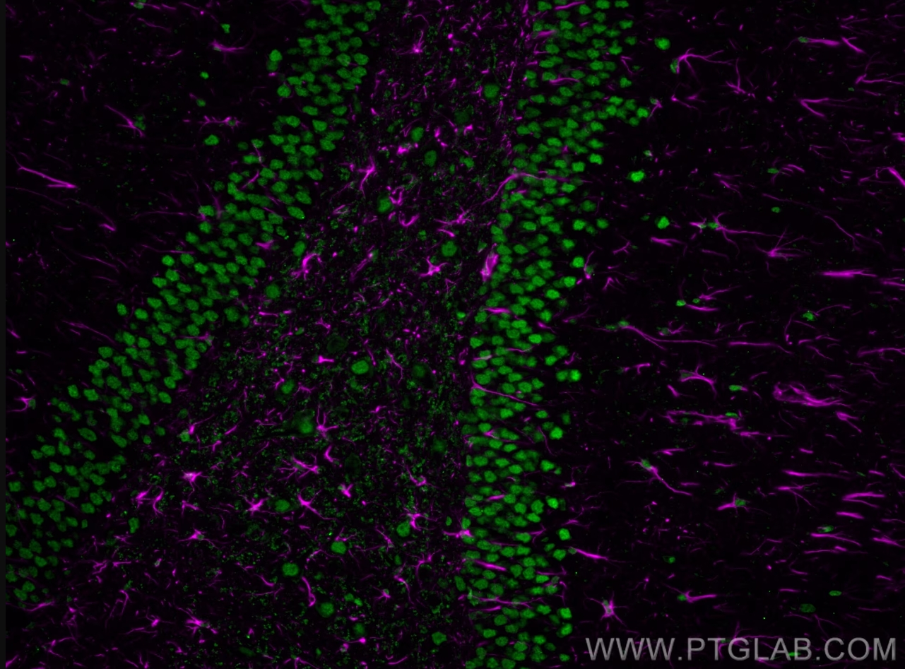
Immunofluorescence analysis of rat brain FFPE section stained with rabbit anti-GFAP polyclonal antibody (16825-1-AP, magenta) and mouse anti-NeuN monoclonal antibody (66836-1-Ig, green). Multi-rAb CoraLite® Plus 647-Goat Anti-Rabbit Secondary Antibody (H+L) (RGAR005, 1:500) and Multi-rAb CoraLite® Plus 488-Goat Anti-Mouse Recombinant Secondary Antibody (H+L) were (RGAM002, 1:500) used for detection.
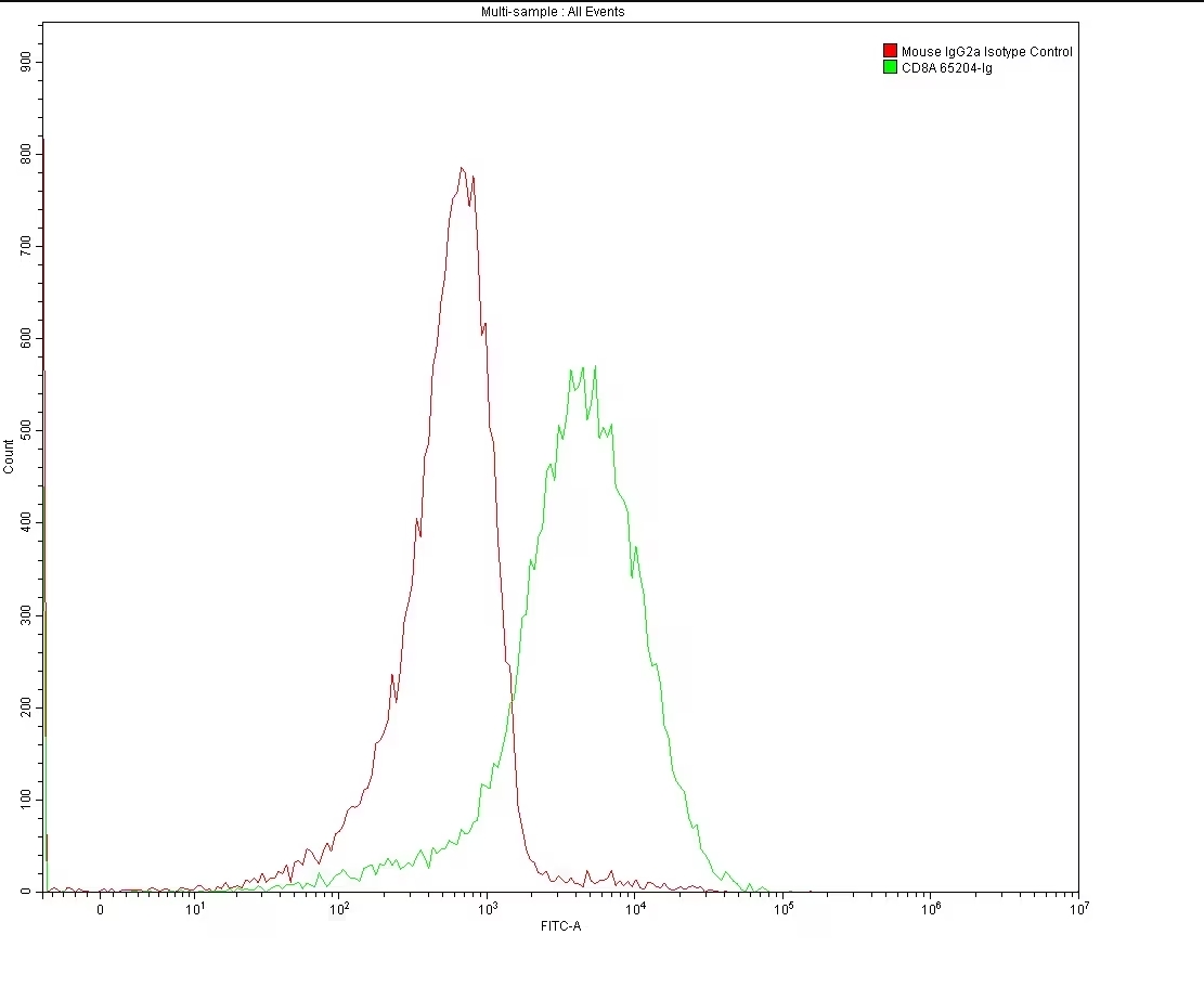
Flow cytometry analysis of 1X10^6 MOLT4 cells surface stained with 0.2 ug anti-Human CD8 antibody (65204-1-Ig, Clone: UCHT4) and mouse IgG2a isotype control antibody (66360-3-Ig). Multi-rAb CoraLite® Plus 488-Goat Anti-Mouse Recombinant Secondary Antibody (H+L) (RGAM002) was used for detection.
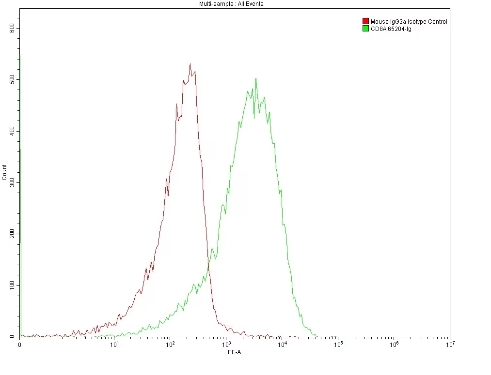
Flow cytometry analysis of 1X10^6 MOLT4 cells surface stained with 0.2 ug anti-Human CD8 antibody (65204-1-Ig, Clone: UCHT4) and mouse IgG2a isotype control antibody (66360-3-Ig). Multi-rAb CoraLite® Plus 555-Goat Anti-Mouse Recombinant Secondary Antibody (H+L) (RGAM003) was used for detection.
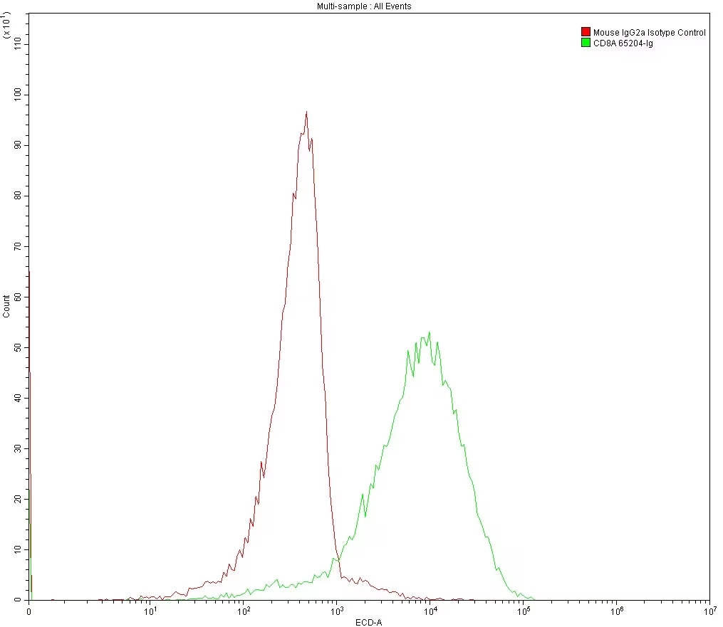
Flow cytometry analysis of 1X10^6 MOLT4 cells surface stained with 0.2 ug anti-Human CD8 antibody (65204-1-Ig, Clone: UCHT4) and mouse IgG2a isotype control antibody (66360-3-Ig). Multi-rAb CoraLite® Plus 594-Goat Anti-Mouse Recombinant Secondary Antibody (H+L) (RGAM004) was used for detection.
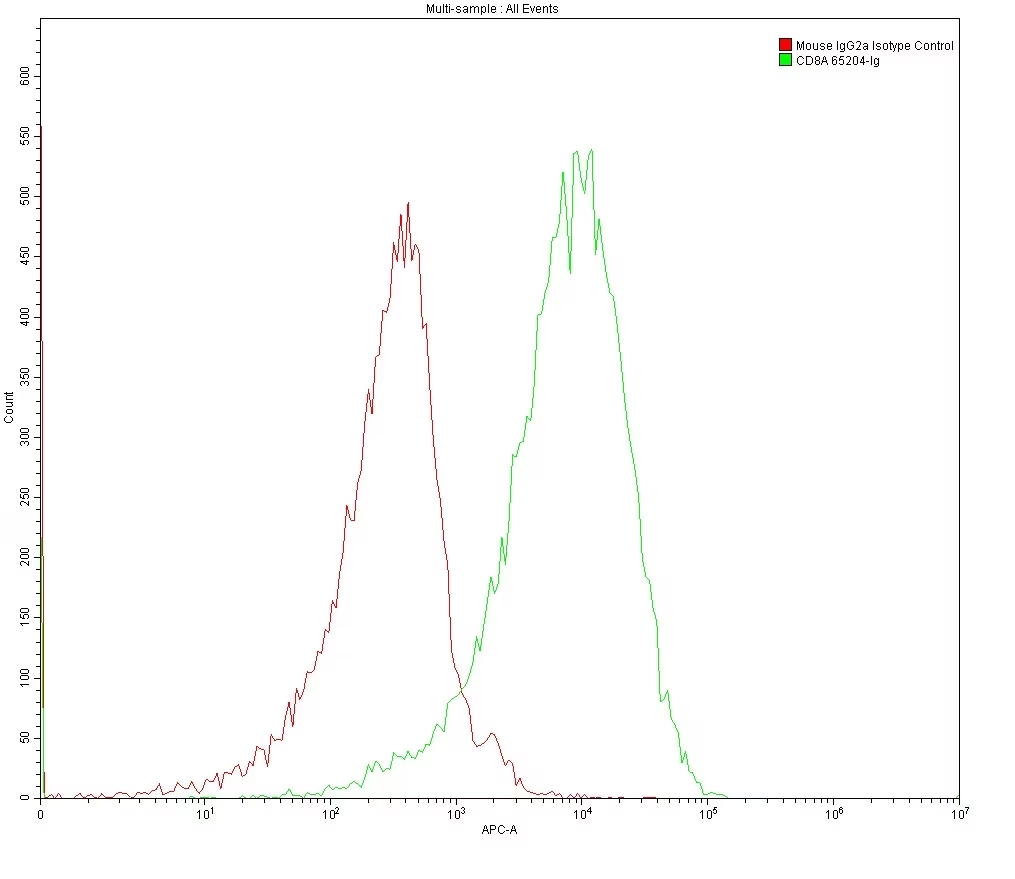
Flow cytometry analysis of 1X10^6 MOLT4 cells surface stained with 0.2 ug anti-Human CD8 antibody (65204-1-Ig, Clone: UCHT4) and mouse IgG2a isotype control antibody (66360-3-Ig). Multi-rAb CoraLite® Plus 647-Goat Anti-Mouse Recombinant Secondary Antibody (H+L) (RGAM005) was used for detection.
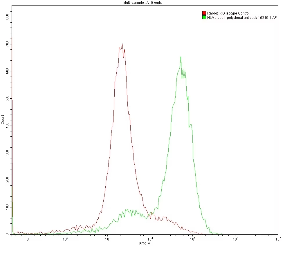
Flow cytometry analysis of 1X10^6 MOLT4 cells surface stained with 0.2 ug anti-HLA class I rabbit polyclonal antibody (15240-1-AP) and rabbit IgG isotype control antibody (30000-0-AP). Multi-rAb CoraLite® Plus 488-Goat Anti-Rabbit Recombinant Secondary Antibody (H+L) (RGAR002) was used for detection.
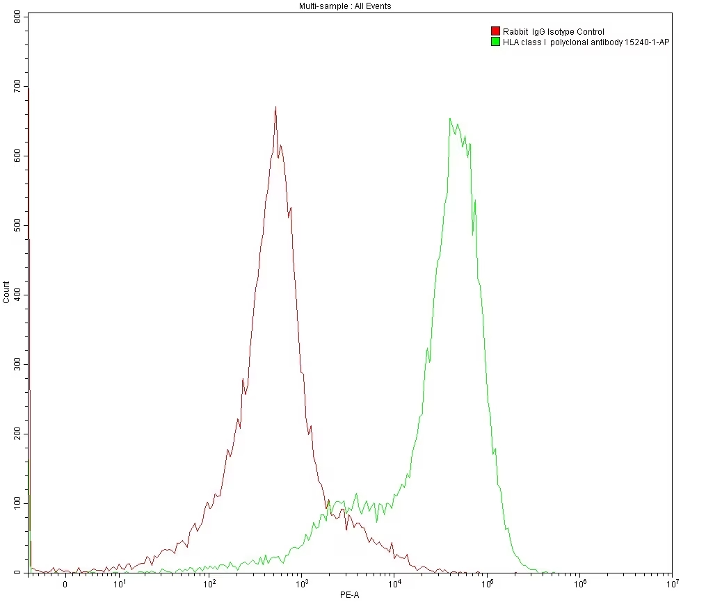
Flow cytometry analysis of 1X10^6 MOLT4 cells surface stained with 0.2 ug anti-HLA class I rabbit polyclonal antibody (15240-1-AP) and rabbit IgG isotype control antibody (30000-0-AP). Multi-rAb CoraLite® Plus 555-Goat Anti-Rabbit Recombinant Secondary Antibody (H+L) (RGAR003) was used for detection.
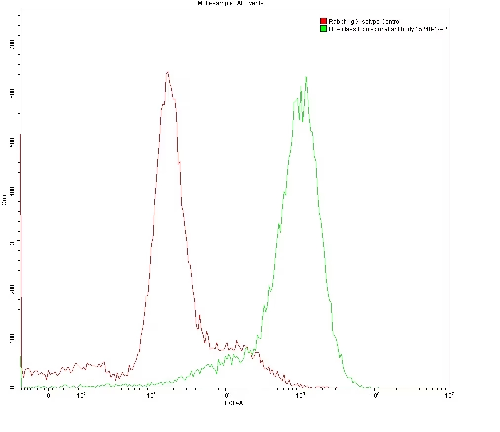
Flow cytometry analysis of 1X10^6 MOLT4 cells surface stained with 0.2 ug anti-HLA class I rabbit polyclonal antibody (15240-1-AP) and rabbit IgG isotype control antibody (30000-0-AP). Multi-rAb CoraLite® Plus 594-Goat Anti-Rabbit Recombinant Secondary Antibody (H+L) (RGAR004) was used for detection.
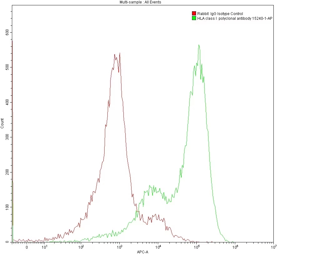
Flow cytometry analysis 1X10^6 MOLT4 cells surface stained with 0.2 ug anti-HLA class I rabbit polyclonal antibody (15240-1-AP) and rabbit IgG isotype control antibody (30000-0-AP). Multi-rAb CoraLite® Plus 647-Goat Anti-Rabbit Recombinant Secondary Antibody (H+L) (RGAR005) was used for detection.
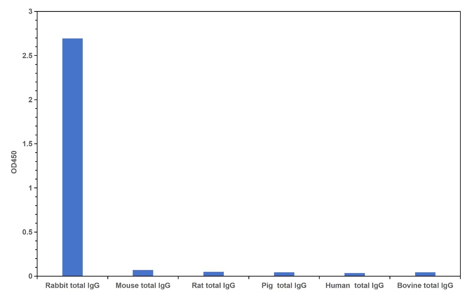
Cross reactivity test using Direct ELISA. Rabbit total IgG, Mouse total IgG, Rat total IgG, Pig total IgG, Human total IgG, Bovine total IgG were coated at 100 ng/well. 0.125 μg/mL of Multi-rAb HRP-Goat Anti-Rabbit Recombinant Secondary Antibody (H+L) (RGAR001) was used for detection. The result indicates that RGAM001 is highly specific for rabbit IgG and does not react with other species tested in the experiment.
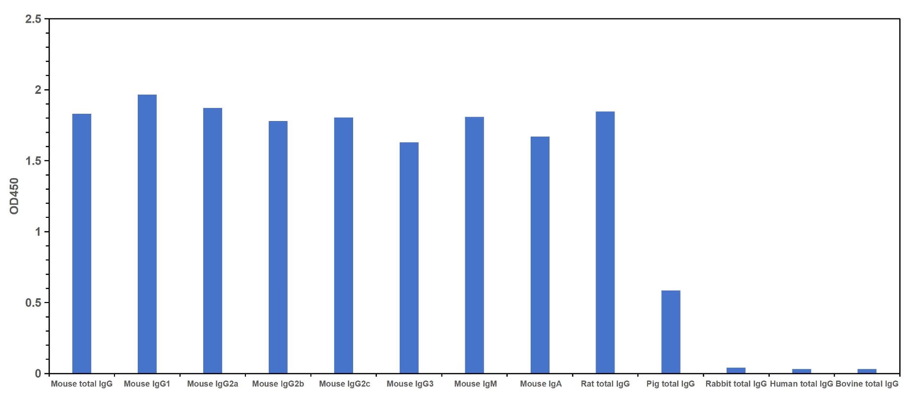
Cross reactivity test using Direct ELISA. Mouse total IgG, Mouse IgG1, IgG2a, IgG2b, IgG2c, IgG3, IgM, IgA monoclonal antibodies, Rat total IgG, Pig total IgG, Rabbit total IgG, Human total IgG, Bovine total IgG were coated at 100 ng/well. 0.125 μg/mL of Multi-rAb HRP-Goat Anti-Mouse Recombinant Secondary Antibody (H+L) (RGAM001) was used for detection. The result indicates that RGAM001 strongly binds to all Mouse IgGs, Mouse IgM and IgA as well as Rat IgG. It shows weak reactivity for pig IgG and does not react with other species tested in the experiment.
二抗的交叉反应性是由于二抗与非预期的IgG 结合。当二抗与样品中的内源性免疫球蛋白结合,或在多重分析中与脱靶抗体结合时,这可能导致高背景或非特异性信号。
Multi-rAb重组二抗是重组单克隆抗体的混合物,经过严格筛选对脱靶物种的IgG具有最小的交叉反应性。因此,使用Multi-rAb重组二抗,您可以获得与高度交叉吸附的传统多克隆二抗相同的高特异性及低背景。
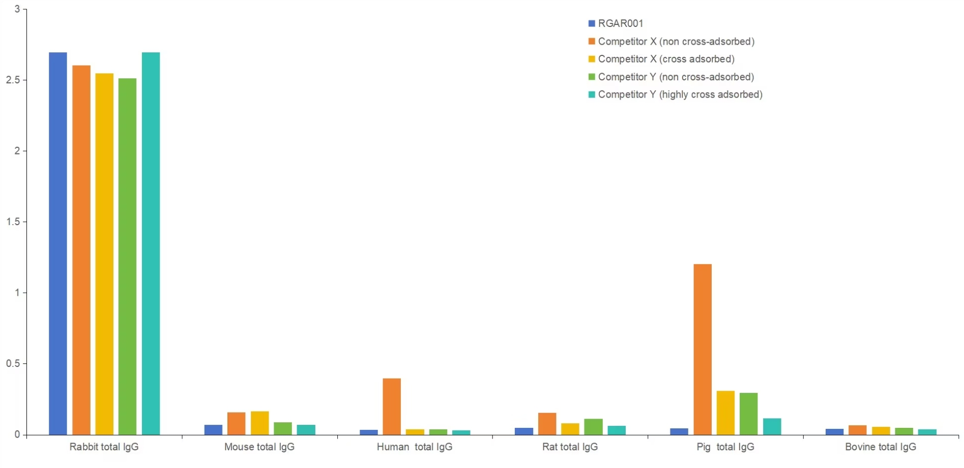
Cross reactivity comparison of RGAR001 with non-cross-adsorbed and cross-adsorbed secondary antibodies from leading competitors using Direct ELISA. Rabbit total IgG, Mouse total IgG, Human total IgG, Rat total IgG, Pig total IgG, and Bovine total IgG were coated at 100 ng/well. 0.125 μg/mL of Multi-rAb HRP-Goat Anti-Rabbit Recombinant Secondary Antibody (H+L) (RGAR001) and non-cross-adsorbed and cross-adsorbed secondary antibodies from two different competitors were used for detection.
每个批次的Multi-rAb重组二抗都是经过几轮严格筛选和验证后,选择同样高性能的重组克隆而生产的。这确保Multi-rAb重组二抗产品具有较高的批次间一致性,从而帮助您在整个项目中获得可重复的结果。
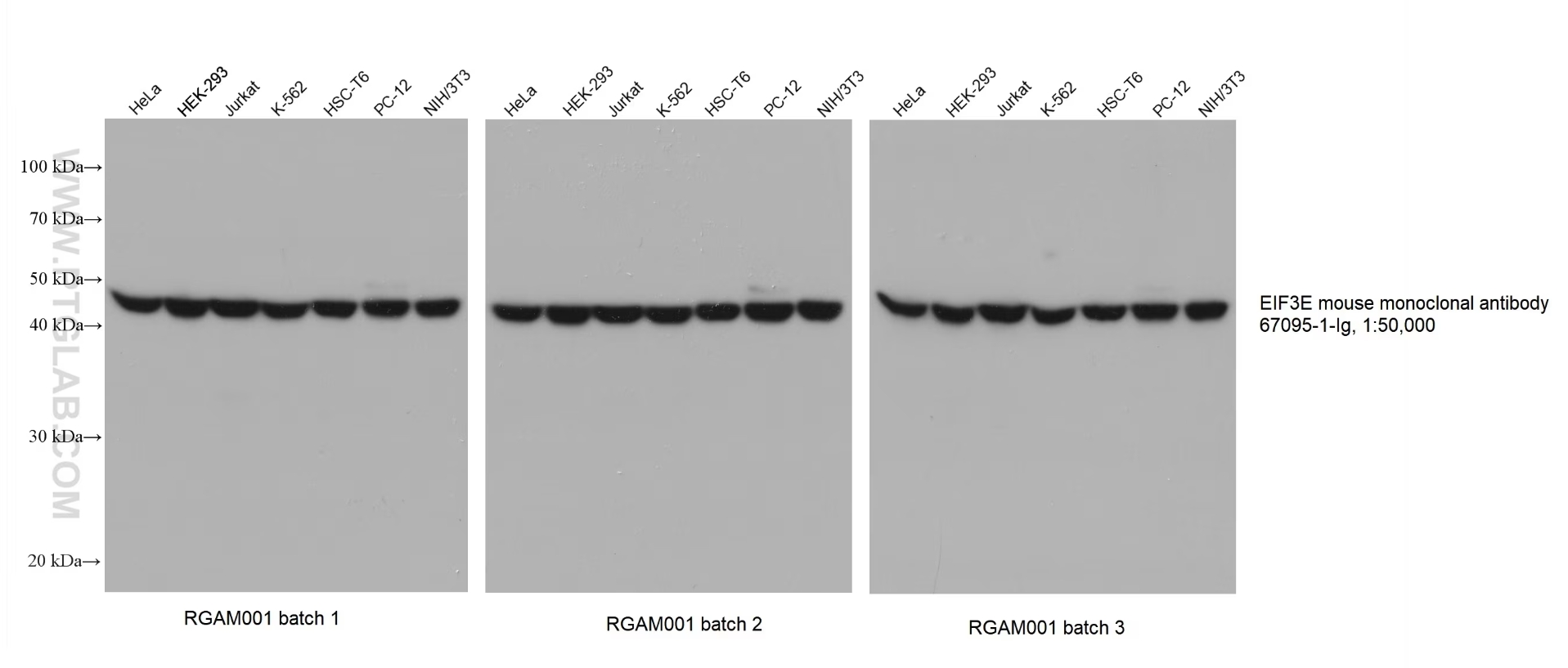
Various lysates were subjected to SDS-PAGE followed by western blot with EIF3E mouse monoclonal antibody (67095-1-Ig) at a dilution of 1:50000. Three separate batches of Multi-rAb HRP-Goat Anti-Mouse Recombinant Secondary Antibody (H+L) (RGAM001) were used at a dilution of 1:20000 for detection.
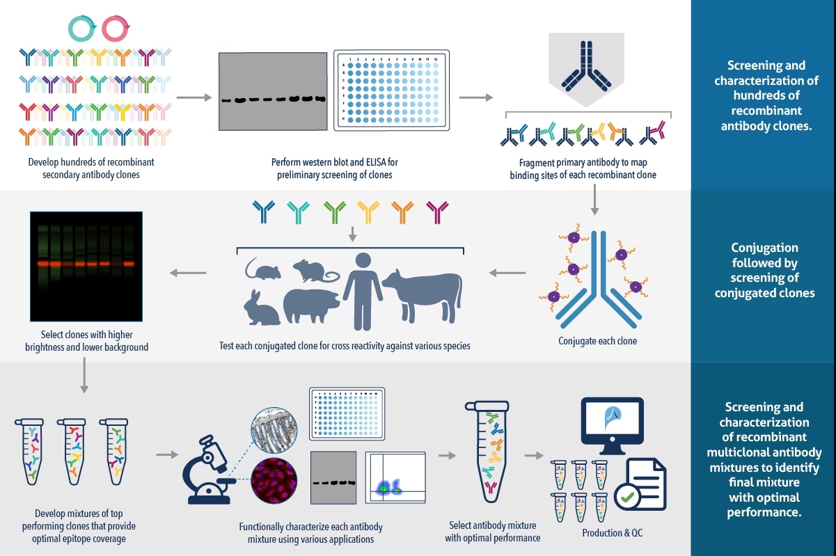
为了确保Multi-rAb重组二抗具有最佳的性能,首先将多个候选克隆与我们先进的CoraLite® Plus 染料偶联,然后分别进行筛选以选择具有最小交叉反应性、低背景和高亮度的克隆。
将选定的克隆以各种组合混合,然后在多个应用中对每个克隆进行功能验证,以选择具有最佳性能的多克隆混合物。Proteintech的CoraLite® Plus染料采用超亲水dPEG®技术开发,极大的提高了荧光亮度和光稳定性 ,点击了解CoraLite® Plus染料。

Various lysates were subjected to SDS-PAGE followed by western blot with anti-beta tubulin rabbit recombinant antibody (80713-1-RR) at a dilution of 1:20000. Multi-rAb CoraLite® Plus 750-Goat Anti-Rabbit Recombinant Secondary Antibody (H+L) (RGAR006) was used at a dilution of 1:10000 for detection.
Multi-rAb重组二抗非常适用于需要多种一抗-二抗组合的多重实验。Multi-rAb重组二抗由高性能重组克隆组成,这些克隆已被鉴定与脱靶物种的IgG交叉反应性极小,确保它们不会与先前步骤中使用的任何一抗结合
因此,Multi-rAb重组二抗可与以下产品结合使用,用于多重实验:
- 未偶联一抗和其他Multi-rAb重组二抗
- 未偶联一抗和传统多克隆二抗
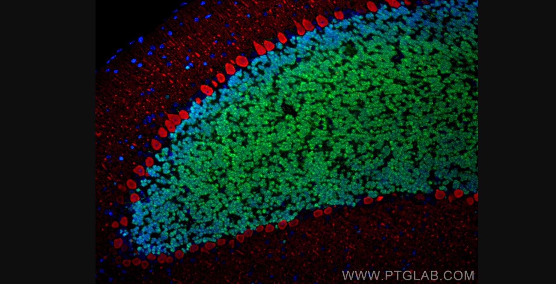
Immunofluorescence analysis of mouse cerebellum FFPE tissue stained with anti-NeuN rabbit polyclonal antibody (26975-1-AP, green) and anti-Calbindin-D28k mouse monoclonal antibody (66394-1-Ig, red). Multi-rAb CoraLite® Plus 488-Goat Anti-Rabbit Recombinant Secondary Antibody (H+L) (RGAR002, 1:500) and Multi-rAb CoraLite® Plus 594-Goat Anti-Mouse Recombinant Secondary Antibody (H+L) (RGAM004, 1:500) were used for detection.
Recent Publications
Lett Appl Microbiol
Xinnaokang improves cecal microbiota and lipid metabolism to target atherosclerosis
R Yang
Mol Med Rep
MAPK inhibitors protect against early?stage osteoarthritis by activating autophagy
Chun-Na Lan
Related
Secondary antibody selection
How to choose the best secondary antibody for your application
Non visible textCoraLite® fluorescent dye-conjugated antibodies
CoraLite dyes have equivalent brightness to the Alexa Fluor® dyes.
Non visible text
