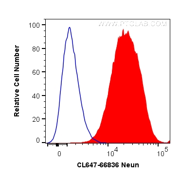
验证数据展示
经过测试的应用
| Positive FC (Intra) detected in | SH-SY5Y cells |
| Positive FC detected in | SH-SY5Y cells |
This antibody is not suitable for staining in frozen section.
推荐稀释比
| 应用 | 推荐稀释比 |
|---|---|
| Flow Cytometry (FC) (INTRA) | FC (INTRA) : 0.20 ug per 10^6 cells in a 100 µl suspension |
| Flow Cytometry (FC) | FC : 0.20 ug per 10^6 cells in a 100 µl suspension |
| It is recommended that this reagent should be titrated in each testing system to obtain optimal results. | |
| Sample-dependent, Check data in validation data gallery. | |
产品信息
CL647-66836 targets NeuN in FC (Intra) applications and shows reactivity with Human, mouse, rat samples.
| 经测试应用 | FC (Intra) Application Description |
| 经测试反应性 | Human, mouse, rat |
| 免疫原 |
CatNo: Ag28016 Product name: Recombinant human NeuN protein Source: e coli.-derived, PET28a Tag: 6*His Domain: 1-100 aa of NM_001082575 Sequence: MAQPYPPAQYPPPPQNGIPAEYAPPPPHPTQDYSGQTPVPTEHGMTLYTPAQTHPEQPGSEASTQPIAGTQTVPQTDEAAQTDSQPLHPSDPTEKQQPKR 种属同源性预测 |
| 宿主/亚型 | Mouse / IgG1 |
| 抗体类别 | Monoclonal |
| 产品类型 | Antibody |
| 全称 | hexaribonucleotide binding protein 3 |
| 别名 | cDNA FLJ56884, FLJ56884, FLJ58356, FOX3, hCG_1776007, Homo sapiens (Human), HRNBP3, NeuN, RBFOX3 |
| GenBank蛋白编号 | NM_001082575 |
| 基因名称 | NeuN |
| Gene ID (NCBI) | 146713 |
| RRID | AB_2935070 |
| 偶联类型 | CoraLite® Plus 647 Fluorescent Dye |
| 最大激发/发射波长 | 654 nm / 674 nm |
| 形式 | Liquid |
| 纯化方式 | Protein G purification |
| UNIPROT ID | A6NFN3 |
| 储存缓冲液 | PBS with 50% glycerol, 0.05% Proclin300, 0.5% BSA, pH 7.3. |
| 储存条件 | Store at -20°C. Avoid exposure to light. Stable for one year after shipment. Aliquoting is unnecessary for -20oC storage. |
背景介绍
NeuN, encoded by FOX3, is a neuron-specific nuclear protein. Anti-NeuN stains exclusively neuronal cells in the central and peripheral nervous systems, especially postmitotic and differentiating neurons, as well as terminally differentiated neurons. Anti-NeuN has been used widely as a reliable tool to detect most postmitotic neuronal cell types. The immunohistochemical staining is primarily localized in the nucleus of the neurons with lighter staining in the cytoplasm.
实验方案
| Product Specific Protocols | |
|---|---|
| FC protocol for CL Plus 647 NeuN antibody CL647-66836 | Download protocol |
| Standard Protocols | |
|---|---|
| Click here to view our Standard Protocols |


