
Proteases for Western Blotting: Choosing the right tools for the job
Proteases are often labeled as “bad guys” that complicate our Western blot (WB) analyses. They cut up target proteins into unrecognizable pieces that do not resemble the expected results.
There are two types of enzymes that can be helpful: phosphatase and glycosidase.
Phosphatase
Let’s start with phosphatase. In the study of signaling pathways, protein phosphorylation and dephosphorylation are ubiquitous. Protein phosphorylation is catalyzed by a protein kinase, and dephosphorylation is catalyzed by phosphatase.
In WB analysis of phosphorylated proteins, there will often be two close bands near the predicted molecular weight. To test whether the upper band is the phosphorylated form, phosphatase can be used. The disappearance or weakening of the upper band – but not the lower band – indicates that the upper band is a phosphorylated form of this protein.
Proteintech always works with phosphatases to verify the specificity of antibodies targeting phosphorylated proteins. An example is our RIPK1 Phospho-S161 monoclonal antibody (66854-1-IG). In the validation data below (Figure 1), the signal in several cell lysates disappeared after phosphatase treatment.
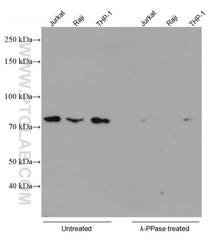 |
A word of caution here: many commercially available phosphatases are inactivated by lysis buffer. We recommend checking with the manufacturer before using them in your experiments.
Glycosidase enzymes
Next, let’s examine glycosidases. We often encounter proteins (especially membrane proteins) in WB where the observed molecular weight is much larger than the calculated value. One possible cause of this discrepancy is glycosylation. To test this hypothesis, glycosidase enzymes can be used. If the molecular weight falls to the calculated value after glycosidase treatment, the target is likely a glycoprotein.
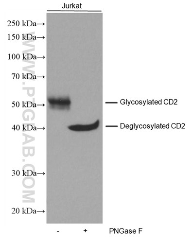
Proteintech does this routinely to validate all antibodies that target glycoproteins. Take our CD2 monoclonal antibody (60005-1-IG), for example. CD2 is composed of 351 amino acids and the calculated size is 39kDa. However, the target migrates to ~50kDa in its glycosylated form. To confirm that the deviation is due to glycosylation, we used the glycosidase PNGase F, which cleaves asparagine-linked glycosyl groups from proteins. Below are the results using Jurkat cells (Figure 2). After PNGase F treatment, the target protein migrates to near the calculated value.
 |

There are three commonly used glycoside enzymes:
- PNGase F – Cuts off almost all types of asparagine-linked carbohydrates.
- EndoH – Cuts high mannose and some hybrid types of N-linked carbohydrates.
- O-Glycosidase – Cuts off O-linked carbohydrates from serine and threonine residues.
Glycosylase experiments can require troubleshooting. Please see our FAQ below:
|
Issue |
Possible causes |
Solution |
|
No change after enzyme digestion |
1. The enzyme chosen is not compatible with the lysis buffer. 2. Insufficient enzyme amount or incubation time. 3. The detection resolution is not enough. 4. The target protein is unmodified. |
1. Ensure compatibility with enzyme manufacturer. 2. Conduct trials with varying amounts of these parameters. 3. Use an appropriate gel concentration to resolve close bands. 4. Combine with other experiments to confirm the presence or absence of modification. |
|
Putative glycosylated species weakens only slightly after digestion |
1. Insufficient enzyme amount or incubation time. 2. The target signal is produced by two proteins or two forms of the same protein. |
1. Increase the amount of enzyme used or extend the incubation time. 2. Combine other experiments to confirm the presence of other modifications or non-related protein interference. |
|
The size of the enzyme after digestion does not match the calculated value |
1. Insufficient enzyme amount or incubation time. 2. There are other modifications to the protein. |
1. Increase the amount of enzyme used or extend the enzyme cutting time. 2. Verify other modifications in conjunction with other experiments. |
Whether you are using phosphatase, glycosidase, or other proteases, each enzyme has its own place in experiments. If the design of the experiments is rational and sound, many enzymes can become a useful tool in your hands!
Here are a few of Proteintech’s enzyme experiments (Figures 3–6):
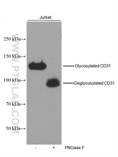 |
Figure 3. Untreated and PNGase F-treated lysates of Jurkat cells were subjected to SDS PAGE followed by WB with 66065-1-IG (CD31 antibody) at a dilution of 1:8000. |
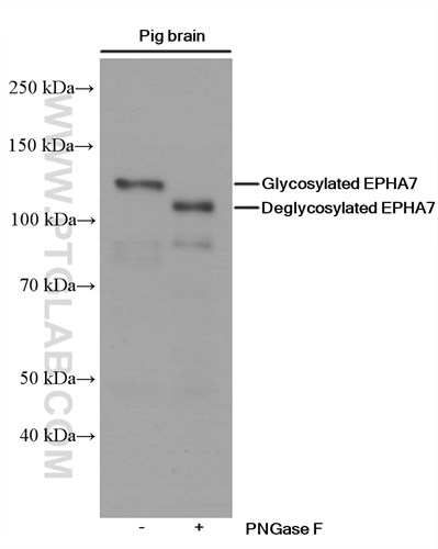 |
Figure 4. Untreated and PNGase F-treated lysates of Pig brain samples were subjected to SDS PAGE followed by WB with 66667-1-IG (EPHA7 antibody) at a dilution of 1:2000. |
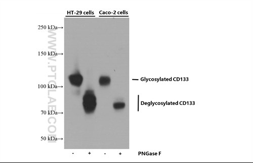 |
Figure 5. Untreated and PNGase F-treated lysates of HT-29 and CACO2 cells were subjected to SDS PAGE followed by WB with 66666-1-IG (CD133 antibody) at a dilution of 1:5000.
|
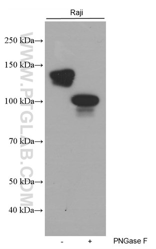 |
Figure 6. Untreated and PNGase F-treated lysates of Raji cells were subjected to SDS PAGE followed by WB with 66103-1-IG (CD22 antibody) at a dilution of 1:20000 |


