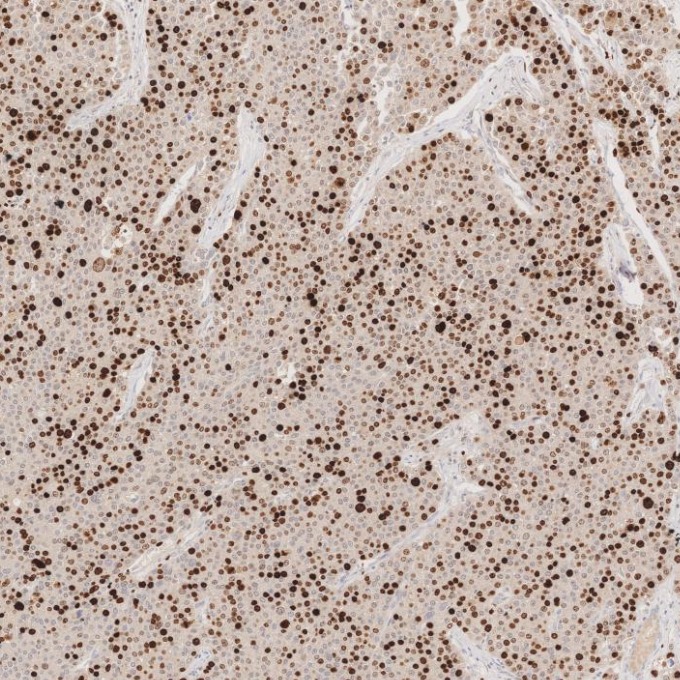
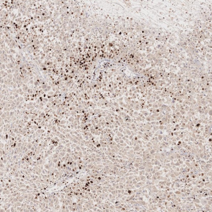
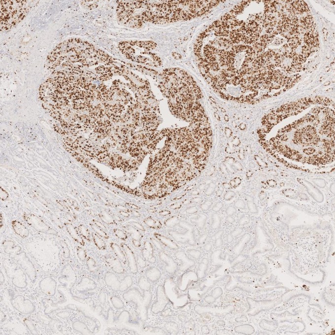
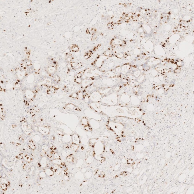
IHC staining of Cholangiocarcinoma using 66555-6-Ig (same clone as 66555-6-PBS)
Immunohistochemical analysis of paraffin-embedded Cholangiocarcinoma tissue slide using 66555-6-Ig (Ki-67 antibody) at dilution of 1:1000. Heat mediated antigen retrieval with Tris-EDTA buffer (pH 9.0). This data was developed using the same antibody clone with 66555-6-PBS in a different storage buffer formulation.
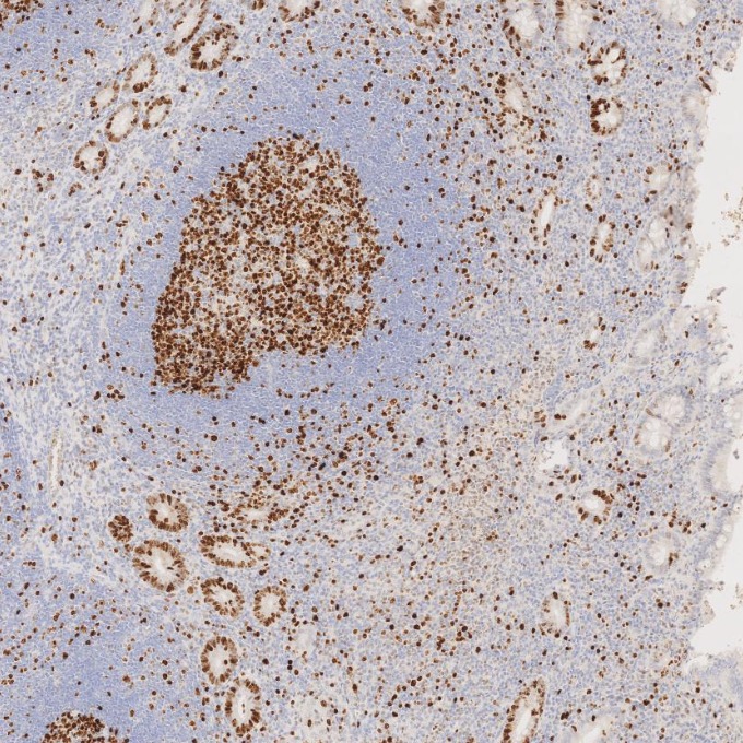
IHC staining of human appendicitis using 66555-6-Ig (same clone as 66555-6-PBS)
Immunohistochemical analysis of paraffin-embedded human appendicitis tissue slide using 66555-6-Ig (Ki-67 antibody) at dilution of 1:1000. Heat mediated antigen retrieval with Tris-EDTA buffer (pH 9.0). This data was developed using the same antibody clone with 66555-6-PBS in a different storage buffer formulation.
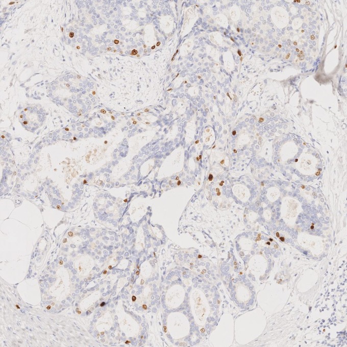
IHC staining of human breast cancer using 66555-6-Ig (same clone as 66555-6-PBS)
Immunohistochemical analysis of paraffin-embedded human breast cancer tissue slide using 66555-6-Ig (Ki-67 antibody) at dilution of 1:1000. Heat mediated antigen retrieval with Tris-EDTA buffer (pH 9.0). This data was developed using the same antibody clone with 66555-6-PBS in a different storage buffer formulation.
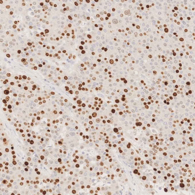
IHC staining of human liver cancer using 66555-6-Ig (same clone as 66555-6-PBS)
Immunohistochemical analysis of paraffin-embedded human liver cancer tissue slide using 66555-6-Ig (Ki-67 antibody) at dilution of 1:1000. Heat mediated antigen retrieval with Tris-EDTA buffer (pH 9.0). This data was developed using the same antibody clone with 66555-6-PBS in a different storage buffer formulation.
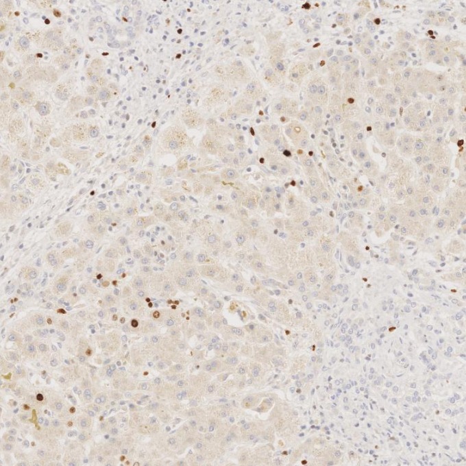
IHC staining of human liver using 66555-6-Ig (same clone as 66555-6-PBS)
Immunohistochemical analysis of paraffin-embedded human liver tissue slide using 66555-6-Ig (Ki-67 antibody) at dilution of 1:1000. Heat mediated antigen retrieval with Tris-EDTA buffer (pH 9.0). This data was developed using the same antibody clone with 66555-6-PBS in a different storage buffer formulation.
IHC staining of human ovary cancer using 66555-6-Ig (same clone as 66555-6-PBS)
Immunohistochemical analysis of paraffin-embedded human ovary cancer tissue slide using 66555-6-Ig (Ki-67 antibody) at dilution of 1:5000 (under 10x lens). Heat mediated antigen retrieval with Tris-EDTA buffer (pH 9.0). This data was developed using the same antibody clone with 66555-6-PBS in a different storage buffer formulation.
IHC staining of human ovary cancer using 66555-6-Ig (same clone as 66555-6-PBS)
Immunohistochemical analysis of paraffin-embedded human ovary cancer tissue slide using 66555-6-Ig (Ki-67 antibody) at dilution of 1:5000 (under 40x lens). Heat mediated antigen retrieval with Tris-EDTA buffer (pH 9.0). This data was developed using the same antibody clone with 66555-6-PBS in a different storage buffer formulation.
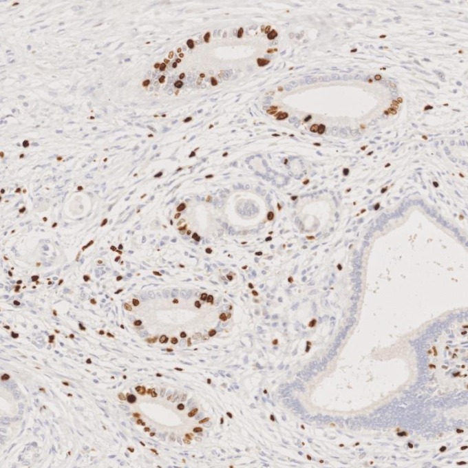
IHC staining of human pancreas cancer using 66555-6-Ig (same clone as 66555-6-PBS)
Immunohistochemical analysis of paraffin-embedded human pancreas cancer tissue slide using 66555-6-Ig (Ki-67 antibody) at dilution of 1:1000. Heat mediated antigen retrieval with Tris-EDTA buffer (pH 9.0). This data was developed using the same antibody clone with 66555-6-PBS in a different storage buffer formulation.
IHC staining of human rectal cancer using 66555-6-Ig (same clone as 66555-6-PBS)
Immunohistochemical analysis of paraffin-embedded human rectal cancer tissue slide using 66555-6-Ig (Ki-67 antibody) at dilution of 1:5000 (under 10x lens). Heat mediated antigen retrieval with Tris-EDTA buffer (pH 9.0). This data was developed using the same antibody clone with 66555-6-PBS in a different storage buffer formulation.
IHC staining of human rectal cancer using 66555-6-Ig (same clone as 66555-6-PBS)
Immunohistochemical analysis of paraffin-embedded human rectal cancer tissue slide using 66555-6-Ig (Ki-67 antibody) at dilution of 1:5000 (under 40x lens). Heat mediated antigen retrieval with Tris-EDTA buffer (pH 9.0). This data was developed using the same antibody clone with 66555-6-PBS in a different storage buffer formulation.
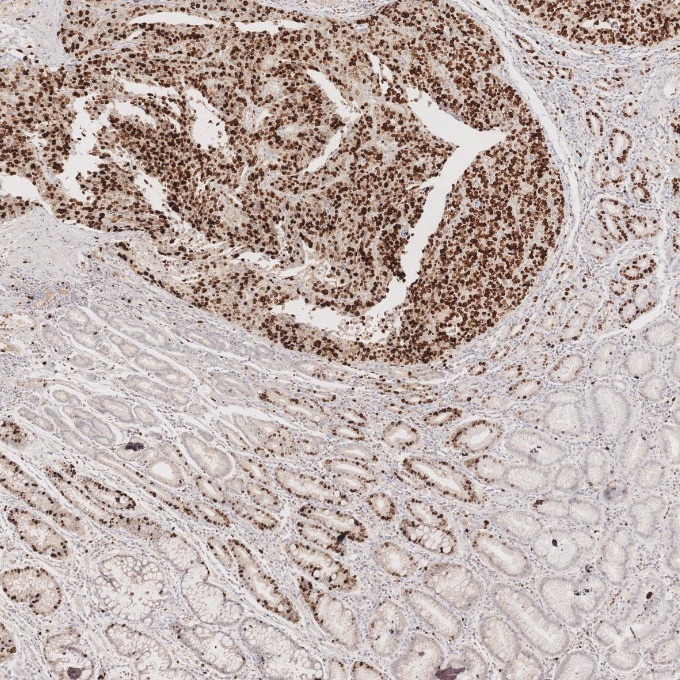
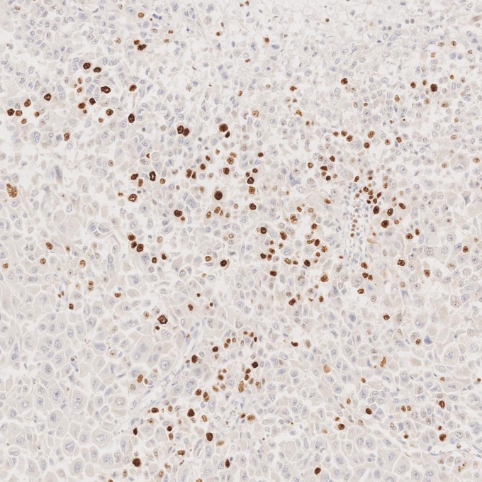
IHC staining of human stomach cancer using 66555-6-Ig (same clone as 66555-6-PBS)
Immunohistochemical analysis of paraffin-embedded human stomach cancer tissue slide using 66555-6-Ig (Ki-67 antibody) at dilution of 1:1000. Heat mediated antigen retrieval with Tris-EDTA buffer (pH 9.0). This data was developed using the same antibody clone with 66555-6-PBS in a different storage buffer formulation.
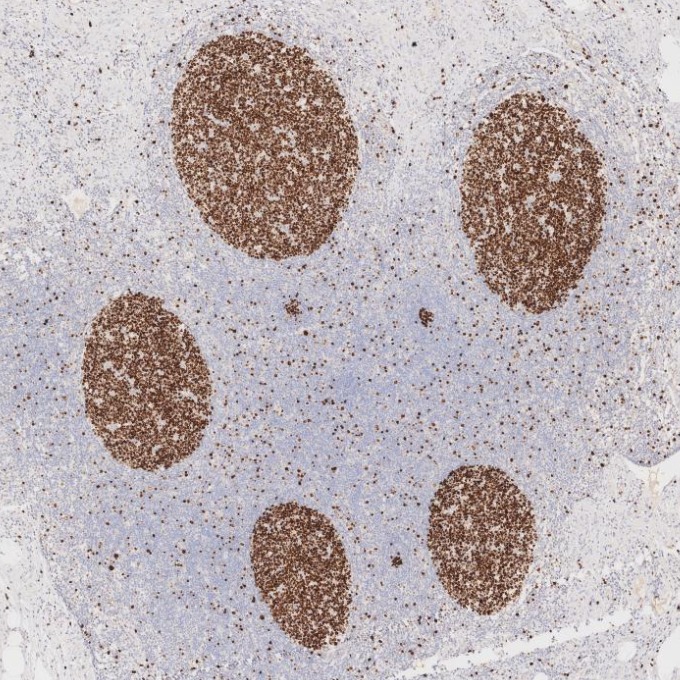
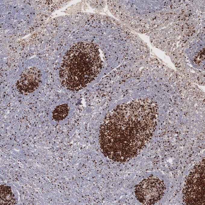
IHC staining of human tonsillitis using 66555-6-Ig (same clone as 66555-6-PBS)
Immunohistochemical analysis of paraffin-embedded human tonsillitis tissue slide using 66555-6-Ig (Ki-67 antibody) at dilution of 1:1000. Heat mediated antigen retrieval with Tris-EDTA buffer (pH 9.0). This data was developed using the same antibody clone with 66555-6-PBS in a different storage buffer formulation.
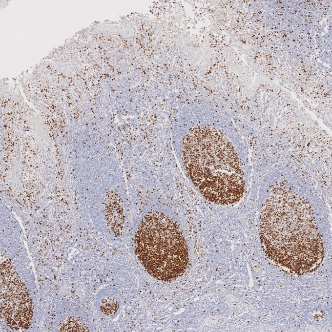
IHC staining of human tonsillitis using 66555-6-Ig (same clone as 66555-6-PBS)
Immunohistochemical analysis of paraffin-embedded human tonsillitis tissue slide using 66555-6-Ig (Ki-67 antibody) at dilution of 1:5000 (under 10x lens). Heat mediated antigen retrieval with Tris-EDTA buffer (pH 9.0). This data was developed using the same antibody clone with 66555-6-PBS in a different storage buffer formulation.
IHC staining of human tonsillitis using 66555-6-Ig (same clone as 66555-6-PBS)
Immunohistochemical analysis of paraffin-embedded human tonsillitis tissue slide using 66555-6-Ig (Ki-67 antibody) at dilution of 1:5000 (under 4x lens). Heat mediated antigen retrieval with Tris-EDTA buffer (pH 9.0). This data was developed using the same antibody clone with 66555-6-PBS in a different storage buffer formulation.
IHC-AUTOSTAINER Figures
IHC staining of human appendicitis using 66555-6-Ig (same clone as 66555-6-PBS)
Immunohistochemical analysis of paraffin-embedded human appendicitis tissue slide using 66555-6-Ig (Ki-67 antibody) at dilution of 1:1000. Heat mediated antigen retrieval with Tris-EDTA buffer (pH 9.0). The staining was performed on an autostainer. This data was developed using the same antibody clone with 66555-6-PBS in a different storage buffer formulation.
IHC staining of human liver cancer using 66555-6-Ig (same clone as 66555-6-PBS)
Immunohistochemical analysis of paraffin-embedded human liver cancer tissue slide using 66555-6-Ig (Ki-67 antibody) at dilution of 1:1000. Heat mediated antigen retrieval with Tris-EDTA buffer (pH 9.0). The staining was performed on an autostainer. This data was developed using the same antibody clone with 66555-6-PBS in a different storage buffer formulation.
IHC staining of human stomach cancer using 66555-6-Ig (same clone as 66555-6-PBS)
Immunohistochemical analysis of paraffin-embedded human stomach cancer tissue slide using 66555-6-Ig (Ki-67 antibody) at dilution of 1:1000. Heat mediated antigen retrieval with Tris-EDTA buffer (pH 9.0). The staining was performed on an autostainer. This data was developed using the same antibody clone with 66555-6-PBS in a different storage buffer formulation.
IHC staining of human tonsillitis using 66555-6-Ig (same clone as 66555-6-PBS)
Immunohistochemical analysis of paraffin-embedded human tonsillitis tissue slide using 66555-6-Ig (Ki-67 antibody) at dilution of 1:1000. Heat mediated antigen retrieval with Tris-EDTA buffer (pH 9.0). The staining was performed on an autostainer. This data was developed using the same antibody clone with 66555-6-PBS in a different storage buffer formulation.
IF-P Figures
IF Staining of human ovary cancer using 66555-6-Ig (same clone as 66555-6-PBS)
Immunofluorescent analysis of (4% PFA) fixed paraffin-embedded human ovary cancer tissue using Ki-67 antibody (66555-6-Ig, Clone: 1B9H2 ) at dilution of 1:400 and CoraLite®594-Conjugated Goat Anti-Mouse IgG(H+L) (SA00013-3). Heat mediated antigen retrieval with Tris-EDTA buffer (pH 9.0). This data was developed using the same antibody clone with 66555-6-PBS in a different storage buffer formulation.
IF/ICC Figures
IF Staining of HeLa using 66555-6-Ig (same clone as 66555-6-PBS)
Immunofluorescent analysis of (4% PFA) fixed HeLa cells using Ki-67 antibody (66555-6-Ig, Clone: 1B9H2 ) at dilution of 1:2000 and Multi-rAb CoraLite® Plus 594-Goat Anti-Mouse Recombinant Secondary Antibody (H+L) (Cat.NO. RGAM004 ). This data was developed using the same antibody clone with 66555-6-PBS in a different storage buffer formulation.
FC (INTRA) Figures
FC experiment of A431 using 66555-6-Ig (same clone as 66555-6-PBS)
1x10^6 A431 cells were intracellularly stained with 0.2 μg Ki-67 Monoclonal antibody (66555-6-Ig, Clone:1B9H2,red) and Multi-rAb CoraLite® Plus 647-Goat Anti-Mouse Recombinant Secondary Antibody (H+L)(RGAM005), Mouse IgG1 isotype control (66360-1-Ig, Clone: 1F8D3, blue) was parallel stained as control. Cells were fixed with 4% PFA. This data was developed using the same antibody clone with 66555-6-PBS in a different storage buffer formulation.
CYTOMETRIC BEAD ARRAY Figures
Cytometric bead array standard curve of MP50237-3
Cytometric bead array standard curve of MP50237-3, KI67 Monoclonal Matched Antibody Pair, PBS Only. Capture antibody: 66555-5-PBS. Detection antibody: 66555-6-PBS. Standard:Ag26266. Range: 0.098-50 ng/mL.