-
 fixed human lung cancer tissue using CoraLite®594 ICAM-1 antibody (CL594-10831) at dilution of 1:200.") ICAM-1/CD54 Polyclonal antibodies (3 total products)
ICAM-1/CD54 Polyclonal antibodies (3 total products)-_- / -_-
-
 at dilution of 1:10000 incubated at room temperature for 1.5 hours. This data was developed using the same antibody clone with 82827-1-PBS in a different storage buffer formulation.") ICAM-1/CD54 Recombinant antibodies (2E23) (2 total products)
ICAM-1/CD54 Recombinant antibodies (2E23) (2 total products)-_- / -_-
-
 fixed human lung cancer tissue using FITC ICAM-1 antibody (FITC-60299, Clone: 2F9A8 ) at dilution of 1:200.") ICAM-1 Monoclonal antibodies (2F9A8) (5 total products)
ICAM-1 Monoclonal antibodies (2F9A8) (5 total products)-_- / -_-
-
 at dilution of 1:5000 incubated at room temperature for 1.5 hours.") ICAM-1/CD54 Recombinant antibodies (230342A2) (2 total products)
ICAM-1/CD54 Recombinant antibodies (230342A2) (2 total products)-_- / -_-
-
 at dilution of 1:10000 incubated at room temperature for 1.5 hours. This data was developed using the same antibody clone with 83696-3-PBS in a different storage buffer formulation.") ICAM-1/CD54 Recombinant antibodies (240725C2) (2 total products)
ICAM-1/CD54 Recombinant antibodies (240725C2) (2 total products)-_- / -_-
-
 or CoraLite® Plus 647 Mouse IgG2a Isotype Control (C1.18.4) (CL647-65208, Clone: C1.18.4). Cells were not fixed. Lymphocytes were gated.") Anti-Human ICAM-1/CD54 (15.2) Recombinant antibodies (4 total products)
Anti-Human ICAM-1/CD54 (15.2) Recombinant antibodies (4 total products)-_- / -_-
-
 at dilution of 1:10000 incubated at room temperature for 1.5 hours.") ICAM-1/CD54 Recombinant antibodies (240249H6) (2 total products)
ICAM-1/CD54 Recombinant antibodies (240249H6) (2 total products)-_- / -_-
-
ICAM-1 Matched Antibody PairsNew
Renewable, reliable, and scalable: rabbit recombinant and mouse monoclonal antibody production.
Multiplex assay ready: PBS only formulation, ready for custom conjugation.
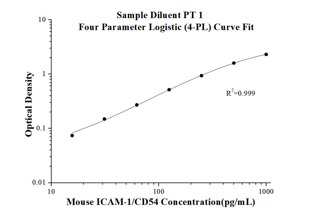
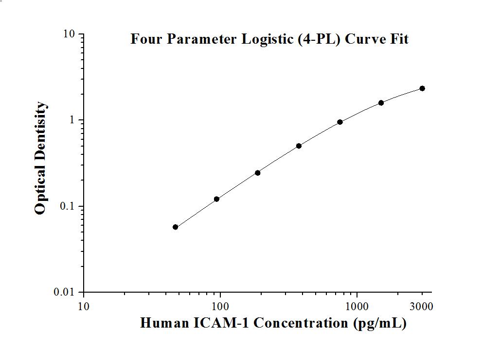
Multiple pairs per target: find the best pair for your assay.Pair Cat No. Capture antibody Detection antibody Target Species reactivity Validated Applications Linear Range MP00678-2 83696-4-PBS 83696-2-PBS ICAM-1 Rat Cytometric bead array, Sandwich ELISA sELISA: 15.6-1000 pg/mL, CBA: 0.156-20 ng/mL MP00028-1 83069-1-PBS 83069-3-PBS ICAM-1 human Cytometric bead array CBA: 0.625-20 ng/mL MP00028-2 83069-1-PBS 83069-4-PBS ICAM-1 human Cytometric bead array CBA: 0.625-20 ng/mL MP00028-3 83069-5-PBS 83069-4-PBS ICAM-1 human Cytometric bead array CBA: 0.625-20 ng/mL MP00028-4 83069-5-PBS 83069-2-PBS ICAM-1 human Sandwich ELISA sELISA: 78.1-5000 pg/mL MP00028-5 83069-1-PBS 83069-2-PBS ICAM-1 human Sandwich ELISA sELISA: 39-2500 pg/mL MP00543-1 82827-6-PBS 82827-3-PBS ICAM-1 Mouse Cytometric bead array CBA: 0.313-40 ng/mL MP00543-2 82827-7-PBS 82827-5-PBS ICAM-1 Mouse Cytometric bead array CBA: 0.313-20 ng/mL MP00543-3 82827-2-PBS 82827-4-PBS ICAM-1 Mouse Cytometric bead array CBA: 0.313-40 ng/mL MP00543-4 82827-9-PBS 82827-5-PBS ICAM-1 Mouse Sandwich ELISA sELISA: 15.6-1000 pg/mL MP00678-1 83696-3-PBS 83696-1-PBS ICAM-1 Rat Cytometric bead array CBA: 0.156-20 ng/mL MP00678-3 83696-3-PBS 83696-2-PBS ICAM-1 Rat Cytometric bead array CBA: 0.156-20 ng/mL MP50085-1 65075-1-PBS 68693-1-PBS ICAM-1 human Sandwich ELISA sELISA: 62.5-4000 pg/mL -
 (65075-1-Ig, Clone:15.2) and APC-Donkey anti-Mouse IgG at dilution 1:1000. Cells were not fixed. Monocytes were gated.") Anti-Human ICAM-1/CD54 (15.2) antibodies (13 total products)
Anti-Human ICAM-1/CD54 (15.2) antibodies (13 total products)-_- / -_-
-
 (red) or 1.00 ug isotype control antibody (blue). Cells were not fixed.") Anti-Mouse ICAM-1/CD54 (YN1/1.7.4) antibodies (5 total products)
Anti-Mouse ICAM-1/CD54 (YN1/1.7.4) antibodies (5 total products)-_- / -_-
 or FcZero-rAb™ PE Rabbit IgG Isotype Control Recombinant Antibody (PE-FcA98136, Clone: 240953C9). Cells were not fixed.")
 at dilution of 1:3000 incubated at room temperature for 1.5 hours.")
 with si-Control and si-ICAM1 transfected Raji cells. .")


 at dilution of 1:1000 incubated at room temperature for 1.5 hours.")
 at dilution of 1:4000 incubated at room temperature for 1.5 hours.")
 at dilution of 1:10000 incubated at room temperature for 1.5 hours.")
.")