Why Human Cell Expressed Proteins Are Superior for Clinical Applications

For Humans...By Human Cells

Why Human Cell Expressed Proteins Are Superior for Clinical Applications

Breakthroughs in molecular biology during the 20th century to write, edit, and erase DNA created new possibilities for medicine. A successful realization of this potential is the use of a tractable living system to make an active human protein. A notable early example was the use of E. coli by Genentech to make recombinant insulin (1).

As biology and medicine have changed, so too have the needs of recombinant proteins. For activity, many proteins require glycosylation and processing that is only available in eukaryotic systems. This led to the development of insect and Chinese hamster ovary (CHO) cells as recombinant protein producers.

As science and medicine have progressed, these systems have struggled to satisfy the growing number of needs. For example, human stem cell technology and CAR-T cell therapy require culture media whose components must be animal component and xenobiotic free, yet retain high activity and scalability.

For human applications and research, a human cell expression system is ideal. All our HumanKine® recombinant proteins are made using HEK293 cells and are perfect for the new needs of clinical research.

Here's why HumanKine® products are a cut above the rest:

1. Animal component and xenobiotic free

Products derived from or by an animal are not used at any point during production.

Benefits of animal component free and xeno free proteins:
  • The final product does not contain any constituent that is either an animal tissue, body fluid or components derived from it.

  • All materials from procurement to final products are stored and handled in dedicated animal free facility.

  • Final product and the process does not involve the use of materials from non- human animal sources or recombinant materials made from non-human animal sources.

2. Tag-free

Expressed and purified without any tags.

Benefits of tag free proteins:
  • No tags are used for expression and purification of proteins.

  • Inclusion of a tag can often result in changes to the structure of the protein of interest.

  • Sometimes a tag interferes with the active site of the protein resulting in altered biological activity.

  • Presence of a tag can increase immunogenicity of some protein, which makes a tag-free recombinant protein more desirable for in vivo applications.

3. Native folding and maturation

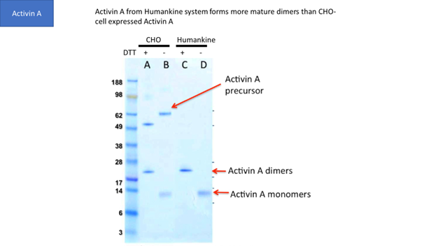
Though CHO and insect cells are eukaryotic, their ability to process human proteins does not match human cells in many cases. For example, Figure 1 shows that human cells generate more mature Activin A dimers than CHO cells.

4. Native human glycosylation

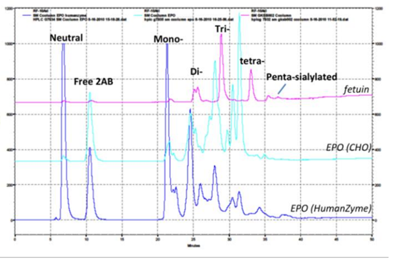
Glycosylation is crucial to stability and activity. CHO and insect cells have vastly different machinery for this process, producing glycosylated species that can be very different from humans. Figure 2 contains an example where glycosylation differs between expression systems.

5. High stability

Due to native glycosylation and maturation, human cell expressed proteins can outperform other systems in terms of stability. Figure 3 shows how HumanKine FGF has greater stability in culture than an E. coli­-derived FGF.

6. High activity

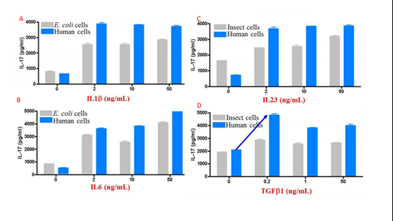
The above features synergize to create proteins that tend to have higher activity than those produced in other expression systems. Figure 4 compares activity for multiple cytokines between eukaryotic systems.

There is a common thread that unites many biotechnology stories: Discover a general principle in bacteria, then work up model organisms until ready to apply to human goals. Protein production follows this pattern and with HumanKine® products, we are finally at the stage of using human cells to make human proteins for human use.

Figure 1
SDS-PAGE gel with Coomassie blue staining of purified Activin A from CHO cell and HumanKine® systems, demonstrating the formation of mature Activin A dimers.
Figure 2
HPLC comparison of glycosylation of human cell expressed (Humanzyme) and CHO EPO, demonstrating significant differences in glycan composition. Fetuin was used as a positive control.
Figure 3
Comparison of stability of HumanKine® (FGF basic-TS, red) and E. coli-derived (FGF basic, blue) FGF incubated in cell culture at 37°C without cells.
Figure 4
Comparison of activity of insect, human, and E. coli cell expressed cytokines (A, IL1B. B, IL6. C, IL23. D, TGFB1), determined by Th17 differentiation of human CDE4+ cells.

 Related resources

  1. Proteintech’s HumanKine® Human cell-expressed cytokines and growth factors are now available in GMP-compliant versions for use in clinical trials and commercial manufacturing.

  2. New GMP laboratory to focus on HumanKine® line of human cell line-derived growth factors and cytokines for use in the pharmaceutical industry

  3. Proteintech announces an agreement to acquire HumanZyme, a leading manufacturer of recombinant human proteins

  4. Proteintech's recombinant proteins product line

  5. HumanKine® Cytokines and Growth Factors

  6. Human serum albumin’s importance in biology and biotech

  7. Recombinant human protein Activin A: An Alpha in the TGF Beta Family

  8. GM-CSF – A modulatory cytokine in autoimmunity & inflammation

  9. FGF Basic TS: Thermostable FGF does not require media changes over the weekend

 See below for a full list of  HumanKine® products:

Product Name Catalog number Activity spec. Purity Citations
Activin A HZ-1138 ≤5 ng/mL EC50 >95% 16
beta NGF HZ-1222 ≤3 ng/mL EC50 >95%  
BMP-2 HZ-1128 ≤60 ng/mL EC50 >95% 21
BMP-4 HZ-1045 ≤10 ng/mL EC50 >95% 41
BMP-7 HZ-1229 ≤100 ng/mL EC50 >95% 6
Cystatin C HZ-1211 ≤5 µM IC50 >95%  
EPO HZ-1168 ≤2.5 ng/mL EC50 >95% 3
FGF Basic TS HZ-1285 ≤0.5 ng.mL EC50 >95% 8
FGF-4 HZ-1218 ≤1.25 ng/mL EC50 >95%  
FGF-7 (KGF) HZ-1100 ≤7.5 ng/mL EC50 >95% 1
FGF-8b HZ-1103 ≤10 ng/mL EC50 >95% 1
FLT3 Ligand HZ-1151 ≤0.8 ng/mL EC50 >95% 10
G-CSF HZ-1207 ≤0.1 ng/mL EC50 >95% 6
GDNF HZ-1311 ≤ 10 ng/mL  EC50 >95%  
GM-CSF HZ-1002 ≤0.5 ng/mL EC50 >95% 10
HGF HZ-1084 ≤20 ng/mL EC50 >95% 8
HGH HZ-1007 ≤0.5 ng/mL EC50 >95% 2
HSA HZ-3001 N/A >95%  
IFN alpha 2A HZ-1066 ≤0.4 ng/mL EC50 >95% 4
IFN alpha 2B HZ-1072 ≤0.12 ng/mL EC50 >95% 2
IFN beta HZ-1298 ≤0.1 ng/mL EC50 >95%  
IFN gamma HZ-1301 ≤0.05 ng/mL EC50 >95% 3
IL-1 beta HZ-1164 ≤0.05 ng/mL EC50 >95% 16
IL-2 HZ-1015 ≤5 ng/mL EC50 >95% 5
IL-3 HZ-1074 ≤2 ng/mL EC50 >95% 10
IL-4 HZ-1004 ≤0.6 ng/mL EC50 >95% 17
IL-6 HZ-1019 ≤0.5 ng/mL EC50 >95% 19
IL-7 HZ-1281 ≤1 ng/mL EC50 >95%  
IL-9 HZ-1240 ≤1 ng/mL EC50 >95% 2
IL-10 HZ-1145 ≤1.5 ng/mL EC50 >95%  
IL-12 HZ-1256 ≤2 ng/mL EC50 >95% 3
IL-17 (IL-17A) HZ-1113 ≤2 ng/mL EC50 >95% 3
IL-17F HZ-1116 ≤10 ng/mL EC50 >95%  
IL-23 HZ-1254 ≤4 ng/mL EC50 >95% 11
IL-27 HZ-1275 ≤12 ng/mL EC50 >95% 1
IL-28A HZ-1235 ≤5 ng/mL EC50 >95% 2
IL-28B HZ-1245 ≤1 ng/mL EC50 >95%
IL-29 HZ-1156 ≤5 ng/mL EC50 >95% 2
Lefty-1 HZ-1109 ≤40 ng/mL EC50 >95% 1
LIF HZ-1292 ≤0.2 ng/mL EC50 >95%  
M-CSF HZ-1192 ≤4 ng/mL EC50 >95% 5
Noggin HZ-1118 ≤15 ng/mL EC50 >95% 5
Oncostatin M HZ-1030 ≤1 ng/mL EC50 >95%
3
PDGF-aa HZ-1215 ≤10 ng/mL EC50 >95%  
PDGFbb HZ-1308  ≤3 ng/mL EC50 >95% 1
Pleiotrophin-PTN HZ-1278 N/A >95%  
pro-IGF-II HZ-1161 ≤50 ng/mL EC50 >95% 1
SCF HZ-1024 ≤25 ng/mL EC50 >95% 9
Sonic Hedgehog (SHH) HZ-1306 ≤350 ng/mL EC50 >90%
 
TGF beta 1 HZ-1011 ≤0.5 ng/mL EC50 >95% 82
TGF beta 2 HZ-1092 ≤0.5 ng/mL EC50 >95%  4
TGF beta 3 HZ-1090 ≤0.5 ng/mL EC50 >95% 8
Thrombin HZ-3010 N/A >95% 1
TNF alpha HZ-1014 ≤0.5 ng/mL EC50 >95%  9
TPO HZ-1248 ≤5 ng/mL EC50 >95% 2
VEGF 121 HZ-1204 ≤15 ng/mL EC50 >95%  14
VEGF 165 HZ-1038 ≤5 ng/mL EC50 >95%  11
Wnt3A HZ-1296 ≤20 ng/mL EC50 >90%  1